


If you’ve never had your eyes checked, or it’s been a while, you’re probably wondering what happens during an eye test and whether there’s anything you need to prepare for. It’s a fair question. Most people picture the letter chart on the wall, but a comprehensive eye examination actually involves a series of targeted checks that assess far more than how clearly you can read line five.
An eye test screens for conditions like glaucoma, macular degeneration, cataracts, and diabetic eye disease, many of which develop without obvious symptoms in their early stages. Catching these problems early can make a real difference to your treatment options and long-term vision. At Bright Sight Eye Specialists in Randwick, Dr Rahul Dubey and Dr Capucine Odouard see firsthand how routine testing leads to earlier diagnoses and better outcomes for patients across Sydney’s Eastern Suburbs.
This guide walks you through each step of a standard eye test in Australia, from the initial history-taking right through to retinal imaging and pressure checks. By the end, you’ll know exactly what to expect, why each test matters, and when to book your next appointment.
Why regular eye tests matter
Your eyes rarely tell you when something is going wrong. Many of the most serious eye diseases develop over months or years without causing pain, blurring, or any noticeable change to your vision. By the time you notice something is off, the condition may already be at a stage where treatment is more difficult. Regular eye tests are the single most effective way to catch these problems before they cause lasting damage.
Most serious eye conditions give no early warning signs
Glaucoma is a prime example. Pressure builds inside the eye and quietly damages the optic nerve, often for years before peripheral vision starts to fade. Most people do not notice anything wrong until a significant portion of their visual field is already lost, and that loss is permanent. The same pattern applies to diabetic retinopathy, where damaged blood vessels at the back of the eye can bleed or leak without you ever sensing a change in your day-to-day sight.
Early detection through a routine eye test remains the most reliable way to protect your vision from conditions that produce no obvious symptoms until significant damage has already occurred.
Age-related macular degeneration follows a similar path. The central part of your vision can deteriorate gradually enough that your brain compensates without you realising it. By the time you notice that faces or fine print look distorted, the macula may already be significantly affected. Catching these changes early, through retinal imaging and other checks that are part of what happens during an eye test, gives your specialist the chance to slow or halt the disease before it reaches that point.
A routine check can detect more than just vision problems
Your eyes offer a direct, unobstructed view of blood vessels, nerve tissue, and the retina, which makes an eye examination a surprisingly useful screening tool for systemic health conditions. Signs of high blood pressure and diabetes are often visible in the retinal blood vessels before a person receives a formal diagnosis through any other channel.
Certain autoimmune conditions, thyroid disease, and some neurological changes can also show up during a thorough eye examination. Your optometrist or ophthalmologist looks at structural details that reflect your overall health, which means a routine visit can sometimes prompt follow-up with your GP for reasons entirely unrelated to your glasses prescription.
How often you should get tested in Australia
The right testing frequency depends on your age, family history, and any existing health conditions. As a general guide, adults with no known eye problems should aim for a full eye examination every two years. Once you pass 65, annual checks become more appropriate because your risk of cataracts, glaucoma, and macular degeneration all increases with age.
People with diabetes, a family history of glaucoma, or other risk factors may need to be seen more frequently than the standard two-year guideline. Your optometrist or specialist will advise you on the right interval for your individual situation. If you wear glasses or contact lenses, updating your prescription at regular intervals also helps you avoid unnecessary eye strain and persistent headaches that can quietly affect your daily quality of life.
What to do before your appointment
A little preparation before your visit makes a real difference. Knowing what information to bring and what to expect helps your optometrist or ophthalmologist use your appointment time efficiently, and it gives you the chance to get answers to questions you’ve been sitting on. Understanding what happens during an eye test starts before you walk through the door.
Gather your health and medication history
Your eye health does not sit in isolation from the rest of your body. Conditions like diabetes, high blood pressure, and autoimmune disease all have direct implications for your eyes, so your practitioner needs to know about them. Write down any diagnosed medical conditions, any medications you’re currently taking including eye drops, vitamins, and supplements, and whether anyone in your immediate family has been treated for glaucoma, macular degeneration, or retinal disease.
A family history of glaucoma significantly raises your personal risk, so telling your practitioner upfront means they can focus additional attention on your optic nerve and eye pressure readings during the examination.
Bring the right items on the day
Turning up with the right things means your appointment runs smoothly and nothing important gets missed. Your current glasses or contact lenses should come with you, along with any previous prescriptions you have on file. If you wear contact lenses, ask your practitioner whether to remove them before your appointment, as some tests produce more accurate readings without lenses in place.
You should also bring your Medicare card and any private health insurance details, as these directly affect your out-of-pocket costs. If you’ve had eye tests or surgery elsewhere, bringing those records gives your practitioner a useful baseline for comparison, particularly if they are monitoring a long-standing condition.
Prepare a few questions in advance
A busy appointment moves quickly, and it’s easy to forget what you wanted to ask once you’re in the room. Write down any symptoms you’ve noticed, no matter how minor they seem, such as occasional blurring, light sensitivity, floaters, or difficulty driving at night. These details help your practitioner direct the examination and decide which additional tests are worth running.
Think about what outcomes matter most to you as well. Whether you want to reduce your dependence on glasses or simply confirm your retina is healthy, sharing your personal goals with your specialist helps them tailor their recommendations to your situation rather than giving you a generic result at the end.
What happens during an eye test step by step
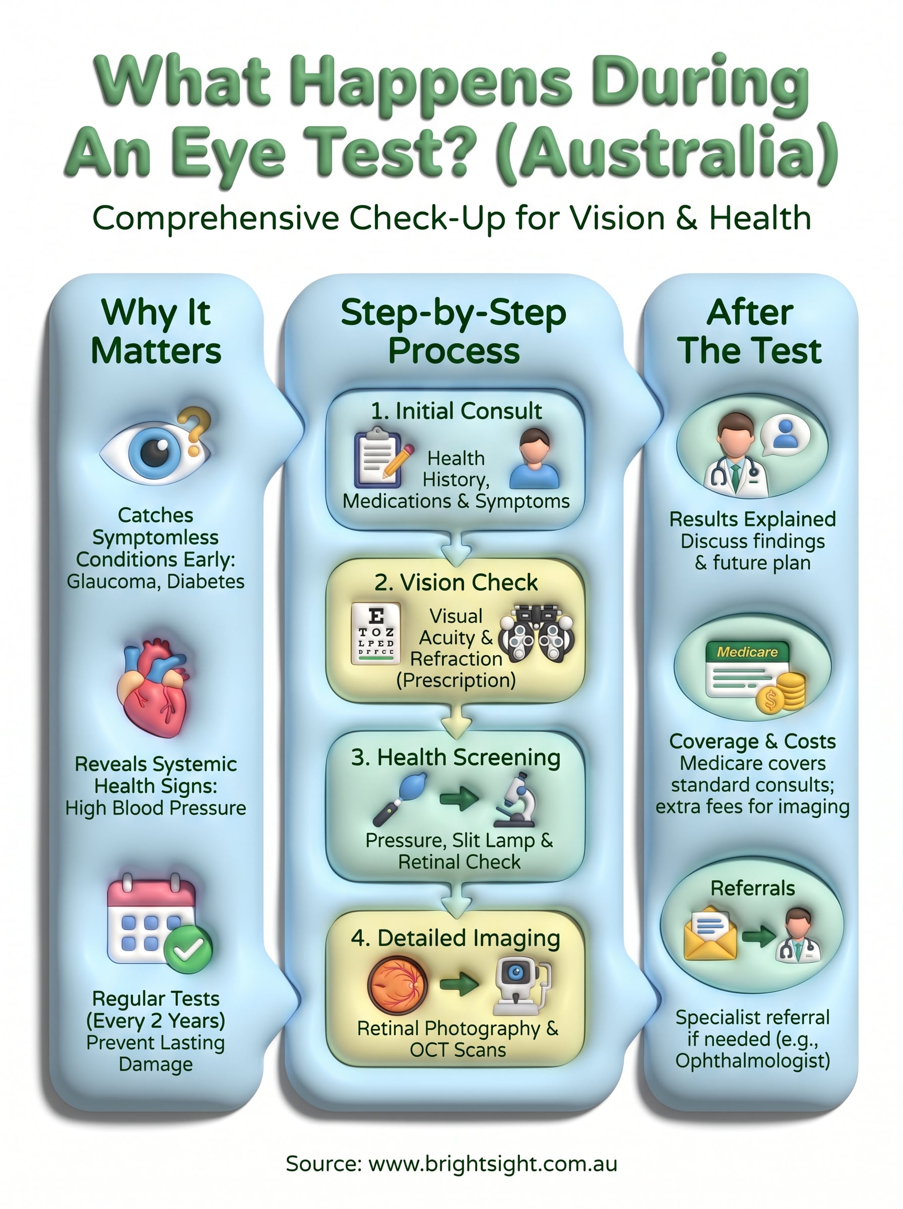
Understanding what happens during an eye test in sequence helps you feel more comfortable during the appointment and makes it easier to follow along when your practitioner explains their findings. The process is straightforward, moves at a steady pace, and involves no pain or invasive procedures. Each step builds on the one before it, so your practitioner assembles a complete picture of your eye health by the end of the session.
Your health history and initial questions
Your practitioner starts by asking about your current vision, any symptoms you’ve noticed, and your broader health history. These answers directly shape which tests get prioritised during the rest of your appointment, so be thorough even if a symptom seems trivial. Common topics covered include:
- Medications and supplements you currently take
- Family history of glaucoma, macular degeneration, or retinal disease
- Any recent changes to your vision such as flashes, floaters, or difficulty at night
- Previous eye conditions, surgery, or injuries
Visual acuity and refraction
Next comes the part most people recognise: reading letters off a chart at a set distance, usually six metres or its equivalent. This measures how sharp your central vision is in each eye separately. Your practitioner then moves into refraction, using a phoropter or a set of trial lenses to narrow down the exact prescription your eyes need. You’ll be asked to compare options, such as "which is clearer, one or two?" until your practitioner locks in the most accurate correction.
The refraction step isn’t just about updating your glasses prescription; it also identifies conditions like astigmatism, short-sightedness, and long-sightedness that may explain persistent headaches or eye strain.
Eye pressure, slit lamp, and retinal check
Measuring intraocular pressure comes next, using a tonometer that either delivers a small puff of air or applies gentle contact after numbing drops. Elevated pressure is a key risk factor for glaucoma. The slit lamp examination then lets your practitioner inspect the front of the eye, including the cornea, iris, and lens, using a high-intensity beam of light that reveals fine structural detail.
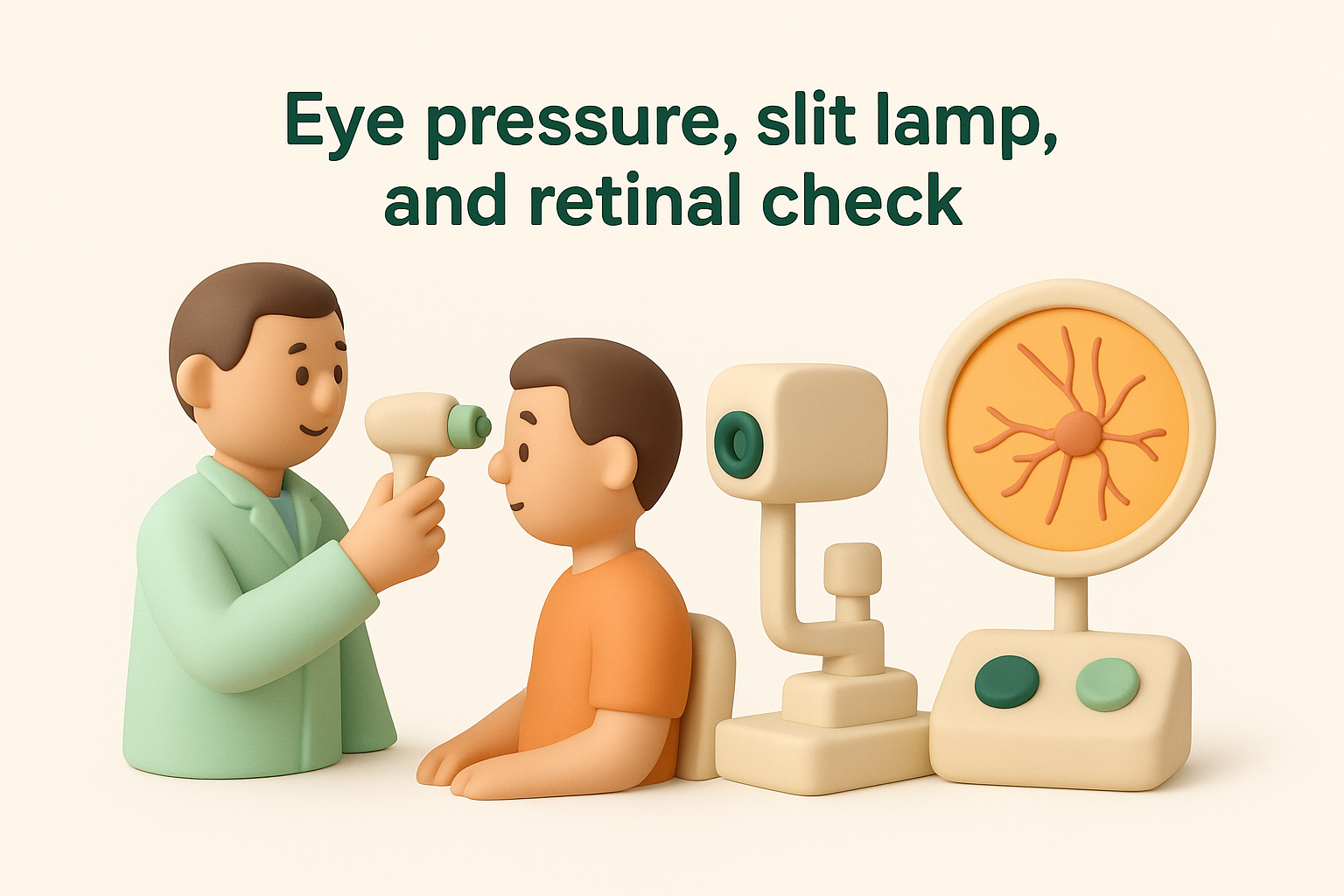
The session closes with an examination of the retina and optic nerve. Your practitioner may use dilating drops to widen your pupil and improve their view of the macula, blood vessels, and optic disc. Retinal photography or optical coherence tomography (OCT) may also capture detailed images that serve as a useful baseline for future visits.
Common tests and what they check for
Beyond the standard steps of what happens during an eye test, your practitioner may run a series of additional targeted tests depending on your age, symptoms, and risk factors. Each test examines a specific aspect of your visual system, and understanding what they check for helps you make sense of your results when they are explained at the end of the appointment.
Visual field testing
A visual field test, also called perimetry, maps how well you can detect objects at the edges of your vision without shifting your gaze. You sit in front of a screen and press a button whenever you spot a small flicker of light appearing at different positions across your visual field. The resulting map highlights any areas where your peripheral vision is reduced or absent.
This test is particularly important for detecting glaucoma, where nerve damage typically starts at the outer edges of your visual field long before it affects your central vision. It can also identify neurological changes along the visual pathway, including those linked to stroke or pressure on the optic nerve.
A single visual field test gives a useful snapshot, but comparing results across multiple visits reveals gradual changes that would otherwise go unnoticed in any one appointment.
Colour vision and eye coordination
Colour vision testing uses plates covered in patterns of coloured dots, known as Ishihara plates, to check whether you can distinguish between different hues. Colour deficiencies are often inherited and picked up in childhood, but acquired changes to colour perception can also signal retinal disease or optic nerve damage in adults.
Your practitioner may also assess how well your two eyes work together using a cover test. One eye is briefly covered while your practitioner watches whether the uncovered eye moves to take up fixation. This reveals problems with eye alignment that can affect depth perception and cause persistent fatigue during close-up tasks like reading or screen use.
Retinal imaging and OCT
Optical coherence tomography (OCT) produces a cross-sectional scan of the retina, showing individual tissue layers in fine detail. Your practitioner uses these images to assess the thickness and integrity of the macula, which is the central zone of the retina responsible for sharp, detailed vision.
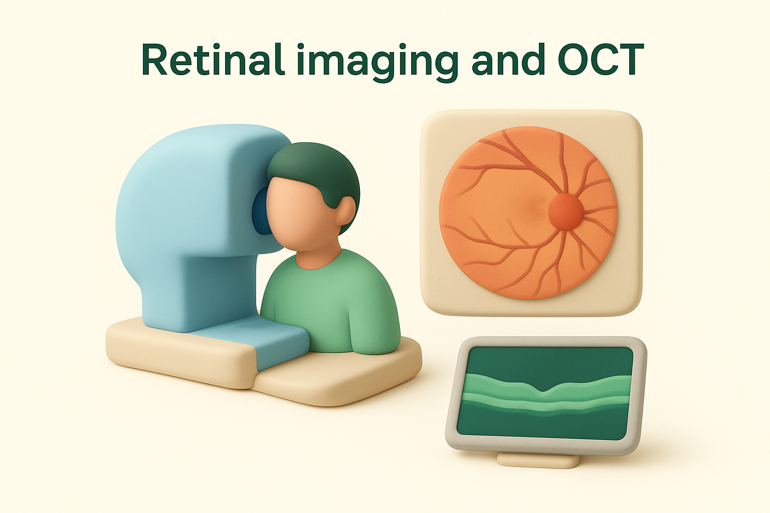
Fundus photography captures a wide-angle image of the retina, optic disc, and blood vessels in a single photograph. These images become a permanent record that lets your specialist track subtle structural changes across visits, which is especially useful for monitoring macular degeneration, diabetic retinopathy, and glaucoma over the long term.
After the test: results, costs and referrals
Once your practitioner finishes the final check, they sit down with you and walk through every finding from your appointment. This is the part of what happens during an eye test where your results come together as a complete picture rather than a series of isolated numbers. Most practitioners explain your vision prescription, eye pressure reading, and any structural findings in plain language, so ask questions if anything is unclear before you leave.
Understanding your results
Your practitioner will tell you whether your vision prescription has changed since your last test and explain what your eye pressure and retinal findings mean for your long-term health. If everything looks healthy, they will confirm a recommended return interval and note your results as a baseline for future comparison. If they identify any concern, such as an elevated pressure reading or an unusual retinal appearance, they will explain what the finding could indicate and what the next step should be.
A single abnormal result does not automatically mean you have a serious condition; many findings require a second assessment or a specialist review before any diagnosis is confirmed.
What you’ll pay and what Medicare covers
Medicare covers the standard consultation fee for a bulk-billed optometry appointment in Australia, which means most routine eye tests cost you nothing out of pocket if your optometrist bulk bills. However, additional tests such as OCT scans or retinal photography are not always included under Medicare and may attract a separate fee. It is worth asking your optometrist upfront which tests carry an extra charge so there are no surprises when you pay.
If you hold private health insurance with extras cover, some or all of those additional test costs may be partially reimbursed depending on your policy limits. Optical items such as glasses or contact lenses are generally not covered by Medicare but may attract a benefit through your insurer.
When your optometrist refers you to a specialist
Your optometrist refers you to an ophthalmologist when a finding requires surgical assessment, specialist imaging, or medical treatment beyond the scope of routine optometry. Common reasons for referral include suspected glaucoma, signs of macular degeneration or diabetic retinopathy, or a cataract that is affecting your daily function. A written referral from your optometrist or GP also allows you to access Medicare rebates on your specialist consultation, which reduces your out-of-pocket costs significantly.
Wrapping up and when to get urgent help
Understanding what happens during an eye test removes the uncertainty and makes it much easier to attend regularly. Each step, from the initial history-taking to retinal imaging, gives your practitioner a detailed picture of your eye health and picks up problems long before they affect your daily life. Staying consistent with your testing schedule is one of the most straightforward things you can do to protect your vision long-term.
Some situations cannot wait for a routine appointment. If you suddenly notice a significant increase in floaters, flashing lights, a dark curtain across your vision, or a rapid drop in sight, seek urgent assessment the same day. These symptoms can indicate a retinal detachment or other serious condition where prompt treatment directly affects the outcome. Do not drive yourself if your vision is significantly affected.
If you are ready to book or have a referral in hand, contact Bright Sight Eye Specialists to arrange your appointment in Randwick.