


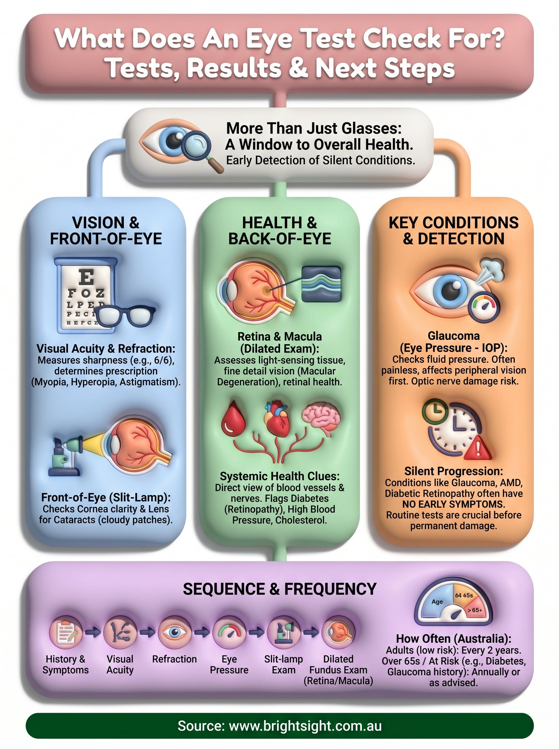
Most people book an eye test expecting a quick check of their vision, read a few letters on a chart, get a prescription, and leave. But a comprehensive eye examination does far more than measure whether you need glasses. So what does an eye test check for, exactly? The answer might surprise you. Your eyes can reveal early signs of conditions that affect not just your sight, but your overall health, from diabetes to high blood pressure.
A thorough eye examination screens for a range of conditions including glaucoma, cataracts, macular degeneration, and retinal disease. Many of these develop without obvious symptoms, which means they can progress silently until real damage is done. Early detection through routine testing gives you the best chance of preserving your vision long-term.
At Bright Sight Eye Specialists in Randwick, Dr Rahul Dubey and Dr Capucine Odouard see firsthand how a detailed eye assessment can change the course of someone’s eye health. Whether it’s catching diabetic retinopathy early or identifying the first signs of a cataract, the right tests at the right time make a measurable difference. This article walks you through every test performed during a comprehensive eye examination, what each one checks for, how to read your results, and what to do next if something needs attention.
Why an eye test matters beyond glasses
Most people think of an eye test as a way to update their glasses prescription. That view undersells what the examination actually delivers. A comprehensive eye exam assesses your visual system and, in many cases, your general health. Understanding what does an eye test check for goes well beyond measuring short or long-sightedness; it includes screening for serious, sight-threatening conditions that often give you no warning at all.
Your eyes as a window to overall health
Your eyes are unique. They are the only place in your body where a clinician can directly examine blood vessels and nerve tissue without any cutting or invasive procedure. During an eye test, your optometrist or ophthalmologist looks through the pupil at the retina and optic nerve, structures that reflect the state of your circulatory and nervous systems. Changes in those structures can point to conditions elsewhere in the body long before you feel any symptoms.
Conditions such as high blood pressure, diabetes, and high cholesterol can all leave visible signs inside the eye. In some cases, an eye test is the first appointment that flags a systemic problem, prompting referral for further investigation. That makes routine eye testing genuinely preventive healthcare, not just an optical service.
Early changes in retinal blood vessels are sometimes the first detectable sign that blood pressure or blood sugar is affecting the body.
Conditions that develop without warning signs
Several of the most serious eye conditions are painless and progress gradually in their early stages. Glaucoma, for example, typically causes no pain and begins by affecting peripheral vision, which most people do not notice until the damage is significant. Macular degeneration follows a similar pattern, slowly affecting central vision while the person compensates without realising it. By the time symptoms become hard to ignore, meaningful and permanent damage has already occurred.
Relying on symptoms alone is not a reliable way to protect your vision. Regular testing catches these conditions at a stage when treatment is most effective. For glaucoma, early intervention can halt progression almost entirely. For macular degeneration, timely treatment can slow or stop the loss of central vision. Waiting for a problem to announce itself costs you options.
Why frequency matters as much as the test itself
A single eye test gives you a useful snapshot. A series of tests over time gives your eye specialist something far more valuable: a baseline and a trend. Changes in your optic nerve, retinal thickness, or eye pressure only become meaningful when compared against previous measurements. An isolated reading may look unremarkable; a shift from your personal normal tells a different story entirely.
Age, family history, and general health all influence how often you should attend. People with diabetes or a family history of glaucoma typically need annual checks, while a healthy adult in their twenties with no risk factors may be fine with testing every two years. Getting that schedule right is just as important as attending each individual appointment, so treat it as an ongoing conversation with your eye care provider rather than a fixed rule applied to everyone.
What happens in a comprehensive eye exam
A comprehensive eye examination follows a structured sequence of tests, each building on the last. Your practitioner gathers information about your history and symptoms first, then works through objective measurements before completing a hands-on clinical assessment. The whole appointment typically takes 30 to 60 minutes, depending on your age, your risk factors, and whether any findings require a closer look. Understanding the flow helps you arrive prepared and get the most from the visit.
Before the tests begin
Your eye care professional starts by asking about your current vision concerns, any recent changes you have noticed, and your personal and family medical history. This background matters because conditions like glaucoma and macular degeneration run in families, and systemic diseases like diabetes directly affect what the clinician looks for inside your eye. Be honest about medications you take and any previous eye injuries or surgeries, since that information shapes which tests are prioritised and how results are interpreted.
The questions at the start of your appointment are not small talk; they guide every clinical decision that follows.
The sequence of clinical testing
After the history is taken, the examination moves through a series of specific assessments. These typically include:
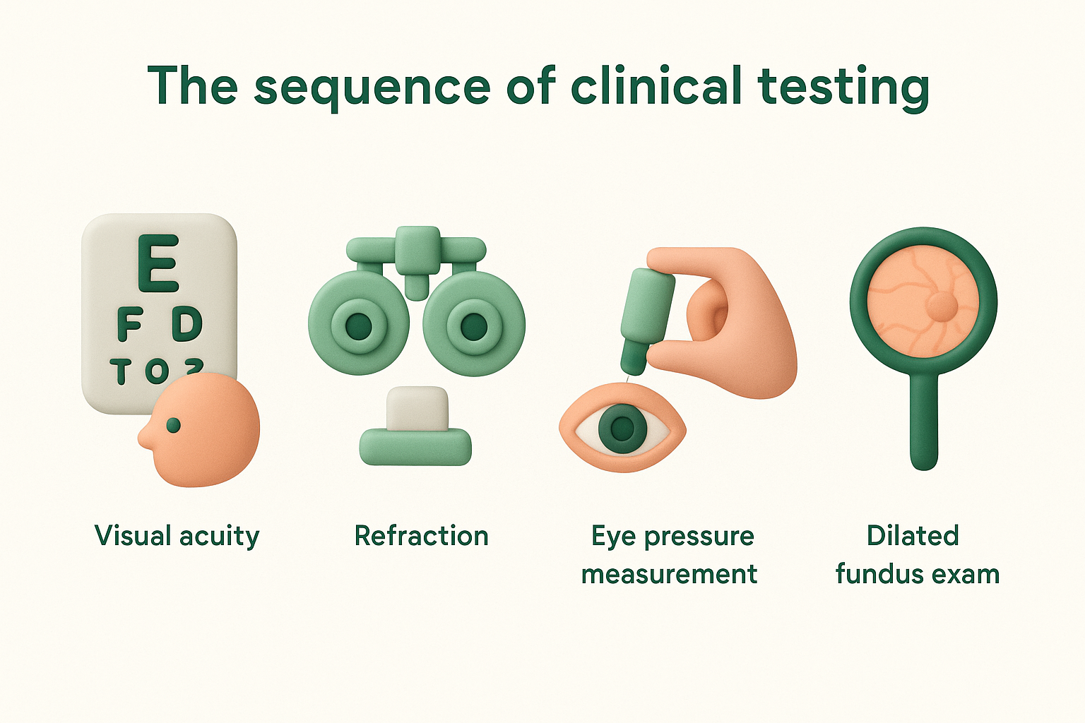
- Visual acuity testing to measure how clearly you see at distance and near
- Refraction to determine your spectacle or contact lens prescription
- Eye pressure measurement to screen for glaucoma
- Slit-lamp examination to inspect the front of the eye under magnification
- Dilated fundus examination to assess the retina, macula, and optic nerve
Not every appointment includes all of these in equal depth. Your practitioner selects and adjusts the tests based on your age, symptoms, and risk profile. A 25-year-old attending for the first time without any family history of eye disease receives a different emphasis than a 65-year-old with diabetes and elevated eye pressure.
Who performs the examination
In Australia, both optometrists and ophthalmologists perform comprehensive eye examinations, but their scope differs. Optometrists manage routine vision testing and prescribe glasses or contact lenses. Ophthalmologists are medical doctors specialising in eye disease and surgery, and they assess and treat conditions like cataracts, glaucoma, and retinal disease. If your optometrist identifies something that needs further investigation, they will refer you to an ophthalmologist. At Bright Sight Eye Specialists, both Dr Rahul Dubey and Dr Capucine Odouard provide specialist-level assessment for complex or high-risk cases.
Vision and refraction tests: how well you see
Vision and refraction tests are usually the part of a comprehensive eye exam that people recognise most readily. These tests measure how sharply you see at different distances and identify whether your eyes focus light correctly onto the retina. When considering what does an eye test check for, this is the foundation: establishing a clear picture of your current visual function before any other assessments begin.
Visual acuity: reading the chart
Visual acuity testing measures how clearly you can see detail at a set distance, typically six metres. You read letters or symbols from a standardised chart, usually called a Snellen chart, with each eye tested separately while the other is covered. The result is expressed as a ratio, such as 6/6, which means you can read at six metres what a person with normal vision reads at the same distance. A result of 6/12 means you need to be at six metres to read what someone with normal vision reads at twelve.
Your practitioner uses this result to identify how much, if any, optical correction you need. Poor acuity on its own does not always indicate disease; it may simply mean your glasses prescription is out of date. However, a significant drop in acuity since your last test prompts a closer look at the structures inside the eye.
Refraction: finding your prescription
Refraction is the process used to determine your spectacle or contact lens prescription. Your eye care professional uses a phoropter, a device with multiple lenses, to trial different combinations while asking you to compare which lens gives you the clearest image. This process identifies whether you are myopic (short-sighted), hyperopic (long-sighted), or astigmatic, and to what degree.
Even small changes in your prescription can cause headaches, eye strain, or difficulty with tasks like reading or driving at night, so keeping your prescription current is worth the appointment.
Some clinics also perform automated refraction using a machine called an autorefractor before the manual test begins. This gives the practitioner a useful starting point but does not replace the clinical judgement involved in fine-tuning the final prescription. Both techniques together produce a more accurate and reliable result than either would alone. If your prescription has changed noticeably since your last test, your optometrist or ophthalmologist will factor that into their overall assessment of your eye health.
Eye pressure and glaucoma checks
Glaucoma is one of the leading causes of preventable blindness in Australia, and eye pressure measurement is the front-line screening tool used to detect it. When people ask what does an eye test check for, glaucoma often comes up because it progresses without pain or obvious symptoms until significant nerve damage has already occurred. Measuring the pressure inside your eye, known as intraocular pressure (IOP), gives your practitioner an important indicator of your glaucoma risk, though it is only one piece of a larger picture.
How eye pressure is measured
The most common method used to measure eye pressure is non-contact tonometry, sometimes called the air-puff test. A brief burst of air is directed at the surface of your eye, and the instrument calculates the pressure based on how your cornea responds. It takes only a second and requires no drops or physical contact. Many people find it mildly startling but not uncomfortable.
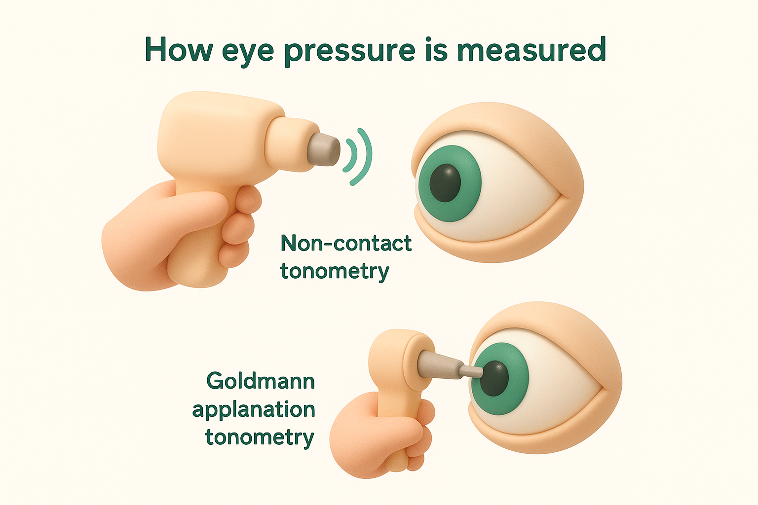
For a more precise reading, your clinician may use Goldmann applanation tonometry, which is considered the gold standard. This involves a small probe making brief contact with your numbed cornea after anaesthetic eye drops are applied. The technique gives a highly accurate measurement and is typically used when there is a concern about elevated pressure or when a more detailed baseline is needed. Both methods measure pressure in millimetres of mercury (mmHg).
Normal intraocular pressure generally falls between 10 and 21 mmHg, though some people develop glaucoma within this range while others tolerate higher pressures without damage.
What the results tell your specialist
Eye pressure alone does not diagnose glaucoma; it identifies risk. Your specialist interprets your IOP reading alongside optic nerve appearance, visual field test results, and your personal history. A pressure reading above 21 mmHg raises the level of scrutiny applied to the rest of the examination, but it does not confirm disease on its own. Equally, a pressure within the normal range does not rule glaucoma out entirely, which is why looking at all findings together matters.
If your results indicate elevated pressure or suspicious changes to the optic nerve head, your specialist may arrange further imaging such as optical coherence tomography (OCT), which maps the layers of the retina and optic nerve in precise detail. At Bright Sight Eye Specialists, this kind of structured, multi-test approach ensures that early glaucoma is not missed simply because one reading appeared borderline.
Front-of-eye checks: cornea and lens
When people ask what does an eye test check for, the front of the eye is often overlooked in the conversation, yet it contains some of the most telling clinical information available. Your cornea (the clear dome at the front of your eye) and lens (the structure behind your pupil that focuses light) both reveal a great deal about your eye health, and problems in either area can significantly affect your vision if left undetected.
The slit-lamp examination
Your specialist examines the front of your eye using an instrument called a slit lamp, also known as a biomicroscope. This device projects a thin beam of light into your eye at high magnification, allowing your clinician to inspect the cornea, iris, conjunctiva, and lens in fine detail. You rest your chin on a support while the clinician moves the light across the surface and internal structures of your eye, adjusting the angle and intensity to assess different layers.
The slit lamp gives your specialist a level of magnification that makes tiny abnormalities visible long before they cause noticeable symptoms.
The examination is non-invasive and painless. It takes only a few minutes but delivers a comprehensive view of structures that would otherwise be impossible to assess without surgery. Your clinician may use eye drops to dilate your pupils beforehand, which improves the view of the lens and deeper structures.
What your specialist looks for in the cornea
The cornea needs to be clear and uniformly curved to focus light accurately onto your retina. During the slit-lamp assessment, your specialist checks for corneal abrasions, ulcers, scarring, and conditions such as keratoconus, where the cornea gradually thins and bulges into a cone shape. Dry eye disease and previous infections can also leave marks on the corneal surface that affect your vision and guide your treatment options.
Checking the lens for cataracts
Your lens sits behind the iris and is normally clear and transparent. A cataract forms when proteins inside the lens begin to break down and clump together, causing the lens to become cloudy. The slit lamp allows your specialist to see exactly where the cloudiness sits within the lens, how dense it is, and how much it is likely to be affecting your functional vision. Cataracts develop slowly, and many people adjust to gradual blurring without realising the extent of the change until a direct comparison is made. Identifying a cataract early means you can plan surgery at the right time, rather than waiting until your vision is severely compromised.
Retina and macula checks: the back of the eye
The retina lines the inside of the back of your eye and converts light into electrical signals that your brain interprets as vision. Your macula sits at the centre of the retina and handles fine detail, colour, and straight-ahead sight. When asking what does an eye test check for, the examination of these structures is among the most clinically significant parts of the whole appointment, because retinal disease can cause permanent vision loss before you notice anything is wrong.
Dilating your pupils for a clearer view
To inspect the retina and macula properly, your specialist typically applies dilating eye drops that widen your pupil, giving a much broader view of the structures at the back of your eye. The drops take around 20 to 30 minutes to take full effect, and your vision will remain blurry and sensitive to light for a few hours afterwards, so arrange someone to drive you home or avoid driving yourself to the appointment.
Dilation is a small inconvenience that gives your specialist a substantially clearer view of structures that would otherwise be partially hidden.
Once your pupils are dilated, your specialist uses an ophthalmoscope or a condensing lens with the slit lamp to examine the retina, optic nerve head, blood vessels, and macula directly. This part of the assessment takes only a few minutes but reveals a remarkable amount of detail about the health of your posterior eye structures.
What the retinal assessment reveals
Your specialist looks for several specific findings during the retinal examination. Retinal detachment appears as a lifting or tearing of the retinal surface and requires urgent surgical attention to prevent permanent vision loss. Signs of age-related macular degeneration (AMD) include drusen deposits beneath the retina and changes to the retinal pigment epithelium, which your specialist can identify and grade even in early stages. Abnormal blood vessel growth, haemorrhages, and fluid accumulation each point to different conditions that require tailored management.
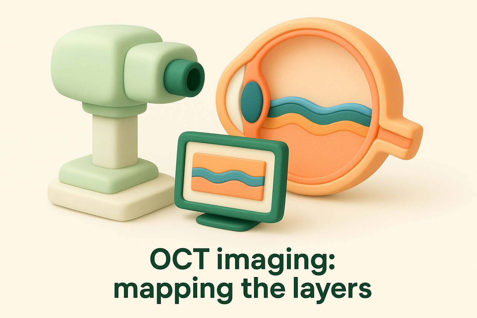
OCT imaging: mapping the layers
Optical coherence tomography, known as OCT, uses light waves to produce cross-sectional images of the retina in fine detail, similar to a high-resolution scan of its internal layers. The scan is painless and non-invasive, taking only seconds to complete. It allows your specialist to measure retinal thickness, detect fluid that is not visible during direct inspection, and track subtle changes between visits. At Bright Sight Eye Specialists, OCT imaging forms a core part of the diagnostic process for conditions affecting the macula and optic nerve.
Other screening: diabetes and health clues
Understanding what does an eye test check for means recognising that the examination extends well beyond the visual system. The structures at the back of your eye, particularly your retinal blood vessels, give your specialist a direct view of what is happening inside your circulatory system. Changes in these vessels can point to systemic conditions that you may not yet know you have, making the eye examination one of the few routine appointments where a diagnosis unrelated to your eyes can emerge.
Diabetic retinopathy: what your specialist looks for
Diabetes affects blood vessels throughout the body, and the vessels inside your retina are among the first to show signs of damage. Diabetic retinopathy develops when elevated blood sugar weakens retinal blood vessels, causing them to leak, swell, or grow abnormally. Your specialist looks for microaneurysms (small bulges in vessel walls), haemorrhages, hard exudates, and new vessel growth, each of which indicates a different stage of the condition.

Detecting diabetic retinopathy early, before symptoms appear, gives you the best opportunity to preserve your central vision through timely treatment.
If you have type 1 or type 2 diabetes, annual retinal screening is not optional; it is a core part of managing your overall health. At Bright Sight Eye Specialists, Dr Rahul Dubey manages diabetic retinal disease with treatments including laser therapy and intravitreal injections, tailored to the severity of what your specialist finds during examination.
Blood pressure and vascular health clues
Hypertension, or high blood pressure, leaves visible marks on the retinal blood vessels over time. Your specialist looks for arteriovenous nicking (where thickened arteries compress veins at crossing points), copper or silver wiring of vessel walls, and in severe cases, haemorrhages or swelling of the optic nerve head. These findings can reflect long-standing, poorly controlled blood pressure that has been placing strain on your cardiovascular system.
Elevated cholesterol can also leave deposits at the retinal vessel bifurcations, appearing as bright yellow plaques called Hollenhorst plaques. Finding these during a retinal assessment may prompt your specialist to recommend follow-up with your general practitioner for cardiovascular risk management. Other rare but detectable conditions include autoimmune and inflammatory diseases that produce characteristic retinal patterns visible under direct examination. Your eye appointment, in other words, has the potential to trigger investigations that protect your overall health, not just your sight.
Understanding your results and what they mean
After your appointment, your eye care professional will walk you through what they found. Understanding your results gives you context for any next steps and helps you take an active role in your eye health rather than simply receiving instructions. Most practitioners summarise findings verbally during the appointment, and many provide a written report you can share with your GP or keep for your own records.
Reading your prescription
Your spectacle prescription is expressed as a series of numbers covering three measurements: sphere, cylinder, and axis. Sphere indicates the degree of short or long-sightedness, expressed in dioptres, with a minus sign for myopia and a plus sign for hyperopia. Cylinder and axis together describe the amount and orientation of astigmatism present. A reading such as -2.50 / -0.75 x 180 means you are moderately myopic with a small degree of astigmatism. Your optometrist or ophthalmologist can explain each value clearly if the numbers feel unfamiliar, so ask before you leave.
What grading and staging mean
For clinical findings beyond refraction, your specialist uses grading scales to describe severity. Cataracts are graded by density and position within the lens. Diabetic retinopathy is staged from mild non-proliferative through to proliferative disease, which involves abnormal new blood vessel growth. Macular degeneration is classified as early, intermediate, or late, with the late stage subdivided into geographic atrophy and neovascular forms. These classifications tell you where you sit on a clinical spectrum and directly influence what treatment, if any, your specialist recommends at this point.
Knowing your grade or stage is not a cause for alarm; it is the information your specialist needs to make a precise and appropriate plan.
When results prompt a referral
Some findings at a routine appointment require specialist input that goes beyond what an optometrist can provide. If your optic nerve appearance is suspicious, your intraocular pressure is elevated, or your retinal assessment reveals changes consistent with macular degeneration or retinal disease, you will receive a referral to an ophthalmologist. Understanding what does an eye test check for becomes especially relevant here: results that appear minor in isolation can carry real clinical weight once your full picture is reviewed by a surgeon with specialist expertise. At Bright Sight Eye Specialists, Dr Rahul Dubey and Dr Capucine Odouard assess referral cases and ensure every patient understands what their findings mean and what the recommended next step involves before leaving the consultation.
How often you need an eye test in Australia
How often you attend for testing is just as important as attending at all, and what does an eye test check for shifts in relevance depending on your stage of life and your personal risk profile. Australia has no single mandatory schedule that applies to everyone, so your optometrist or ophthalmologist sets a recall interval based on your individual findings, history, and risk factors. Following that recommended interval, rather than waiting until something goes wrong, is the most practical way to protect your vision over the long term.
General frequency by age group
Your age group is the starting point for determining a sensible testing interval. Children need assessment early, since uncorrected vision problems affect learning and development in ways that are not always obvious. Adults with healthy eyes and no risk factors can generally attend every two years. Once you pass 65, annual testing becomes the standard recommendation because the risk of glaucoma, cataracts, and macular degeneration rises sharply with age.
| Age group | Recommended interval |
|---|---|
| Children (0 to 5 years) | At birth, at 6 months, before starting school |
| Children (school age) | Every 2 years, or sooner if symptoms arise |
| Adults (18 to 64, low risk) | Every 2 years |
| Adults (65 and over) | Annually |
Your specialist’s recommended recall interval takes priority over any general guideline, so follow their specific advice for your situation.
When you need more frequent checks
Certain risk factors reduce your testing interval significantly. If you have been diagnosed with diabetes, your retina needs assessment at least once a year because diabetic retinopathy can develop and progress quickly without symptoms. A family history of glaucoma places you at higher genetic risk, and your ophthalmologist will likely want to see you annually to track your optic nerve and pressure over time.
Existing eye conditions also change the schedule. If you are already being managed for macular degeneration, glaucoma, or a retinal condition, your specialist determines your follow-up frequency based on how stable or active that condition is, sometimes as often as every few months. Patients who have had cataract or retinal surgery at Bright Sight Eye Specialists receive a structured follow-up plan from Dr Rahul Dubey or Dr Capucine Odouard to ensure recovery stays on track and any complications are caught early.
Next steps for your eye health
Now that you understand what does an eye test check for, the most useful thing you can do is act on that knowledge. A comprehensive eye examination covers far more than a glasses prescription; it screens for glaucoma, cataracts, retinal disease, and systemic conditions like diabetes that leave early, detectable signs inside your eye. Many of these conditions cause no symptoms in their early stages, which means routine testing is genuinely your best protection against preventable vision loss.
Your next step is straightforward: book an appointment with a qualified eye care professional and attend at the interval your specialist recommends. If you have risk factors such as diabetes, a family history of glaucoma, or any recent changes in your vision, do not wait for your next scheduled check. Book a consultation with Bright Sight Eye Specialists in Randwick to get a thorough assessment from Dr Rahul Dubey or Dr Capucine Odouard.