


Sudden vision loss or a shower of dark floaters can be alarming, and in many cases, these symptoms point to bleeding inside the eye. Understanding vitreous bleeding causes is an important first step, whether you’ve just received a diagnosis or you’re trying to make sense of what’s happening with your sight. The vitreous is the clear, gel-like substance that fills the centre of your eye, and when blood leaks into it from damaged retinal vessels, the result is a condition called vitreous haemorrhage.
Several underlying conditions can trigger this bleeding. Some, like diabetic retinopathy and retinal tears, are relatively common. Others, such as retinal vein occlusions or trauma-related injuries, may come on without warning. The cause matters because it directly shapes which treatment path will protect your vision, and how urgently you need to act. Without a proper examination, it’s difficult to distinguish between causes that may resolve on their own and those that require prompt surgical intervention.
At Bright Sight Eye Specialists in Randwick, Sydney, Dr Rahul Dubey brings fellowship training from Moorfields Eye Hospital and extensive experience in retinal and vitreous surgery, including the management of vitreous haemorrhage from all causes. This article breaks down the most common triggers of vitreous bleeding, the symptoms to watch for, and the treatment options available, so you can have a clearer picture before your next consultation.
What vitreous bleeding is and where the blood comes from
The vitreous humour is the transparent, gel-like substance that occupies roughly 80% of the interior of your eye. It sits between the lens at the front and the retina at the back, and under normal conditions it remains completely clear. Vitreous haemorrhage occurs when blood from nearby retinal blood vessels leaks or bursts into this gel, clouding what was once transparent tissue. Even a relatively small volume of blood can scatter light dramatically, which is why vision changes can feel sudden and severe.
The anatomy behind the bleed
Your retina is the thin layer of light-sensitive tissue that lines the back wall of the eye. It relies on a dense network of small blood vessels to deliver oxygen and nutrients to the photoreceptor cells that process what you see. These vessels sit on the retinal surface and, in some cases, grow abnormally into the vitreous itself. When one of these vessels ruptures or is torn, blood spills directly into the vitreous cavity, where it has nowhere to drain and no mechanism for rapid absorption.
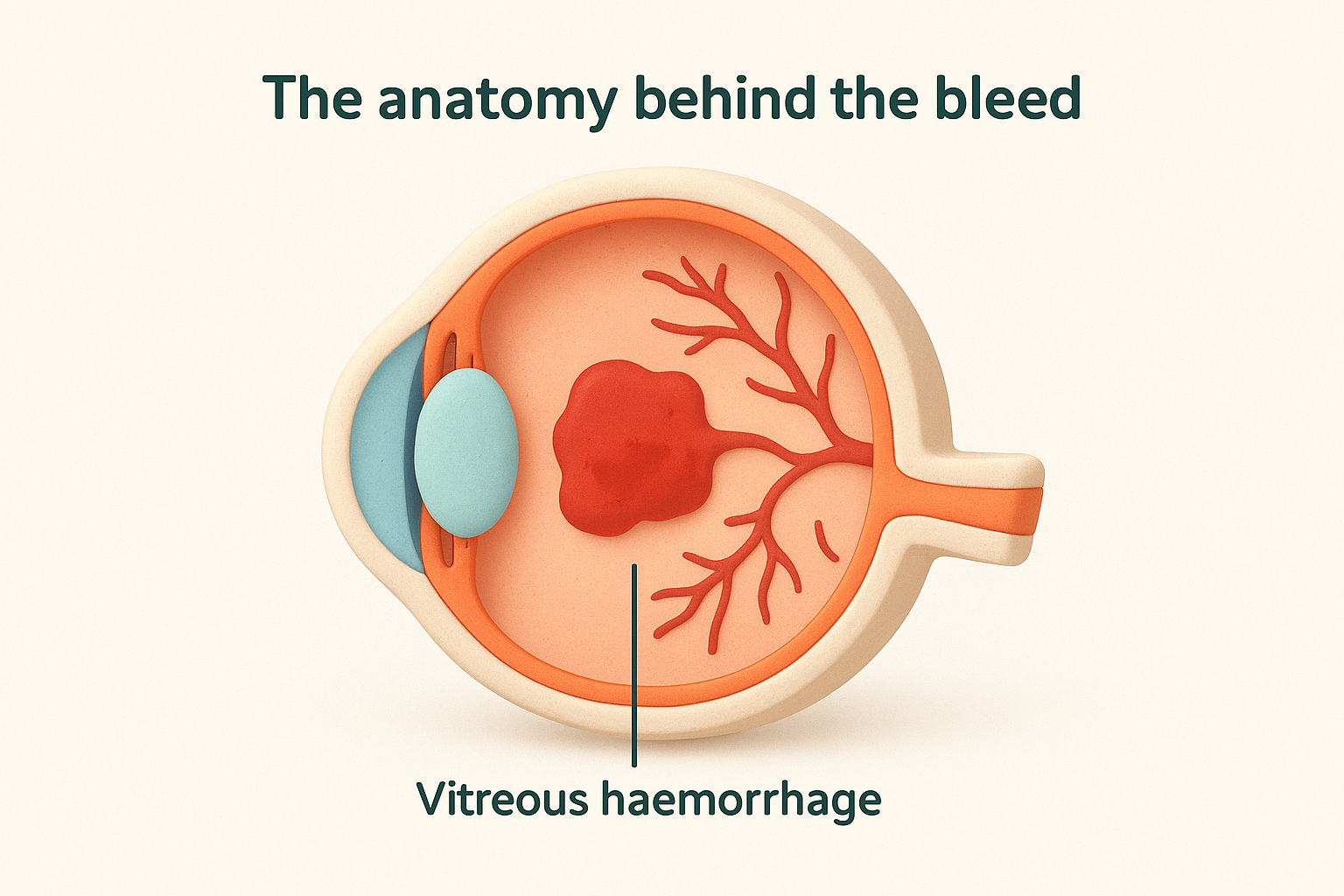
The retina is the only structure in the human body where blood vessels can be observed directly and non-invasively, which is why retinal examination is such a powerful diagnostic tool.
Understanding this anatomy helps explain why so many different vitreous bleeding causes can lead to the same end result. Whether the trigger is a weakened vessel from diabetes, a torn retina, or a sudden pressure change, the final pathway is the same: blood enters the vitreous and impairs vision.
Why the vitreous has no rapid clearance mechanism
Unlike your bloodstream, the vitreous is an avascular structure, meaning it carries no blood vessels of its own and relies on slow diffusion for any exchange of nutrients or waste. When blood enters this compartment, the body cannot flush it out quickly. Red blood cells gradually break down over days to weeks, releasing haemoglobin and other breakdown products that further cloud the gel. In heavier bleeds, this process can take months, and in some cases the blood never fully clears without surgical intervention.
This slow clearance is clinically significant. The position you rest in can actually affect where blood settles within the vitreous. Lying flat allows blood to spread across the central visual axis, while sitting upright lets it settle inferiorly, away from the main line of sight. This is why doctors sometimes advise propped-up rest in the early stages after a bleed.
Where the blood originates in the eye
Blood does not appear in the vitreous from nowhere. It always comes from a specific vascular source, and identifying that source is the central goal of any diagnostic workup. The most common origins include:
- New abnormal vessels growing on the retinal surface (neovascularisation), typically associated with diabetic retinopathy or retinal vein occlusion
- Torn retinal vessels at the site of a retinal break or detachment
- Ruptured retinal vessels following blunt or penetrating trauma to the eye
- Bleeding from established retinal vessels due to sudden intraocular pressure changes or vascular disease
Each of these sources carries different implications for treatment and urgency. A bleed from neovascularisation in an eye with proliferative diabetic retinopathy, for example, often signals that the underlying disease has reached an advanced and active stage, while a small bleed associated with a posterior vitreous detachment may resolve without intervention. Knowing where the blood comes from is the foundation of managing the condition effectively.
Why vitreous bleeding matters and when it is urgent
Vitreous haemorrhage is never just a visual inconvenience. While some bleeds do resolve on their own, the underlying condition causing the bleeding can continue to damage your eye even as the blood slowly clears. Several vitreous bleeding causes are progressive diseases that will worsen without treatment, which means that waiting to see if your vision improves on its own carries real risk. The sooner a cause is identified, the better your chances of protecting your long-term sight.
Vision loss that can become permanent
When blood fills the vitreous, it blocks light from reaching the retina, so your vision loss reflects the density and location of the bleed rather than any direct retinal damage. A dense central bleed can reduce your vision to nothing more than light perception. However, the deeper concern is what happens to the retina itself during this time. If the underlying cause is a retinal tear or detachment, the retina may be losing its blood supply while the blood in the vitreous prevents anyone, including your ophthalmologist, from seeing clearly inside your eye.
Retinal detachment is a surgical emergency. Every hour of delay increases the risk of permanent central vision loss if the macula becomes involved.
Proliferative retinal diseases, such as advanced diabetic retinopathy, can also progress rapidly if left unmanaged. New abnormal blood vessels continue to grow and bleed, and scar tissue can form, pulling the retina away from the back wall of the eye. This is why identifying the cause promptly is not just good practice; it is essential for preserving your sight.
Warning signs that require same-day assessment
Not every bleed looks the same, and some presentations are more urgent than others. Certain symptoms should prompt you to seek care on the same day, rather than waiting for a routine appointment. These include:
- A sudden, dense black curtain or shadow across part of your visual field
- Complete or near-complete loss of vision in one eye
- Flashing lights combined with a large number of new floaters, which can signal a retinal tear
- Any vision change following a direct blow or injury to the eye or head
- Rapid worsening of symptoms over hours rather than days
If you experience any of these, contact an eye specialist immediately. Even if the cause turns out to be less serious than feared, a same-day examination with ultrasound imaging can rule out the conditions that cannot wait.
The three most common causes in adults
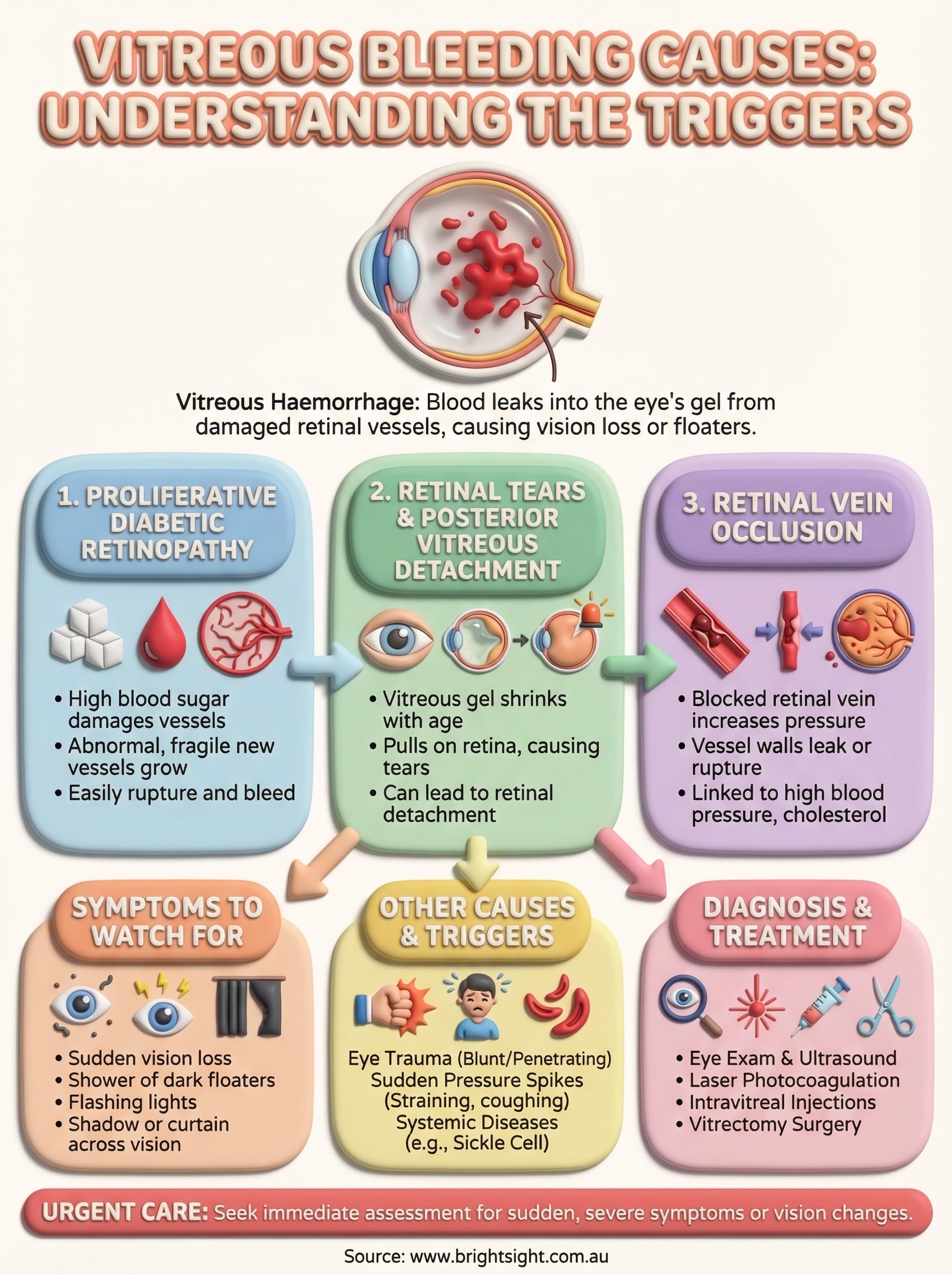
Understanding the most frequent vitreous bleeding causes gives you a clearer sense of what your ophthalmologist is looking for during an examination. While many conditions can lead to blood entering the vitreous, three account for the vast majority of cases in adults: proliferative diabetic retinopathy, retinal tears associated with posterior vitreous detachment, and retinal vein occlusion. Each one follows a distinct disease pathway, but all three produce bleeding by damaging the delicate vascular network that serves the retina.
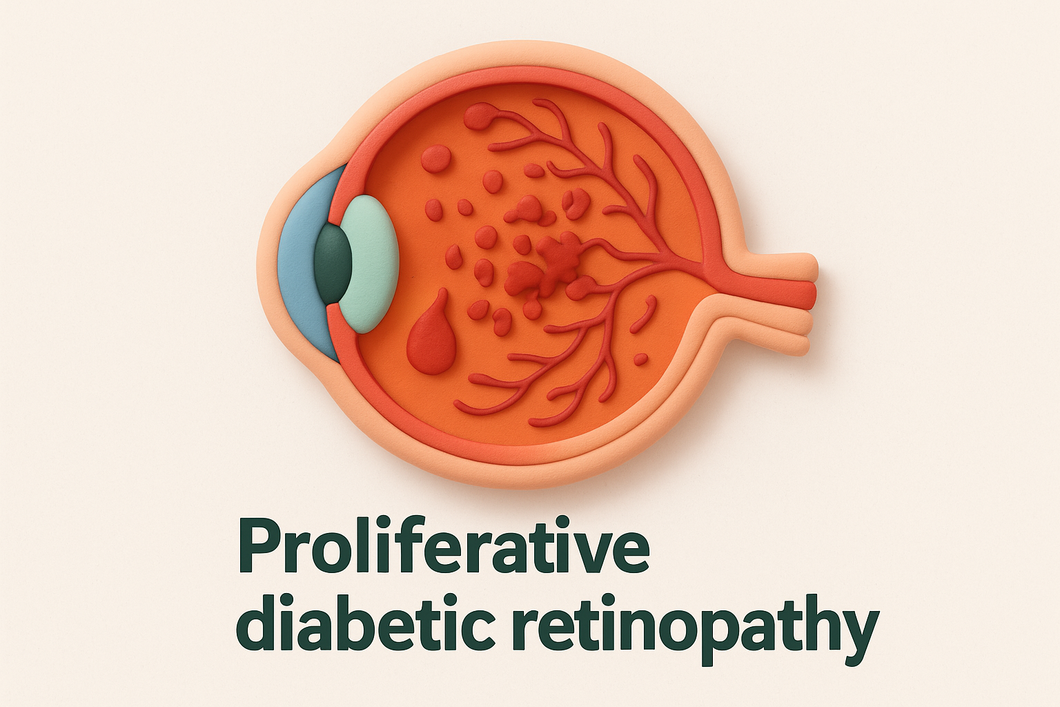
Proliferative diabetic retinopathy
Diabetic retinopathy is the leading cause of vitreous haemorrhage in working-age adults. When blood sugar levels remain elevated over time, the small blood vessels supplying the retina become damaged and eventually close off. In response, the eye releases growth signals that stimulate the formation of new fragile blood vessels, a process called neovascularisation. These new vessels are structurally weak and prone to rupturing, often with little or no warning. Because they grow directly on or into the vitreous, even minor physical activity or a change in blood pressure can trigger a bleed. Controlling blood sugar and blood pressure significantly reduces your risk of reaching this stage, but once neovascularisation is present, active treatment is usually required.
Retinal tears and posterior vitreous detachment
As you age, the vitreous gel gradually shrinks and liquefies, eventually pulling away from the retinal surface in a process called posterior vitreous detachment (PVD). In most people this separation happens without consequence, but in some cases the vitreous tears a small blood vessel as it detaches, causing a bleed. More critically, the same pulling force can create a full-thickness break in the retina, which is a serious risk factor for retinal detachment. A sudden shower of new floaters combined with flashing lights is a classic sign that a PVD may have torn a retinal vessel or created a break. This presentation requires urgent assessment, not watchful waiting.
If you notice a sudden increase in floaters alongside flashing lights, treat it as an urgent symptom regardless of how well you can still see.
Retinal vein occlusion
Retinal vein occlusion occurs when one of the veins draining blood away from the retina becomes blocked. The resulting pressure buildup causes the vessel walls to leak or rupture, sending blood into the surrounding retinal tissue and, in more severe cases, into the vitreous cavity. This condition is closely linked to cardiovascular risk factors including high blood pressure, diabetes, high cholesterol, and glaucoma. A central retinal vein occlusion affects the entire retina, while a branch occlusion involves only one segment, though both can produce significant vitreous haemorrhage in advanced cases.
Other medical causes you should know about
Beyond the three most common triggers, several other conditions can produce vitreous haemorrhage. Recognising these less frequent but clinically significant vitreous bleeding causes matters because they each carry distinct treatment implications. If you have a systemic health condition or a family history of vascular disease, these causes are worth understanding before your ophthalmologist raises them in a consultation.
Sickle cell retinopathy
Sickle cell disease affects the shape of red blood cells, making them rigid and prone to blocking small blood vessels throughout the body, including those that supply the retina. Over time, blocked retinal vessels lead to areas of oxygen deprivation, which triggers the same neovascularisation response seen in diabetic retinopathy. These fragile new vessels are prone to rupturing, and in advanced cases, the bleeding can extend into the vitreous cavity. People of African, Caribbean, Mediterranean, or Middle Eastern descent carry a higher genetic risk of sickle cell-related eye disease, so regular retinal screening is particularly important if you have a confirmed diagnosis.
Age-related macular degeneration
Wet age-related macular degeneration (AMD) involves the growth of abnormal blood vessels beneath and into the retina through a process called choroidal neovascularisation. These vessels are leaky and fragile, and while they most commonly cause bleeding within or beneath the retinal layers, a severe episode can break through into the vitreous cavity. This is a less common presentation of AMD, but it does occur, particularly in eyes with longstanding, untreated disease. If you are over 60 and notice a sudden, dense loss of central vision alongside floaters, wet AMD should be considered as a potential cause.
Any unexplained vitreous haemorrhage in a person over 60 warrants imaging of the macula to rule out underlying wet AMD, even when the bleeding initially obscures the view.
Terson syndrome and systemic bleeding disorders
Terson syndrome refers to vitreous or intraocular bleeding caused by a sudden, severe rise in intracranial pressure, most commonly from a subarachnoid haemorrhage or other intracranial bleed. The mechanism involves pressure transmitted along the optic nerve sheath, which ruptures retinal vessels. Patients who survive a brain haemorrhage may present with simultaneous vision loss, and Terson syndrome is often identified during recovery. Separately, systemic bleeding disorders such as leukaemia, severe anaemia, or anticoagulant therapy can reduce the blood’s ability to clot normally, making retinal vessels more susceptible to rupture from minor stress. Your treating ophthalmologist will ask about any blood-thinning medications or haematological conditions as part of a thorough diagnostic workup.
Trauma and pressure spikes that can trigger bleeding
Not all vitreous bleeding causes stem from systemic disease or slow retinal deterioration. Physical injury and sudden changes in intraocular pressure can rupture retinal vessels just as effectively as years of uncontrolled diabetes, and these events often strike without any prior warning signs. Understanding how trauma and pressure spikes contribute to vitreous haemorrhage helps you recognise situations where seeking immediate assessment is non-negotiable.
Blunt and penetrating trauma to the eye
Direct impact to the eye or surrounding orbit is one of the most straightforward pathways to vitreous haemorrhage. A forceful blow, whether from a sports collision, a fall, or an assault, creates a sudden compression and expansion of the globe that can shear retinal blood vessels at the point of impact or at structurally vulnerable sites such as the vitreous base. You do not need a penetrating injury for this to occur; blunt trauma alone generates sufficient hydraulic force to tear vessels and drive blood into the vitreous cavity.
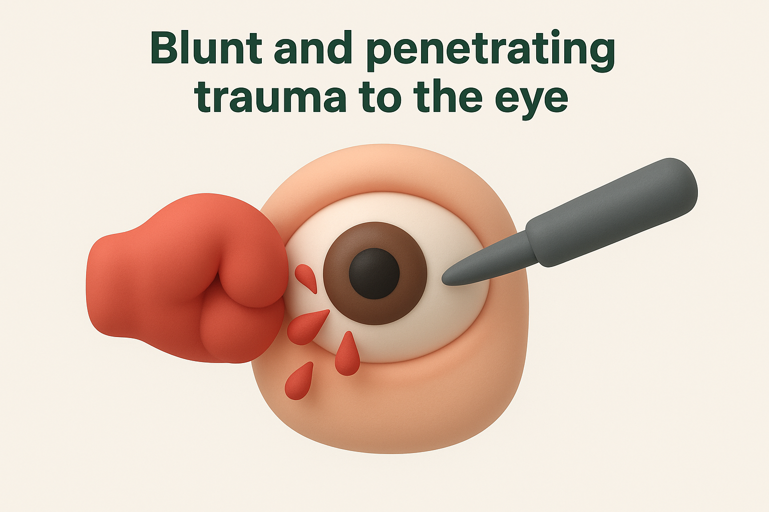
Even if your eye appears externally undamaged after a significant blow, internal bleeding can be progressing without any visible signs, which is why a prompt dilated examination is essential.
Penetrating injuries, such as those from sharp objects or high-velocity projectiles, carry the additional risk of introducing infection and foreign material into the eye alongside the bleeding. In these cases, surgical intervention is typically needed to address both the source of haemorrhage and any structural damage to the eye wall.
Sudden spikes in intraocular pressure
Rapid increases in intraocular pressure can stress retinal vessels to the point of rupture, particularly in eyes where those vessels are already weakened by underlying disease. Activities that dramatically raise venous pressure in the head, such as heavy straining, severe coughing episodes, or vomiting, can translate that pressure into the eye and trigger a bleed. This mechanism is more likely to produce significant haemorrhage if you already have fragile neovascular vessels from diabetic retinopathy or a previous retinal vein occlusion.
Severe acute angle-closure glaucoma is another pressure-related trigger worth noting. During a glaucomatous crisis, intraocular pressure can rise sharply within hours, placing the retinal vasculature under extreme stress. If you have a known risk of angle-closure glaucoma, regular monitoring and preventive laser treatment can significantly reduce this risk. Any sudden eye pain combined with halos around lights, nausea, and vision changes requires urgent assessment.
Symptoms that point to the underlying cause
The way your symptoms present can give your ophthalmologist important clues about which of the vitreous bleeding causes is responsible, even before any imaging or formal examination takes place. Floaters, flashes, and patterns of vision loss each carry diagnostic weight, and the combination you experience, along with how quickly your symptoms developed, helps narrow the list of possible triggers considerably.
Floaters and what their appearance suggests
The type and density of floaters you notice can hint strongly at the underlying cause. A sudden shower of small dark specks or dots that all appear at once often points to a ruptured retinal vessel, as occurs with a posterior vitreous detachment or a small retinal tear. A single large floater or a web-like shape drifting across your visual field may suggest a more substantial bleed, such as one from proliferative diabetic retinopathy or a retinal vein occlusion. If your floaters are accompanied by a diffuse red or brownish tint across your vision, the bleed is likely denser and sitting more centrally within the vitreous cavity.
Try to note when your floaters started, how many you can see, and whether they are increasing or staying the same, since this information is valuable at your first appointment.
Flashing lights and what they signal
Photopsia, the perception of flashing lights or sparks at the edges of your vision, usually indicates that the vitreous is pulling against or tearing the retinal surface. When flashes accompany new floaters, the combination is a strong signal that a retinal break or detachment may be involved, and this pairing warrants same-day assessment rather than a wait-and-see approach. Flashes without floaters, particularly in older adults, may relate to a posterior vitreous detachment alone, which carries a lower but still real risk of retinal tearing.
Pain, pressure, and visual field changes
A vitreous haemorrhage on its own does not cause eye pain, which makes pain a diagnostically useful symptom when it does appear alongside a bleed. Pain combined with vision loss points more toward acute angle-closure glaucoma or a severe inflammatory condition rather than a straightforward haemorrhage. Separately, a curtain or shadow across part of your visual field, particularly if it spreads upward from below, is a classic warning sign of retinal detachment and requires immediate attention regardless of how mild your other symptoms appear to be. Reporting these symptom combinations accurately gives your ophthalmologist the clearest possible starting point for identifying the cause.
How doctors find the cause in an eye exam
Pinpointing the specific vitreous bleeding causes behind a haemorrhage requires a systematic approach, because the blood itself often obscures the very structures your ophthalmologist needs to examine. A thorough diagnostic workup combines direct visualisation, imaging technology, and your medical history to build a complete picture of what triggered the bleed and how urgent the situation is.
Dilated fundus examination and slit-lamp assessment
Dilating your pupils with eye drops is the first step in any meaningful retinal examination. Dilation widens the pupil enough to give your ophthalmologist a broad view of the peripheral retina, where many tears and areas of neovascularisation develop. Using a slit-lamp with a condensing lens, your specialist can examine the vitreous gel itself for blood distribution, clots, and visible retinal pathology at the posterior pole.
During the examination, your ophthalmologist will assess several key features:
- Blood density and location within the vitreous cavity
- Whether the retinal surface remains visible through the bleed
- Any visible neovascularisation or retinal breaks at the periphery
Ultrasound imaging when the view is blocked
B-scan ultrasound is the most important diagnostic tool when a haemorrhage is too dense for optical examination. By transmitting sound waves through the eye, ultrasound can detect retinal detachment, vitreous membranes, and foreign bodies even when blood completely blocks the view. Your specialist will look specifically for a detached retina or traction bands, since these findings change both the urgency and the type of treatment required.
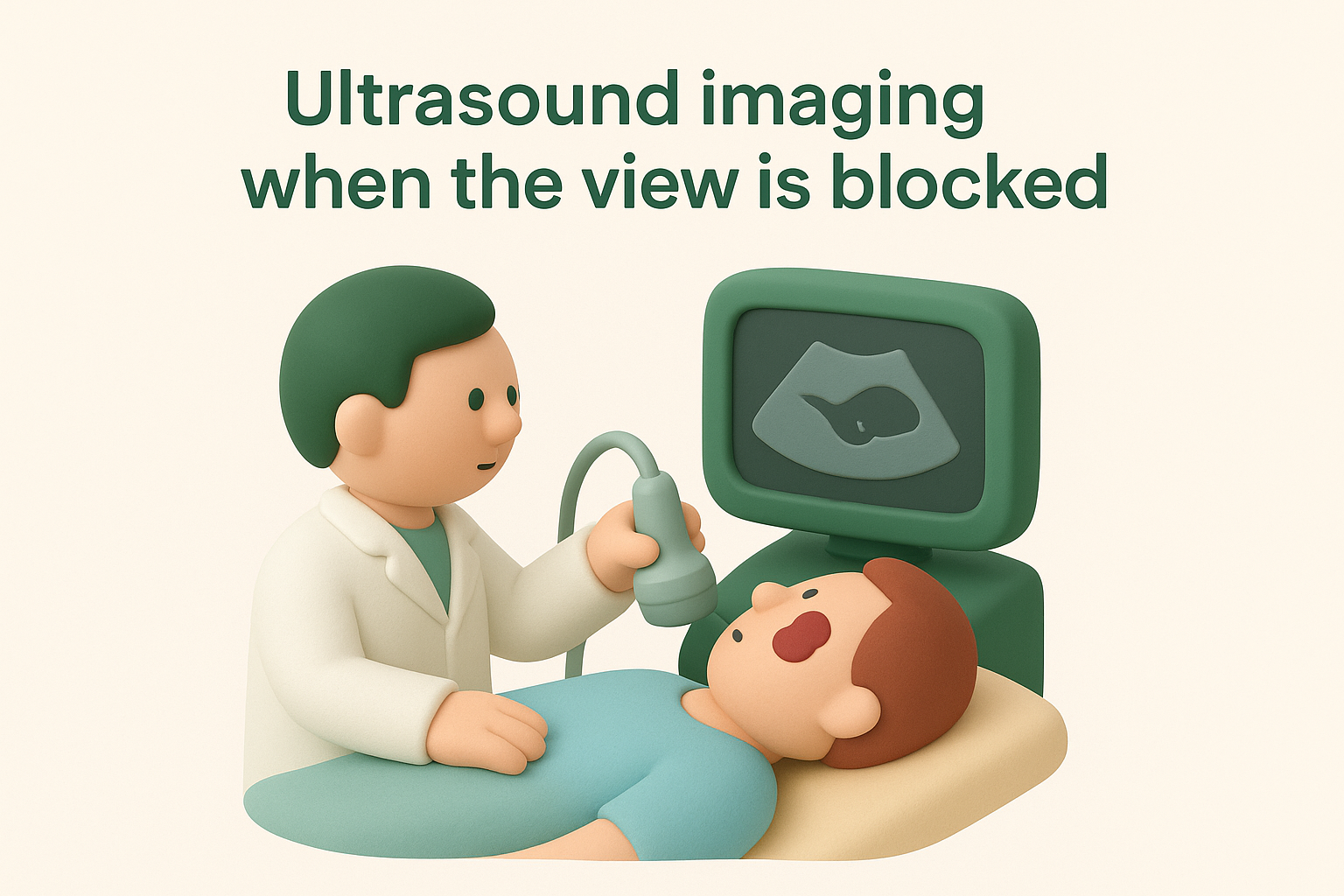
A normal ultrasound result does not eliminate all risk, and repeat imaging is often arranged within days if your symptoms worsen or vision deteriorates further.
Ultrasound also helps assess whether the posterior vitreous has already separated from the retina, information that directly influences surgical planning if a vitrectomy becomes necessary.
Other diagnostic tests your specialist may use
Fluorescein angiography involves injecting a dye into your arm and photographing how it circulates through the retinal vessels, revealing areas of leakage, blocked vessels, and abnormal new vessel growth. This test is typically reserved for cases where the vitreous has cleared sufficiently to allow optical imaging. Optical coherence tomography (OCT) provides cross-sectional images of the retinal layers and is particularly useful for assessing macular involvement, helping your ophthalmologist determine whether central vision has already been compromised by the disease driving the bleed.
Combining these investigations gives your specialist a precise, layered understanding of both the haemorrhage and its underlying cause, which is essential for planning targeted and effective treatment.
Treatment options based on the cause
Because vitreous bleeding causes vary so widely, there is no single treatment protocol that applies to every case. The approach your ophthalmologist recommends will depend directly on what triggered the bleed, the density of the haemorrhage, whether the retina is intact, and how quickly your vision is deteriorating. Some cases call for urgent surgical intervention, while others are managed conservatively while the underlying condition is stabilised.
Managing the underlying disease first
For bleeds driven by systemic conditions such as diabetic retinopathy or retinal vein occlusion, controlling the disease itself is as important as treating the eye directly. If your blood sugar, blood pressure, or cholesterol levels are poorly managed, ongoing damage to retinal vessels will continue regardless of what your ophthalmologist does surgically. Your treating team will coordinate with your GP or endocrinologist to ensure that systemic risk factors are addressed alongside any ocular intervention. In cases of anticoagulant-related bleeding, your haematologist or cardiologist may need to review your medication before any surgical procedure on the eye proceeds.
Laser photocoagulation and intravitreal injections
Laser photocoagulation targets areas of abnormal new vessel growth by applying precise thermal energy to the retina, causing neovascular tissue to regress. This treatment is particularly effective for proliferative diabetic retinopathy and certain presentations of retinal vein occlusion, where reducing the stimulus for new vessel growth prevents future bleeds from recurring. In many cases, laser is combined with intravitreal anti-VEGF injections, which block the chemical signals that drive neovascularisation. Injections are administered directly into the vitreous cavity under local anaesthetic and typically need to be repeated over several months to achieve a sustained effect.
Anti-VEGF injections have significantly changed the management of proliferative retinal disease, reducing the rate of recurrent vitreous haemorrhage in treated eyes.
Vitrectomy surgery
When the haemorrhage is too dense to clear on its own, or when ultrasound reveals an underlying retinal detachment or tractional membrane, vitrectomy surgery becomes necessary. During this procedure, your surgeon removes the blood-filled vitreous gel and replaces it with a clear fluid or gas, restoring optical transparency and allowing direct treatment of the retina. Vitrectomy also enables your surgeon to address retinal tears, peel membranes, and apply laser during the same procedure, making it a highly versatile intervention when multiple problems are present. Recovery following vitrectomy depends on whether a gas bubble was used, since certain activities and positions may be restricted until the gas dissipates.
Recovery timeline and what to do while it clears
Recovery from vitreous haemorrhage varies considerably depending on the density of the bleed and the underlying vitreous bleeding causes involved. A small bleed from a simple posterior vitreous detachment may clear within a few weeks, while a denser haemorrhage linked to proliferative diabetic retinopathy can take several months to absorb, and may never fully resolve without surgery. Understanding what to expect during this period helps you manage your recovery with realistic expectations rather than unnecessary anxiety.
How long recovery typically takes
Most minor bleeds begin to show improvement within four to six weeks, as the body gradually breaks down the red blood cells and absorbs the debris. You will likely notice the floaters becoming less dense before your central vision fully clears. Dense haemorrhages, particularly those filling the central vitreous, can require three to six months for meaningful clearance, and your ophthalmologist may recommend vitrectomy surgery if no progress occurs within that window. Recurrent bleeding from an untreated underlying cause can reset the recovery clock entirely, which is why managing the source of the bleed is just as important as waiting for the blood to clear.
If your vision deteriorates significantly or new symptoms develop during the recovery period, contact your ophthalmologist promptly rather than waiting for your next scheduled review.
Steps you can take to support clearance
During the early recovery phase, sleeping with your head elevated on two or more pillows allows blood to settle downward in the vitreous, away from the central line of sight. This does not speed up absorption, but it does help you maintain more functional vision while the bleed clears. You should also avoid strenuous exercise, heavy lifting, or any activity that raises venous pressure in your head, since these can provoke fresh bleeding, particularly if fragile new vessels are present. Your ophthalmologist will advise you on when it is safe to resume your normal activity level based on the specific cause and severity of your bleed.
When to go back to your doctor
Regular follow-up appointments are essential, not optional, during recovery. Your specialist needs to monitor whether the bleed is clearing as expected, whether the retina remains attached, and whether the underlying condition is responding to treatment. You should return sooner than scheduled if you notice a sudden increase in floaters, a new shadow across your vision, or a sharp drop in the clarity you had been regaining. These changes can indicate a fresh bleed or a developing retinal detachment, both of which require prompt assessment.
Conclusion
Vitreous haemorrhage is a condition where the underlying cause matters as much as the bleed itself. From proliferative diabetic retinopathy and retinal tears to vein occlusions, trauma, and rarer systemic conditions, each of the vitreous bleeding causes covered in this article follows its own disease pathway and requires a tailored response. Early diagnosis gives your ophthalmologist the best chance of treating the source before permanent damage occurs, and understanding your symptoms means you are far better placed to act at the right time.
Whether you are managing a slow-clearing haemorrhage or recovering from surgery, regular specialist review and good control of any underlying systemic conditions remain the most effective tools for protecting your long-term vision. If you are experiencing sudden floaters, flashing lights, or unexplained vision changes, do not wait. Book an appointment with Bright Sight Eye Specialists to get a thorough assessment from an experienced retinal surgeon.