


Most vision conditions don’t announce themselves with sudden, dramatic changes. They develop gradually, a slight blur here, a bit of glare there, until everyday tasks like reading or driving become noticeably harder. Recognising the early signs makes a real difference in protecting your long-term sight.
Whether you’re dealing with a new symptom or simply want to understand what might affect your eyes as you get older, knowing the basics helps you ask the right questions and act sooner rather than later. From refractive errors like short-sightedness to progressive diseases such as macular degeneration and glaucoma, each condition has its own set of warning signs, causes, and treatment paths.
At Bright Sight Eye Specialists in Randwick, Dr Rahul Dubey and Dr Capucine Odouard diagnose and treat the full range of eye conditions covered in this guide, from straightforward cataract surgery to complex retinal procedures. Below, we break down seven of the most common vision conditions, what causes them, how to spot them early, and what treatment options are available.
1. Cataracts
Cataracts are the leading cause of treatable blindness in Australia, affecting most people to some degree by their 70s. Early detection helps you plan the right treatment before vision loss starts to interfere with daily life.
What cataracts are and how they affect vision
The natural lens inside your eye sits just behind the iris and focuses light precisely onto the retina. Over time, proteins within the lens break down and clump together, making it progressively cloudy or opaque. This cloudiness scatters incoming light rather than focusing it cleanly, producing the blurred, washed-out vision that characterises the condition.
A cataract typically develops slowly over years, so many people adjust their habits without realising their vision has changed significantly until a routine eye test flags the problem.
Symptoms people notice first
Early symptoms are easy to dismiss, but a few key signs are worth taking seriously:
- Blurred or hazy vision that doesn’t improve with updated glasses
- Increased glare sensitivity, especially from oncoming headlights
- Faded or yellowed colours
- Halos around lights at night
- More frequent changes to your prescription
Causes and risk factors
Age is the primary driver, but UV exposure, smoking, diabetes, corticosteroid use, and previous eye trauma all speed up lens clouding. These vision conditions also tend to run in families, so your personal and family history matters when assessing your risk.
How an eye specialist diagnoses cataracts
A dilated eye examination, using a slit-lamp microscope, is the standard way to assess both the density and position of a cataract before recommending treatment.
Your specialist will also measure your visual acuity and check the health of your retina and optic nerve. This broader assessment ensures the team plans any surgical approach around your complete eye health, not just the cataract alone.
When cataracts need surgery vs monitoring
Not every cataract needs immediate treatment. Monitoring with regular reviews is appropriate when vision remains functional. Surgery becomes the right step once your cataract affects driving, reading, or working safely, regardless of how it appears clinically. Your specialist will base the timing on your functional needs rather than the cataract’s appearance alone.
Lens implant choices: monofocal, toric, EDOF, multifocal
| Lens type | What it corrects | Glasses still needed? |
|---|---|---|
| Monofocal | One focal distance | Usually for near or distance |
| Toric | Distance + astigmatism | Often for reading |
| EDOF | Extended range of vision | Minimal for most tasks |
| Multifocal | Near, intermediate, distance | Rarely |
Your surgeon will recommend the most suitable lens based on your lifestyle goals and eye measurements.
Recovery, follow-up, and common concerns
Most patients notice clearer vision within days of surgery. Full recovery takes two to four weeks, with scheduled follow-up appointments to confirm healing. Posterior capsule opacification, a secondary cloudiness that can develop months later, is treatable with a simple laser procedure called YAG capsulotomy and does not require a return to theatre.
2. Refractive errors: myopia, hyperopia and astigmatism
Refractive errors are among the most widespread vision conditions affecting Australians across all age groups. They occur when the shape of your eye prevents light from focusing correctly on the retina, producing blurred or distorted vision.

What refractive errors are and why they happen
The eye works like a camera: the cornea and lens bend incoming light to land precisely on the retina. When the eye’s length or curvature is off, light lands in front of or behind the retina instead. Genetics, prolonged near work, and screen exposure all contribute to how refractive errors develop over time.
Myopia: how it changes distance vision
Myopia, or short-sightedness, makes distant objects appear blurry while near vision stays clear. It develops when the eyeball grows too long front to back, shifting the focal point ahead of the retina. Myopia typically progresses through childhood and stabilises in early adulthood.
Hyperopia: how it affects near focus and eye strain
Hyperopia, or long-sightedness, occurs when the eye is too short, causing light to focus behind the retina. Mild cases are often compensated by the eye’s focusing muscles, but this sustained effort leads to headaches, eye strain, and difficulty holding near tasks comfortably.
Astigmatism: why vision looks blurred or distorted
Astigmatism results from an irregularly shaped cornea or lens, which scatters light across multiple focal points rather than one. This produces blurring or distortion at all distances and commonly coexists with myopia or hyperopia.
Undetected astigmatism is a common cause of persistent eye strain and headaches, even when you are already wearing glasses or contact lenses.
How optometrists and ophthalmologists test for refractive errors
A refraction test determines the exact correction your eye needs. Your clinician will also assess corneal curvature and axial eye length, which matters most if laser surgery or premium lens implants are being planned.
Treatment options: glasses, contacts, laser vision correction, lens surgery
Most refractive errors respond well to glasses or contact lenses as a first-line correction. For a longer-term result, several surgical options are available:
- LASIK, PRK, and SMILE: laser procedures that reshape the cornea directly
- Refractive lens exchange: replaces your natural lens with a premium implant matched to your prescription and lifestyle
3. Glaucoma
Glaucoma describes a group of eye diseases that damage the optic nerve, most often linked to raised pressure inside the eye. It is one of the leading causes of irreversible vision loss in Australia, which makes early detection essential.
What glaucoma does to the optic nerve
The optic nerve carries visual signals from the retina to the brain. When intraocular pressure rises beyond what the nerve can tolerate, it compresses and destroys nerve fibres progressively, beginning in your peripheral visual field long before central vision is affected.
Symptoms and why glaucoma often stays silent
Most people with glaucoma notice no symptoms during the early stages. Peripheral vision deteriorates so gradually that your brain compensates, masking the loss until the damage is already significant.
Regular screening catches glaucoma before noticeable symptoms appear, which is why routine eye checks remain the most reliable form of early protection.
Risk factors and who needs earlier checks
Your risk increases if any of the following apply:
- Age over 50
- Family history of glaucoma
- Raised intraocular pressure
- African or Asian ancestry
- Thin corneas or high myopia
If several of these factors apply to you, your specialist will recommend more frequent monitoring than the standard screening interval.
Tests used to diagnose and track glaucoma
Diagnosis combines tonometry for pressure measurement, OCT imaging of the optic nerve, visual field testing, and corneal thickness assessment. Together, these tests map both the current extent of damage and how quickly the condition is progressing over time.
Treatment options: eye drops, laser, surgery
Prescribed eye drops that lower intraocular pressure remain the most common starting point. Selective laser trabeculoplasty offers a longer-term alternative, while surgical procedures such as trabeculectomy are reserved for cases where drops and laser do not achieve adequate control.
Why follow-up matters and what good control looks like
Glaucoma has no cure, so the treatment goal is stabilisation, not reversal. Consistent follow-up confirms that your pressure targets are being met and that nerve damage has stopped progressing, giving you the best chance of protecting the vision you have.
4. Age-related macular degeneration
Age-related macular degeneration, or AMD, is one of the most common vision conditions affecting Australians over 50. It progressively damages the macula, the small central area of the retina responsible for sharp, detailed sight.
What AMD is and how it affects central vision
The macula handles tasks that require fine detail, such as reading, recognising faces, and driving. When the cells in this area break down, your central vision becomes blurred, distorted, or blank while your peripheral vision typically remains intact.
Early symptoms and functional changes
You might first notice straight lines appearing wavy or find that reading requires much more light than before. Colours can seem less vivid, and central blind spots may gradually expand if the condition goes untreated.
If you notice sudden distortion or a new blind spot in your central vision, seek assessment from an eye specialist on the same day.
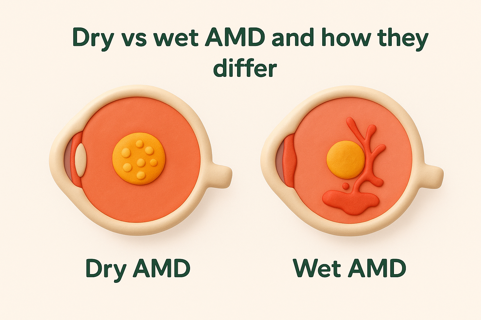
Dry vs wet AMD and how they differ
Dry AMD accounts for around 85 to 90 per cent of cases and progresses slowly as retinal cells thin and accumulate deposits called drusen. Wet AMD is less common but far more aggressive, developing when abnormal blood vessels grow beneath the macula and leak fluid, causing rapid central vision loss.
How imaging confirms AMD and monitors progression
Optical coherence tomography, or OCT, gives your specialist a detailed cross-section of the retina to detect fluid, structural changes, and drusen deposits. Fluorescein angiography may also be used to assess blood vessel leakage in wet AMD.
Treatment options: injections, lifestyle changes, supplements
Wet AMD responds to intravitreal anti-VEGF injections, which reduce abnormal vessel growth and protect remaining vision. For dry AMD, AREDS2 nutritional supplements can slow progression in intermediate cases, alongside quitting smoking and managing cardiovascular risk factors.
Sudden vision changes that need urgent assessment
Any rapid change in central vision, such as new distortion, a sudden blank area, or noticeably worsened blur, should prompt same-day specialist review. Early intervention in wet AMD significantly improves the chance of preserving functional sight.
5. Diabetic retinopathy
Diabetic retinopathy is one of the most serious vision conditions linked to a systemic health condition, affecting around one in three Australians living with diabetes. Early detection and regular monitoring are essential, because damage to the retina can progress significantly before you notice any visual change.
How diabetes damages the retina over time
Consistently elevated blood glucose weakens the small blood vessels that supply the retina. Over time, these vessels leak, swell, or grow abnormally, starving retinal tissue of oxygen and causing progressive structural damage to the light-sensitive cells you rely on for clear sight.
Symptoms and stages, including diabetic macular oedema
Early diabetic retinopathy often produces no noticeable symptoms, which is why so many people discover it during a routine eye examination. As the condition advances, you may notice blurred vision, floaters, or dark patches across your visual field. Diabetic macular oedema, where fluid accumulates beneath the central retina, can cause significant central vision loss even in earlier stages.
Screening timelines and how often to get checked
The earlier diabetic retinopathy is detected, the more treatment options are available and the better your chance of preserving functional vision.
If you have type 1 diabetes, annual retinal screening should begin five years after diagnosis. People with type 2 diabetes should start screening at diagnosis and repeat it annually, or more frequently if changes are already present.
Tests and scans used to assess diabetic eye disease
A dilated fundus examination and OCT imaging together give your specialist the clearest picture of retinal health. Fluorescein angiography can confirm whether abnormal vessel growth or active leakage is present.
Treatment options: diabetes control, laser, injections, surgery
Managing your blood glucose, blood pressure, and cholesterol reduces the rate of progression and remains central to any treatment plan. Anti-VEGF intravitreal injections address macular oedema and abnormal vessel growth effectively, laser photocoagulation targets leaking vessels, and vitrectomy surgery is available for advanced cases.
Warning signs that need same-day care
Sudden vision loss, a sharp increase in floaters, or a shadow across your visual field all require urgent specialist assessment that same day. These symptoms can indicate a vitreous haemorrhage or retinal detachment, both of which need prompt treatment to limit permanent damage.
A clear plan for what to do next
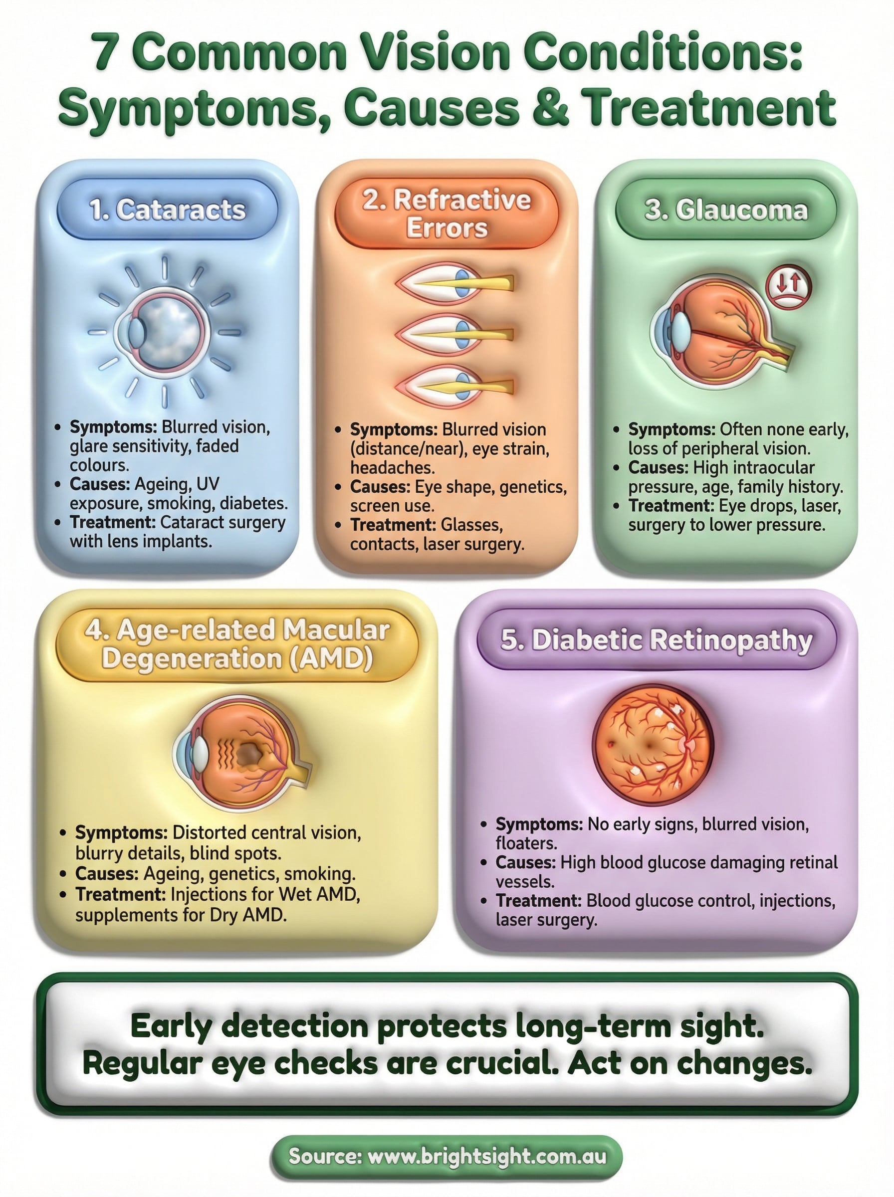
The vision conditions covered in this guide share one important characteristic: outcomes are consistently better when problems are caught early. Whether you’re managing an existing diagnosis, noticing new symptoms, or simply overdue for a check, the most useful step you can take is booking a thorough assessment with a specialist who has the imaging and surgical experience to act on whatever they find.
At Bright Sight Eye Specialists in Randwick, Dr Rahul Dubey and Dr Capucine Odouard offer comprehensive consultations across the full range of conditions discussed here, from cataracts and refractive errors through to retinal disease. Both surgeons bring subspecialty training and a clear, practical approach to helping you understand your options and make confident decisions about your eye health. Telehealth is also available for patients in regional areas.
Book a consultation at Bright Sight Eye Specialists and take the first step towards protecting your sight.