


Seeing a tiny black dot drift across your vision can be unsettling, especially if you’ve never experienced it before. That dark speck floating when you look at a bright sky or white wall is usually what eye specialists call a floater. It’s a small particle suspended in the vitreous humour, the clear gel filling the space inside your eyeball. While most floaters are harmless remnants of the natural aging process, they can sometimes signal a serious problem that requires urgent attention from an ophthalmologist.
This article explains what causes those black dots and how to tell the difference between ordinary floaters and dangerous warning signs. You’ll learn when to simply monitor the situation and when to seek immediate care from an eye specialist. We’ll cover who faces higher risk of complications, how eye specialists diagnose the underlying cause, and what treatment options exist. You’ll also discover practical steps to protect your eyesight and prevent potential complications. By the end, you’ll know exactly how to respond if you notice a tiny black dot in your vision and when that dot demands prompt medical attention.
Why a tiny black dot in your vision matters
Your eyes provide constant feedback about their internal health, and a tiny black dot floating through your field of view acts as one of those signals. Most people experience floaters at some point, particularly as they age, and in the majority of cases these spots pose no immediate threat to your sight. However, that same symptom can occasionally indicate a retinal tear, vitreous haemorrhage, or even early retinal detachment. Because these conditions can lead to permanent vision loss if left untreated, you need to understand when that harmless-seeming dot demands urgent evaluation from an ophthalmologist.
The difference between harmless and dangerous
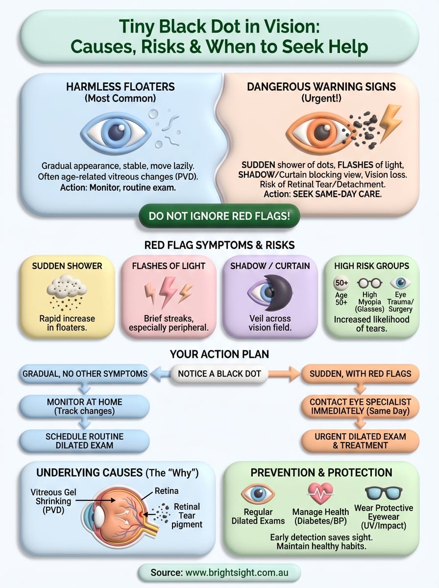
Benign floaters typically appear gradually over time and drift slowly across your vision without any accompanying symptoms. You might notice one or two specks that have been present for months or years, moving lazily when you shift your gaze. Dangerous floaters, by contrast, arrive suddenly and in large numbers, often accompanied by flashes of light or a shadow creeping across your peripheral vision. This distinction matters because it tells you whether the vitreous is simply aging normally or whether it has torn your retina in the process.
A sudden shower of floaters requires same-day assessment, while a single stable speck can usually be monitored at your next routine eye exam.
What your vision is telling you about your eye health
When you notice a tiny black dot in vision, your eye is showing you physical changes happening inside the vitreous gel. These changes reveal how your vitreous humour is contracting and pulling away from the retina, a process that accelerates with age, trauma, or underlying conditions like high myopia. Recognising these visual cues allows you to act quickly when needed and avoid unnecessary worry when the floaters are benign, giving you control over your eye health outcomes.
How to respond when you see a tiny black dot
Your first reaction to noticing a tiny black dot in vision should be calm observation, not panic. Most floaters are benign, but your response depends entirely on the pattern and timing of what you’re experiencing. You need to ask yourself specific questions about the floater’s appearance, how many you can see, whether any other symptoms accompany it, and how quickly it appeared. This immediate assessment determines whether you need urgent care or can simply monitor the situation.
Assess the situation immediately
Stop what you’re doing and test both eyes separately by covering one eye at a time. Check whether the black dot appears in one eye or both, because floaters affecting only one eye typically originate from vitreous changes in that specific eye. Note the size, shape, and number of dots you can see. A single small speck that drifts lazily across your vision differs significantly from a sudden shower of dozens of tiny particles. Pay attention to how long the floater has been present, too. If you’ve noticed it gradually over weeks or months, the situation differs from a dot that appeared within the last few hours.
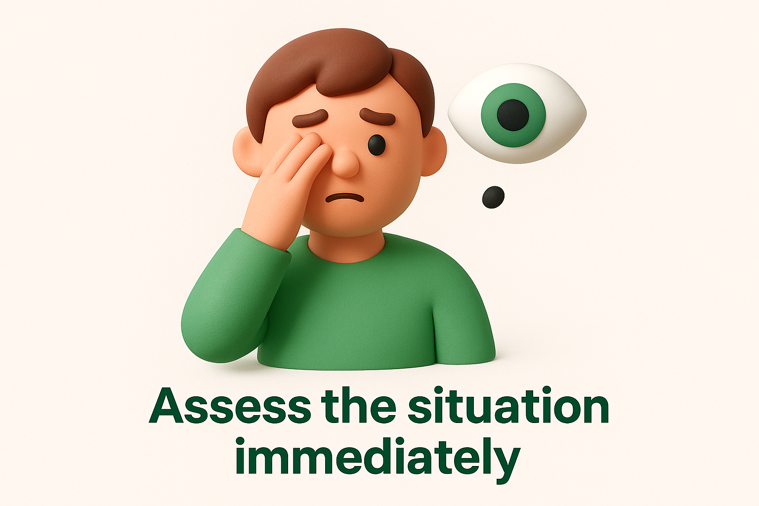
Look for any accompanying symptoms that might signal a more serious problem. Ask yourself whether you can see flashes of light, like brief lightning streaks in your peripheral vision. Check whether any part of your visual field appears shadowed or blocked, as though a curtain is drawing across your sight. These additional symptoms transform a simple floater into a potential emergency that requires immediate evaluation from an ophthalmologist.
When to seek same-day care
You must contact an eye specialist within hours if you notice any of these warning signs alongside your floater:
- A sudden increase in floaters, especially if they appear as a shower or cloud of spots
- Flashes of light in the same eye as the floater
- A shadow or curtain moving across any part of your vision
- A noticeable decrease in your visual clarity or field of vision
These symptoms suggest possible retinal tear or detachment, conditions that can cause permanent vision loss without rapid treatment. Ring your ophthalmologist immediately or visit an emergency eye clinic if your regular specialist isn’t available.
Even if symptoms occur on a weekend or after hours, retinal emergencies cannot wait until Monday morning.
What to monitor at home
If you see one or two stable floaters without any warning signs, you can track them at home while scheduling a routine dilated eye exam within the next few weeks. Keep a simple diary noting when you see the floater, its appearance, and whether it changes in size or number. Document any new symptoms that develop, particularly flashes of light or shadows. This information helps your ophthalmologist understand whether your vitreous is simply aging normally or whether the floaters signal an evolving retinal problem that needs intervention.
What a tiny black dot in vision usually means
That tiny black dot in vision almost always turns out to be an eye floater, a harmless shadow cast on your retina by microscopic debris floating in the vitreous humour. This clear gel fills the space between your lens and retina, helping maintain your eyeball’s round shape while allowing light to pass through. When tiny clumps of protein or collagen fibres form within this gel, they block small amounts of light and cast shadows that your brain interprets as floating specks. These shadows drift because they’re suspended in a liquid-like substance, moving as your eye moves and appearing to dart away when you try to look directly at them.
The role of vitreous humour in your eye
Your vitreous humour consists of 98 per cent water mixed with collagen fibres and hyaluronic acid, giving it a jelly-like consistency. This gel sits behind your lens and in front of your retina, occupying about two-thirds of your eyeball’s volume and maintaining constant pressure to keep your eye’s shape. When you’re young, the vitreous remains uniformly thick and transparent, allowing light to pass cleanly through to your retina without casting any shadows.
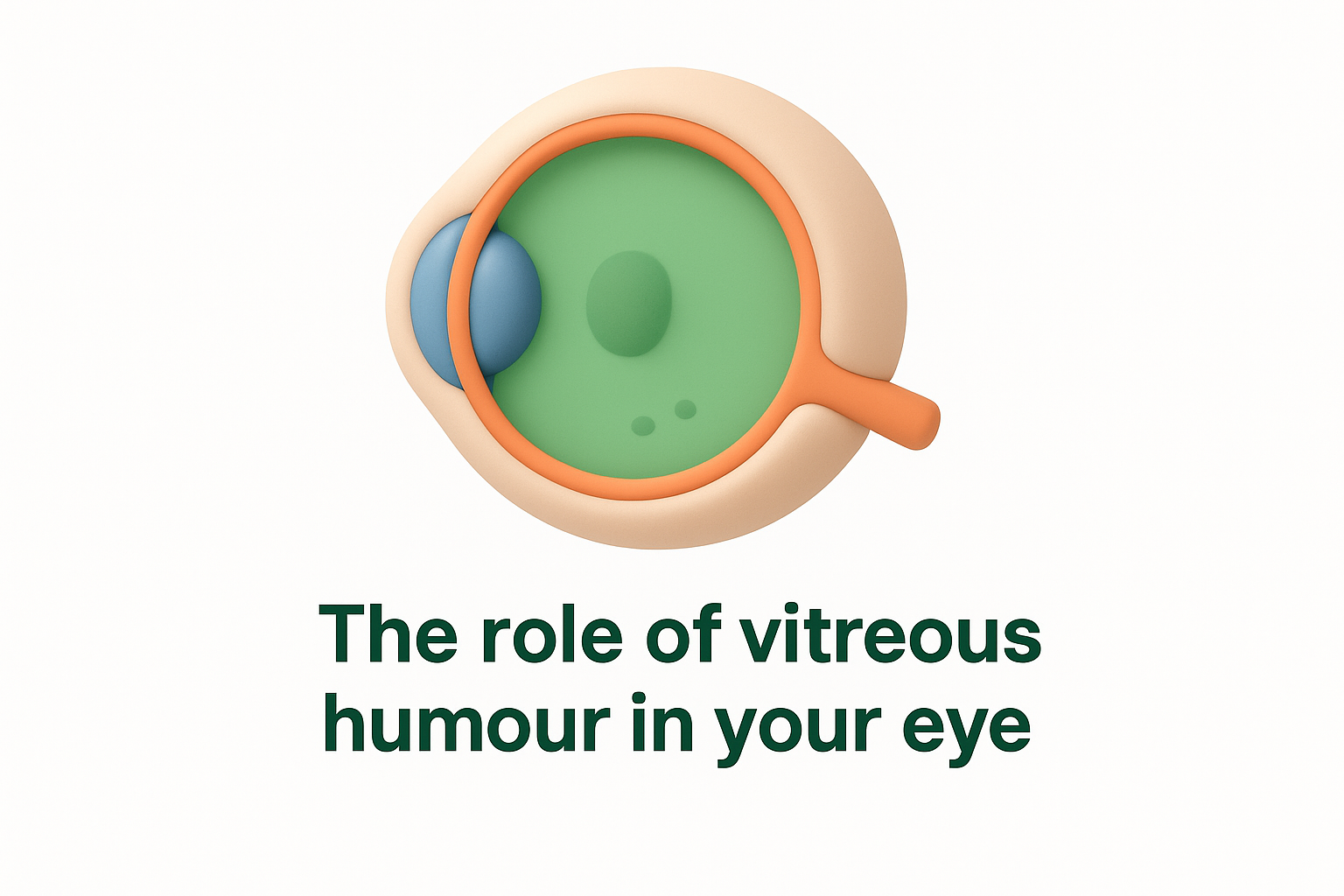
As decades pass, the vitreous gradually liquefies and contracts, breaking down from a firm gel into a more watery substance with floating strands of collagen. These changes are completely normal and occur in virtually everyone over the age of 50, though they can happen earlier in people who are severely short-sighted or who have experienced eye trauma.
Posterior vitreous detachment: the most common cause
Posterior vitreous detachment occurs when the vitreous gel pulls away from your retina’s surface, a natural consequence of the aging process that happens to most people by their seventies. As the vitreous shrinks and detaches, it releases tiny fragments of collagen and cellular debris into the liquid centre of your eye. These particles cast shadows on your retina that appear as dots, threads, or cobweb-like shapes drifting across your vision.
Most posterior vitreous detachments are benign events that require monitoring rather than treatment, though they can occasionally trigger retinal tears that need urgent attention.
Why floaters become more noticeable with age
You notice floaters more as you age because the vitreous changes texture and develops more substantial clumps that cast darker shadows. Younger people rarely see floaters because their vitreous remains uniformly clear, with any debris too small to create visible shadows. After age 40, the vitreous begins liquefying, and by age 60, most people have at least a few permanent floaters that they’ve learned to ignore through neural adaptation, where your brain filters out these repetitive visual distractions.
Serious causes and warning signs to never ignore
While most floaters remain harmless throughout your life, certain serious conditions can announce themselves through a sudden appearance of black dots in your vision. These conditions threaten your sight and demand immediate evaluation from an ophthalmologist, not a wait-and-see approach. Understanding the difference between benign aging changes and vision-threatening emergencies protects you from permanent vision loss that might otherwise have been prevented with rapid treatment.
Retinal tear and its progression risk
A retinal tear occurs when your shrinking vitreous pulls forcefully on the retina, creating a full-thickness break in this delicate tissue. This tearing typically happens at weak spots where the vitreous remains firmly attached to your retina, particularly near the edges. The tear itself allows fluid from the vitreous cavity to seep underneath your retina, separating it from the underlying support tissue that provides oxygen and nutrients. Without prompt treatment, this fluid accumulation can progress to complete retinal detachment within days or even hours.
You often notice a retinal tear through a sudden increase in floaters, particularly if they appear as a shower of tiny specks rather than one or two isolated dots. These floaters represent blood cells or pigment released when the retina tears, creating hundreds of small shadows that drift across your visual field. Flashing lights accompany many retinal tears because the mechanical traction on your retina stimulates photoreceptor cells, which your brain interprets as brief lightning-like streaks in your peripheral vision.
Retinal detachment as a medical emergency
Retinal detachment represents one of the most urgent eye emergencies you can experience, requiring surgical repair within hours to days to preserve your sight. Once your retina separates from its underlying layer, the detached portion stops functioning properly because it loses access to essential blood supply. The longer your retina remains detached, the greater your risk of permanent vision loss in the affected area, even after successful surgery.
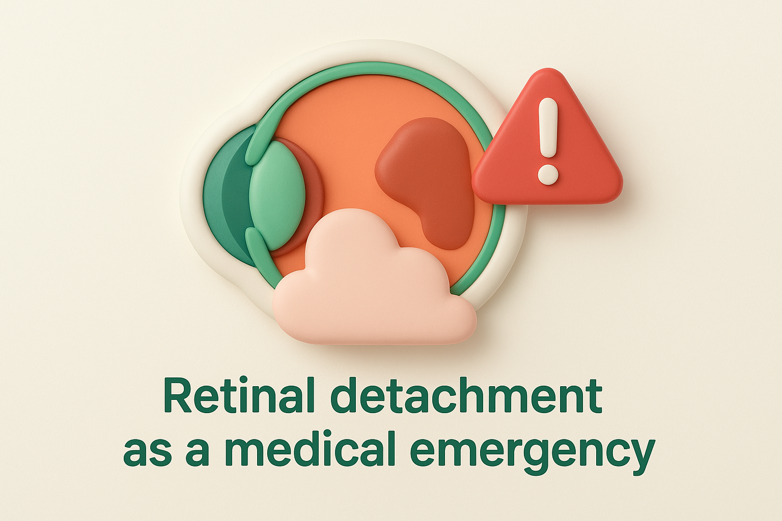
If you notice a shadow or curtain moving across your vision alongside floaters, you must seek emergency eye care immediately, regardless of the time or day.
The classic symptoms of retinal detachment include a curtain-like shadow sweeping across your visual field, often starting from the periphery and moving toward the centre. This shadow represents the area where your detached retina can no longer transmit visual information to your brain. Many people also experience a dramatic increase in floaters and persistent flashing lights just before or during the detachment.
Vitreous haemorrhage and other urgent conditions
Vitreous haemorrhage occurs when blood leaks into your vitreous cavity, creating what appears as a cloud of black or red-tinged floaters. This bleeding can result from a torn retinal blood vessel, advanced diabetic retinopathy, or trauma to your eye. The blood cells floating in your vitreous cast dense shadows that can significantly blur your vision, sometimes described as looking through a red haze or seeing hundreds of tiny dots.
Other serious causes include posterior uveitis, where inflammation in your eye releases white blood cells into the vitreous that appear as floaters. Infections, autoimmune conditions, or inflammatory diseases can trigger this inflammation. Any sudden change in floaters accompanied by eye pain, redness, or light sensitivity warrants urgent evaluation.
Red flag symptoms that demand immediate action
You must contact an ophthalmologist the same day if you experience any combination of these warning signs alongside a tiny black dot in vision:
- Sudden onset of multiple new floaters within hours
- Flashes of light, particularly if they persist or increase in frequency
- A shadow, veil, or curtain blocking any part of your vision
- Rapid decrease in visual clarity or sharpness
- Loss of peripheral vision on one side
These symptoms indicate that your vitreous or retina has undergone a sudden change that threatens your sight, requiring immediate dilated examination to determine whether surgery or other urgent treatment is necessary.
Who is at higher risk of floaters and retinal tears
Certain groups face significantly higher odds of developing floaters that progress beyond harmless aging changes into serious retinal complications. Understanding your personal risk factors helps you decide whether to schedule routine monitoring or seek more frequent eye examinations. Age, eye structure, medical history, and underlying health conditions all influence your likelihood of experiencing a tiny black dot in vision that signals something more serious than simple vitreous debris.
Age and natural vitreous changes
Your risk of posterior vitreous detachment and subsequent floaters increases dramatically after age 50, when the vitreous humour begins its natural liquefaction process in most people. By age 60, approximately two-thirds of adults have experienced some degree of vitreous detachment, with that number rising to nearly 100 per cent by age 80. While these age-related changes usually remain benign, older adults face higher rates of retinal tears during the detachment process because their vitreous often pulls more forcefully on weakened retinal tissue.
High myopia and elongated eyes
Severe short-sightedness stretches your eyeball into an elongated shape, thinning both the vitreous gel and the retinal tissue. This anatomical difference means your vitreous experiences greater traction on your retina when it shrinks and detaches, creating increased risk of tears compared to people with normal vision. If you require glasses stronger than minus six dioptres, you need more frequent dilated eye examinations regardless of your age.
People with high myopia can develop floaters and retinal complications decades earlier than the general population, sometimes as early as their thirties or forties.
Previous surgery and trauma
Any history of eye surgery or significant trauma raises your risk profile for vitreous complications and retinal tears. Cataract surgery, while generally safe, can accelerate vitreous liquefaction and detachment. Blunt trauma to your eye disrupts the delicate vitreous-retinal interface, creating weak spots where tears develop months or even years after the injury.
How eye specialists diagnose and treat black dots
When you report a tiny black dot in vision, your ophthalmologist follows a systematic diagnostic approach to determine whether your floaters are benign or signal a serious retinal problem. The evaluation begins with a detailed discussion about your symptoms and progresses through specialized examinations that reveal what’s happening inside your eye. Treatment depends entirely on the underlying cause discovered during this assessment, ranging from simple observation to urgent surgical intervention.
Comprehensive eye history and examination
Your ophthalmologist starts by asking specific questions about when you first noticed the black dot, how quickly it appeared, and whether you experience any accompanying symptoms like flashes or shadows. They’ll inquire about your medical history, including diabetes, high blood pressure, previous eye surgeries, and any recent trauma. This conversation helps narrow the possible causes before any physical examination begins.
During the initial examination, your specialist checks your visual acuity and eye pressure, then uses a slit lamp to inspect the front structures of your eye. They look for signs of inflammation, bleeding, or other abnormalities that might explain your floaters. This preliminary assessment determines how urgently you need a dilated retinal examination.
Dilated retinal examination
The dilated exam provides your ophthalmologist with a clear view of your vitreous humour and retina, essential for identifying the source of your floaters. Special drops widen your pupils, allowing your specialist to use an ophthalmoscope or indirect lens to examine the back of your eye in detail. This process takes about 20 to 30 minutes once the drops take full effect.
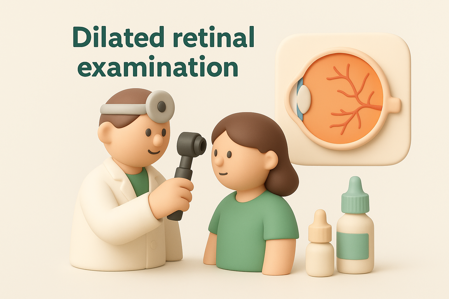
Your ophthalmologist carefully inspects your vitreous for debris, blood, or inflammatory cells while examining your retina for any tears, holes, or areas of detachment. They pay particular attention to the peripheral retina, where tears most commonly develop. The dilated exam remains the gold standard for detecting retinal problems that cause floaters.
A thorough dilated examination can identify retinal tears before they progress to detachment, potentially saving your sight with simple laser treatment.
Advanced imaging techniques
Your specialist may order additional tests if the dilated exam reveals concerns or if clouding in your vitreous prevents a clear view of your retina. Optical coherence tomography (OCT) creates detailed cross-sectional images of your retina, revealing subtle changes in its layers that might not be visible during standard examination. B-scan ultrasound uses sound waves to map the structures inside your eye when blood or inflammation obscures the view, helping detect retinal detachments or vitreous haemorrhages.
Treatment options based on diagnosis
Treatment varies dramatically depending on what your ophthalmologist discovers during the diagnostic process. If you have benign floaters from posterior vitreous detachment without retinal tears, your specialist typically recommends observation with follow-up examinations to ensure no complications develop. Your brain usually adapts to these floaters within weeks or months, making them less noticeable over time.
When imaging reveals a retinal tear, your ophthalmologist performs laser photocoagulation or cryotherapy to seal the break and prevent fluid from tracking underneath your retina. These procedures create scar tissue that bonds your retina to the underlying layer, stopping progression to detachment. Both treatments work best when performed within days of discovering the tear.
Retinal detachment requires surgical repair through vitrectomy, scleral buckle, or pneumatic retinopexy, depending on the location and extent of the detachment. Vitrectomy involves removing the vitreous gel and using gas or silicone oil to reattach your retina while it heals. Success rates exceed 90 per cent when surgery occurs promptly, though you may need multiple procedures in complex cases.
Protecting your eyesight and preventing complications
You can take specific steps to reduce your risk of developing serious complications from a tiny black dot in vision, whether you already experience floaters or want to prevent them from becoming problematic. Prevention focuses on maintaining healthy eye structures and catching problems early, before they progress to vision-threatening conditions. Regular monitoring, lifestyle modifications, and prompt attention to new symptoms create the foundation for long-term eye health and reduce your likelihood of retinal tears or detachments.
Schedule regular dilated eye examinations
Your ophthalmologist can detect early warning signs of retinal problems months or years before you notice any symptoms yourself. Book a dilated eye examination at least once every two years if you’re under 50 with no risk factors, increasing to annual examinations after age 50 or if you have high myopia. People with diabetes, previous eye surgery, or a family history of retinal detachment need even more frequent monitoring, typically every six to twelve months depending on your specialist’s recommendations.
Regular dilated examinations allow your ophthalmologist to identify and treat retinal tears before they progress to detachment, often preventing surgery altogether.
Keep track of any changes in your floaters between scheduled appointments, noting when new dots appear, whether existing ones increase in number, or if you experience any flashes of light. Report these changes at your next examination or contact your specialist immediately if symptoms suggest a potential emergency.
Manage underlying health conditions
Diabetes and high blood pressure damage the delicate blood vessels in your retina, increasing your risk of vitreous haemorrhage and other complications that appear as floaters. Control your blood sugar levels within the target range your doctor recommends, and maintain your blood pressure below 130/80 mmHg to protect your retinal circulation. Schedule regular check-ups with your general practitioner or endocrinologist to monitor these conditions and adjust medications as needed.
Protect your eyes from injury and UV damage
Wear protective eyewear during activities that pose impact risks to your eyes, including sports, construction work, and yard maintenance with flying debris. Choose sunglasses that block 100 per cent of UVA and UVB rays whenever you’re outdoors, reducing long-term damage to your vitreous and retina. Quit smoking if you currently smoke, as tobacco accelerates vitreous degeneration and increases your risk of posterior vitreous detachment occurring earlier in life with greater complications.
Next steps for your eye health
You now understand the difference between harmless floaters and symptoms that require urgent attention, giving you the knowledge to respond appropriately when you notice a tiny black dot in vision. Take action immediately if you experience a sudden shower of floaters, flashing lights, or shadow across your vision by contacting an eye specialist the same day. Schedule routine dilated examinations according to your age and risk factors, allowing your ophthalmologist to catch potential problems before they threaten your sight.
Keep a simple record of any visual changes you notice, including when they started and how they’ve progressed. This information helps your eye specialist make accurate diagnoses and determine whether treatment is necessary. If you’re in Sydney’s Eastern Suburbs and need expert assessment of floaters or other retinal concerns, Bright Sight Eye Specialists offers comprehensive retinal examinations and advanced surgical solutions under the care of Dr. Rahul Dubey, who trained at Moorfields Eye Hospital. Your sight deserves specialist attention.