


A small dark spot drifting across your vision can appear out of nowhere. One moment you’re reading or looking at the sky, the next you notice a tiny shadow moving with your eyes. These spots, commonly known as floaters, are usually harmless bits of debris casting shadows on your retina. Most result from natural ageing changes in the gel inside your eye. Yet sometimes they signal urgent problems like retinal tears or detachment.
This guide explains what causes small dark spots in your vision and helps you decide when to watch and wait versus when to seek immediate care. You’ll learn about benign floaters and serious retinal conditions, discover red flag symptoms that demand same-day attention, and understand how Australian eye specialists diagnose and treat these visual disturbances. By the end, you’ll know exactly what steps to take to protect your sight.
Why a small dark spot in vision matters
A small dark spot in your vision affects more than your comfort. These floaters can distract you while driving, reading, or working on screens, pulling your focus away from tasks that demand concentration. You might find yourself constantly trying to look around the spot or blinking repeatedly, hoping it will disappear. Beyond the nuisance, these dark specks sometimes signal urgent retinal problems that threaten your sight if left untreated.
Impact on your daily activities
Persistent floaters interfere with precision tasks where clear vision matters most. When you’re threading a needle, reading fine print, or watching your grandchildren play, that drifting shadow can block critical details. Bright backgrounds like white walls or blue skies make floaters more noticeable, so outdoor activities and computer work become frustrating. Your quality of life suffers when you constantly adjust your gaze or squint to see past the spots, and anxiety about what’s causing them adds mental strain.

Even harmless floaters deserve attention if they disrupt your day-to-day comfort and independence.
Early warning signs of serious conditions
Most floaters stem from benign age-related changes, but sudden new spots can indicate retinal tears or detachment. Your retina lacks pain receptors, so serious damage often presents with visual symptoms alone before permanent vision loss occurs. Catching tears early means simple laser treatment may prevent detachment, while delayed diagnosis often requires complex surgery with lower success rates. That single dark spot you notice today could be your retina’s only way of alerting you to trouble, making prompt evaluation essential for preserving your sight.
How to respond when you first notice a dark spot
Spotting a small dark spot in vision can trigger worry, but your first response shapes how well you protect your sight. You don’t need to panic, yet you shouldn’t ignore new visual changes either. Taking systematic steps to assess and document what you’re experiencing helps you and your eye specialist determine whether you need urgent care or simple monitoring.
Immediate steps to take
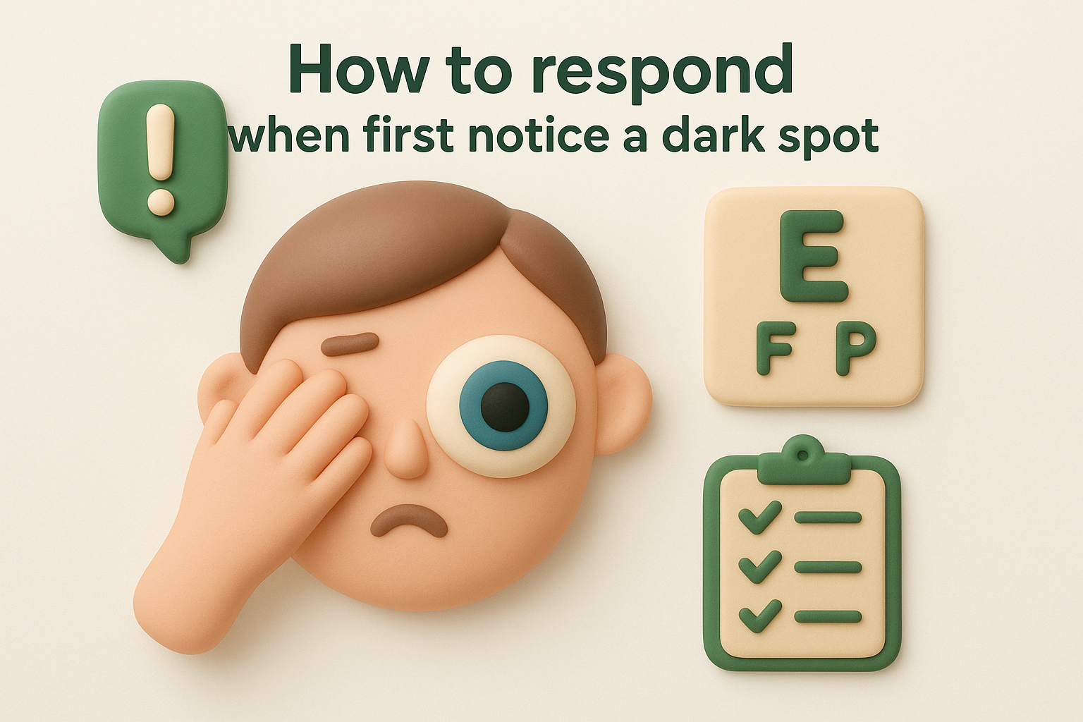
Stop what you’re doing and test each eye separately. Cover one eye with your palm, then look around slowly to check if the spot appears in the uncovered eye. Switch to the other eye and repeat. This tells you whether the problem affects one eye or both, which is crucial for diagnosis. Next, move your eyes up, down, left, and right to see if the dark spot drifts or stays fixed in your vision. Floaters typically move with your gaze and settle when your eyes stop, while retinal problems may create stationary shadows.
What to observe and document
Pay close attention to the spot’s characteristics and any accompanying symptoms. Note its shape (dot, thread, cobweb, or ring), size, and whether it appears alone or alongside multiple new floaters. Write down the exact date and time you first noticed the spot, along with what you were doing when it appeared. Record whether you see flashes of light, experience a curtain-like shadow across your vision, or notice any sudden changes in your sight’s clarity or field.
Your detailed observations give your ophthalmologist the information needed to assess urgency and plan appropriate tests.
When to seek same-day care
Contact an eye specialist immediately if you experience a sudden shower of floaters, especially dozens of new spots appearing within minutes or hours. Flashes of light in the same eye as the dark spot, particularly repeated flashes in your peripheral vision, demand same-day assessment. Any sensation of a curtain or shadow moving across your field of view signals possible retinal detachment, requiring emergency care. Even without these red flags, you should schedule an examination within a few days if the dark spot persists or bothers you during daily activities.
Common harmless causes of small dark spots
Most small dark spots you see floating across your vision stem from natural ageing processes inside your eye. These benign changes affect the vitreous humour, the clear gel filling the space between your lens and retina. While the spots can annoy you, they rarely threaten your sight. Understanding these harmless causes helps you distinguish routine floaters from symptoms that need urgent assessment, saving you unnecessary worry while keeping you alert to genuine problems.
Posterior vitreous detachment
Posterior vitreous detachment (PVD) is the most common benign cause of floaters and accounts for the majority of small dark spots people notice after age 50. As you age, the vitreous gel inside your eye gradually liquefies and shrinks, pulling away from the retina’s surface. This separation releases tiny collagen fibres and cellular debris into the vitreous cavity, where they cast shadows on your retina that appear as drifting spots, threads, or cobwebs.
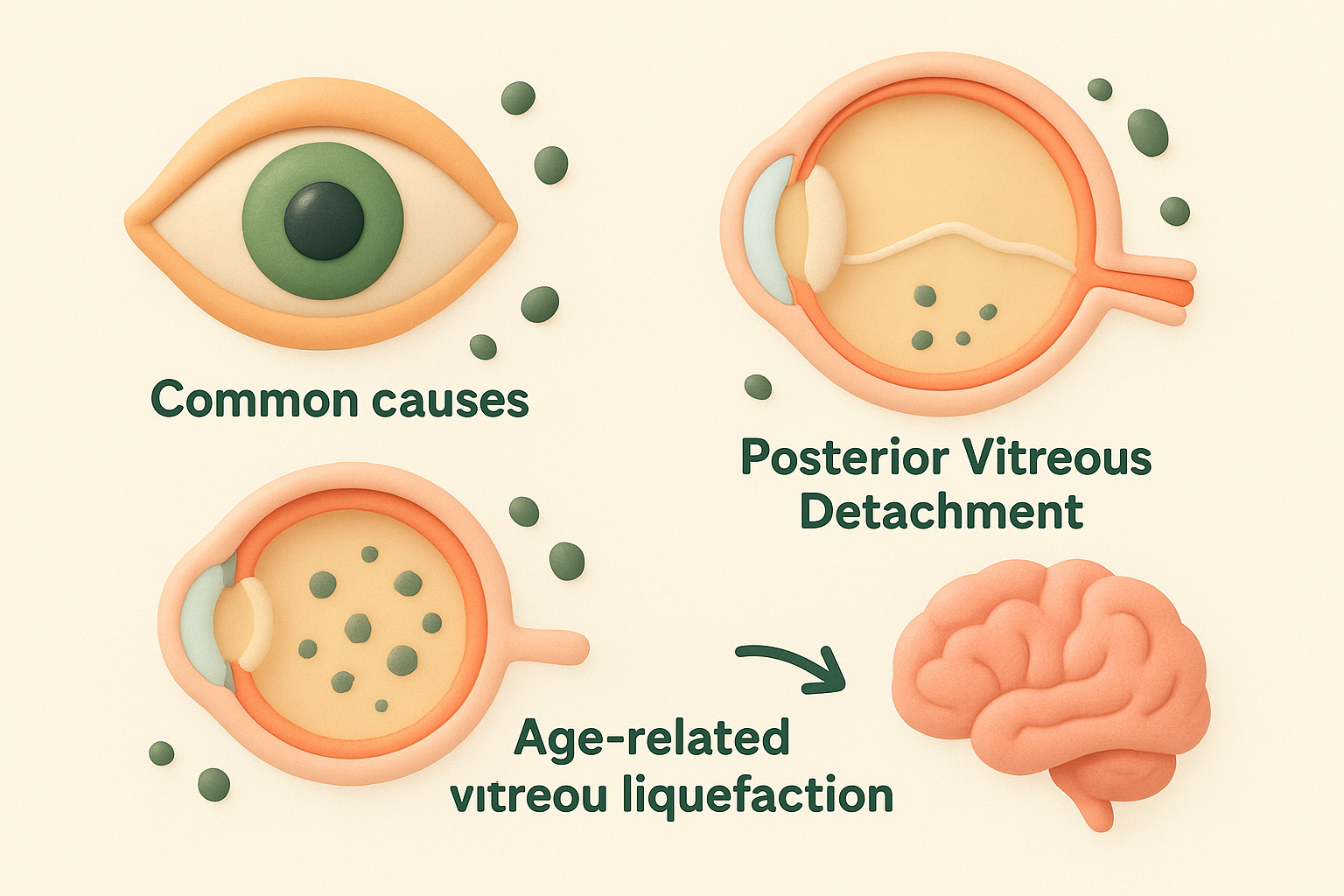
PVD develops naturally in nearly everyone as they reach middle age and beyond. You’re more likely to experience it earlier if you’re nearsighted, have had cataract surgery, or experienced eye trauma. The process typically takes weeks to months, during which you may notice new floaters appearing gradually or in small groups.
While PVD itself is harmless, the tugging motion as the vitreous separates can occasionally create retinal tears, which is why sudden new floaters always warrant examination.
Age-related vitreous liquefaction
Even without complete detachment, your vitreous gel undergoes structural changes throughout your life. The collagen network that gives the gel its structure slowly breaks down and clumps together, forming small aggregates suspended in the increasingly liquid vitreous. These clumps block light and create the floating shadows you perceive as spots or threads.
This liquefaction process accelerates after age 40 and continues gradually for decades. Your genetics, nutrition, and overall eye health influence how quickly the vitreous ages, but everyone experiences some degree of this change eventually.
How your brain adapts over time
Your brain possesses a remarkable ability to filter out unchanging visual information, including persistent floaters. Within weeks to months of noticing a small dark spot in vision, most people find they stop paying attention to it during daily activities. This process, called neural adaptation, doesn’t remove the floater but makes it less intrusive as your visual system learns to ignore the shadow. Floaters become most noticeable against bright, uniform backgrounds like white walls or clear skies, but fade into the background during complex visual scenes where your brain focuses on more important information.
Serious causes and red flag symptoms
While most small dark spots in vision result from harmless floaters, certain causes demand immediate medical attention to prevent permanent sight loss. Your retina contains no pain receptors, so serious damage often announces itself through visual symptoms alone before irreversible changes occur. Recognising these warning signs and acting quickly can mean the difference between a simple outpatient procedure and complex surgery with uncertain outcomes.
Retinal tears and detachment
Retinal tears occur when the shrinking vitreous gel tugs on weak points in your retina, creating a full-thickness break in the tissue. These tears allow fluid to seep underneath the retina, separating it from the nourishing layer beneath. Once detached, that portion of your retina stops functioning properly, creating blind spots in your vision. Without prompt surgical repair, the detachment can spread across your entire retina, leading to permanent vision loss.

Your risk increases if you’re highly nearsighted, have had previous eye surgery or trauma, or have a family history of retinal problems. The tears themselves produce sudden showers of floaters, often described as hundreds of tiny specks appearing within minutes. You might also see repeated flashes of light in your peripheral vision as the vitreous pulls on your retina.
Retinal detachment is a medical emergency that requires same-day assessment and urgent surgical intervention to preserve your sight.
Vitreous haemorrhage
Bleeding inside your eye, known as vitreous haemorrhage, releases blood cells into the clear gel filling your eyeball. These cells appear as numerous dark spots, sometimes accompanied by a red haze across your vision. Common causes include diabetic retinopathy, retinal tears, trauma, or abnormal blood vessel growth. Unlike typical floaters, vitreous haemorrhage often produces dense clouds of spots that significantly impair your vision and may worsen over hours or days.
Diabetic patients face higher risk, particularly if blood sugar control has been poor. The bleeding can signal severe retinal damage requiring immediate treatment to prevent further complications and permanent vision loss.
Red flag symptoms requiring immediate care
Certain symptoms signal urgent retinal problems that need same-day evaluation by an ophthalmologist. Seek immediate care if you notice a sudden shower of new floaters appearing within minutes to hours, especially dozens or hundreds of tiny specks. Flashes of light in your peripheral vision, particularly repeated flashes in the same eye as a small dark spot in vision, indicate vitreous traction on your retina.
Any sensation of a curtain or shadow moving across your field of view suggests retinal detachment in progress. This shadow typically starts at the edge of your vision and advances towards the centre, creating an expanding blind area. You should also seek urgent care if you experience sudden vision loss, significantly decreased visual clarity, or eye pain accompanying new floaters. These symptoms warrant emergency department assessment outside normal clinic hours, as delays in treatment substantially reduce the chances of preserving your full vision.
How doctors diagnose small dark spots in vision
When you report a small dark spot in vision, your eye specialist follows a systematic diagnostic process to determine whether you’re experiencing harmless floaters or a serious retinal problem. This evaluation combines your medical history, a thorough physical examination of your eyes, and potentially advanced imaging technology. The entire process typically takes 30 to 60 minutes and gives your doctor the information needed to recommend appropriate treatment or monitoring. Understanding what to expect during this assessment can ease your anxiety and help you provide the detailed information your specialist needs.
Medical history and symptom assessment
Your ophthalmologist begins by asking specific questions about your symptoms and health background. They’ll want to know exactly when you first noticed the dark spot, whether it appeared suddenly or gradually, and if you’ve experienced any accompanying symptoms like flashes of light, shadows, or vision loss. You’ll be asked about your medical history, including diabetes, high blood pressure, previous eye surgeries, trauma, or family history of retinal disease.
Be prepared to describe the spot’s appearance in detail. Your doctor needs to know its shape, size, and movement pattern, as well as how many spots you see and whether the number has increased. This information helps distinguish between benign posterior vitreous detachment and potentially serious conditions requiring urgent intervention.
Dilated eye examination
The cornerstone of diagnosis is a dilated fundus examination, where special drops widen your pupils to give your specialist a clear view of your retina and vitreous. The dilation takes 15 to 20 minutes to work fully, and the effects last several hours. You’ll experience temporary blurred vision and light sensitivity, so bringing sunglasses and arranging transport home is sensible.
Using a slit lamp microscope and specialised lenses, your ophthalmologist examines the vitreous gel for floaters, debris, or blood. They’ll carefully inspect your retina for tears, holes, thinning areas, or signs of detachment. The examination is painless and allows your doctor to see the full extent of any changes inside your eye.
A thorough dilated examination remains the most reliable way to detect retinal problems before they cause permanent vision loss.
Advanced imaging techniques
In certain cases, your specialist may order additional diagnostic tests to supplement the clinical examination. Optical coherence tomography (OCT) creates detailed cross-sectional images of your retina, revealing subtle abnormalities that aren’t visible during standard examination. This technology proves particularly useful for detecting macular problems or early detachment.
B-scan ultrasound becomes necessary when blood, dense floaters, or other opacities prevent clear visualisation of your retina. The ultrasound waves pass through these obstructions to create images of structures at the back of your eye, helping your doctor identify tears or detachment that would otherwise remain hidden. These imaging tools provide objective documentation that guides treatment decisions and allows precise monitoring of any changes over time.
Treatment options for floaters and retinal disease
Treatment for a small dark spot in vision depends entirely on what’s causing it and how much it affects your daily activities. Your eye specialist will recommend an approach ranging from simple observation to complex surgery, balancing the risks of intervention against the impact on your quality of life. Benign floaters rarely need active treatment, while retinal tears and detachment demand urgent surgical repair to prevent permanent vision loss. Understanding your options helps you make informed decisions about your eye care and know what to expect during recovery.
Observation and watchful waiting
Most harmless floaters caused by posterior vitreous detachment require no treatment at all. Your brain naturally adapts within weeks to months, filtering out the visual distraction so you barely notice the spot during daily activities. Your ophthalmologist will schedule follow-up examinations at intervals suited to your risk profile, typically at three months, six months, and then annually if the floaters remain stable. During this watchful waiting period, you’ll continue normal activities while monitoring for any red flag symptoms that signal complications requiring immediate attention.
Simple observation proves the safest and most appropriate choice when floaters don’t impair your vision or quality of life.
Laser treatment for persistent floaters
Laser vitreolysis offers a minimally invasive option for bothersome floaters that disrupt your work or leisure activities. Your specialist uses a YAG laser to target and vaporise the collagen clumps casting shadows on your retina, breaking them into tiny fragments that become less noticeable or disappear completely. The outpatient procedure takes 20 to 30 minutes and causes minimal discomfort, though you’ll need several treatment sessions spaced weeks apart for optimal results. Success depends on floater location and your eye’s anatomy, with best outcomes when the spots float at least two millimetres away from your retina’s surface.
Vitrectomy surgery
Vitrectomy becomes necessary when floaters severely impair your vision or you develop vitreous haemorrhage that won’t clear on its own. Your surgeon makes tiny incisions in your eye, removes the vitreous gel along with all debris or blood, and replaces it with a clear saline solution that your eye gradually converts to natural fluid. Recovery takes several weeks, during which you may need to maintain specific head positioning if gas or oil was used to support your retina. Risks include cataract formation, retinal detachment, and infection, but the procedure delivers complete relief from persistent floaters that conservative measures can’t resolve.
Surgical repair for retinal tears and detachment
Retinal problems demand prompt surgical intervention to preserve your sight. Small tears can be sealed with laser photocoagulation or cryopexy (freezing treatment), performed as outpatient procedures that prevent fluid from tracking under your retina. Established detachment requires more extensive surgery, either scleral buckle (placing a silicone band around your eye) or vitrectomy with gas or oil injection to reposition and secure your retina. Success rates approach 90 per cent when treatment occurs promptly, though recovery spans weeks to months and may involve temporary vision restrictions while your eye heals.
How to lower your risk and protect your vision
While you can’t prevent all causes of a small dark spot in vision, you can take concrete steps to reduce your risk of serious retinal problems and catch changes early when treatment works best. Protecting your eyes involves a combination of regular professional care, managing health conditions that affect your retina, and making daily choices that support long-term vision health. These strategies work together to keep your eyes functioning well and help you spot potential problems before they threaten your sight.
Regular comprehensive eye examinations
Scheduling routine dilated eye examinations gives your ophthalmologist the chance to detect early warning signs before you notice any symptoms. Your specialist can spot retinal thinning, small tears, or vascular changes during these check-ups, often years before they cause vision problems. These examinations become your first line of defence against conditions that might otherwise progress silently until permanent damage occurs.
Book appointments every one to two years if you’re under 50 with no risk factors, annually after age 50, and more frequently if you have diabetes, high myopia, or previous eye surgery. Don’t wait for symptoms to appear, as many serious retinal conditions develop gradually without producing noticeable changes until advanced stages when treatment options become limited.
Managing underlying health conditions
Keeping diabetes and high blood pressure under tight control protects the tiny blood vessels in your retina from damage that leads to bleeding, swelling, and scarring. Poor blood sugar regulation causes diabetic retinopathy, while uncontrolled hypertension weakens vessel walls and increases haemorrhage risk. Both conditions accelerate the likelihood of vitreous complications that produce persistent floaters or more serious problems.
Work closely with your general practitioner to maintain your HbA1c and blood pressure within target ranges. Regular monitoring and medication adjustments keep these risk factors under control, significantly reducing your chances of developing sight-threatening complications.
Consistent management of systemic diseases delivers better vision outcomes than any treatment available after retinal damage occurs.
Protective measures and lifestyle choices
Wear sunglasses with UV protection whenever you’re outdoors to shield your eyes from radiation that may accelerate vitreous ageing. Choose wrap-around styles that block light from the sides, and look for lenses marked UV400 or 100 per cent UV protection. Protective eyewear during sports or activities with injury risk prevents trauma that can tear your retina or cause vitreous haemorrhage.
Maintain a diet rich in leafy greens, colourful vegetables, and omega-3 fatty acids to support retinal health. Stop smoking, as tobacco damages blood vessels throughout your body, including those nourishing your retina. These simple habits compound over decades, giving you the best chance of preserving clear, comfortable vision well into your later years.

Next steps for your eye health
Noticing a small dark spot in vision demands thoughtful action rather than panic. Schedule a comprehensive eye examination if you’ve experienced any new floaters, particularly if they appeared suddenly or increased in number. Your optometrist can perform an initial assessment and refer you to a specialist if needed. Between appointments, monitor your symptoms daily and note any changes in the spot’s size, number, or behaviour.
Contact an ophthalmologist immediately if you develop flashes of light, a curtain-like shadow, or a sudden shower of new spots. These symptoms signal potential retinal emergencies that require same-day evaluation to prevent vision loss. Don’t wait for your regular appointment when warning signs appear.
Bright Sight Eye Specialists offers comprehensive retinal assessments and treatments, combining Dr Rahul Dubey’s Moorfields-trained expertise with advanced diagnostic technology. Book your eye examination at Bright Sight Eye Specialists to ensure your vision receives the specialist care it deserves.