


You look at a bright wall and notice a tiny dark speck drifting across your vision. You try to focus on it but it slips away. This small black spot is usually a floater—a shadow cast on your retina by clumps of protein or cells floating inside the clear gel that fills your eye. Most floaters are harmless and become less noticeable over time. But sometimes they signal something serious like a retinal tear or detachment that needs immediate treatment.
This article explains what causes small black spots in your vision and when you should worry. You’ll learn how to respond when you first notice one, what separates routine floaters from emergency warning signs, and who faces higher risks. We’ll also cover how eye specialists diagnose these spots, your treatment options, and practical steps to protect your vision. By the end you’ll know exactly when to monitor safely at home and when to seek urgent care.
Why a small black spot in vision matters
Your eyes provide crucial information about threats to your sight that need quick action. A small black spot in vision might seem minor, but it can reveal problems inside your eye that progress rapidly without treatment. The vitreous gel that fills your eyeball naturally changes as you age, often creating harmless floaters that drift through your field of view. However, the same ageing process can pull on your retina with enough force to tear it, allowing fluid to seep underneath and separate it from the nourishing tissue beneath. Once that separation starts, you have hours to days before permanent vision loss sets in.
The difference between harmless and dangerous spots
Harmless floaters usually appear gradually over months or years. You notice one or two spots that move slowly when you shift your gaze, then settle down when your eyes stop moving. They tend to fade from your attention as your brain learns to filter them out. Dangerous spots, by contrast, arrive suddenly as a shower of new specks accompanied by flashes of light or a dark curtain sweeping across your peripheral vision. The sudden onset tells you something has changed inside your eye that demands immediate assessment.

Timing makes the difference between a simple office procedure and complex surgery with risk of lasting impairment.
What happens if you ignore warning signs
Delaying treatment for a retinal tear allows fluid from the vitreous to track through the break and collect under your retina. This detachment spreads gradually, creating a larger blind spot that cannot recover even after successful reattachment surgery. The photoreceptor cells in the detached area lose their blood supply and begin to die within hours. If you wait days or weeks, you risk permanent central vision loss that no surgery can restore. Acting quickly preserves your sight.
How to respond when you first notice a black spot
The moment you spot something drifting across your vision, pause what you’re doing and take a few minutes to assess the situation. Don’t panic, but don’t dismiss it either. Your initial observations will help you decide whether this needs same-day attention or routine monitoring. Most black spots turn out to be harmless floaters, but the way they appear and what accompanies them determines your next steps.
Stop and observe the spot carefully
Look at a plain, light-coloured surface such as a white wall or clear sky. This makes any small black spot in vision stand out clearly. Move your eyes slowly up and down, then side to side, watching how the spot behaves. Does it drift smoothly and settle when your eyes stop moving? That pattern suggests a typical floater. Note the spot’s shape (dot, thread, cobweb), size, and how many you see. If it’s just one or two familiar shapes you’ve noticed before, you’re probably safe to continue monitoring.
Try to remember when you first saw it. A spot that appeared gradually over weeks differs from one that showed up within the past few hours. Write down the time, what you were doing, and any other visual changes you noticed. This information proves valuable if you need to contact a specialist.
Check for emergency warning signs
Scan your entire field of vision for anything unusual beyond the spot itself. Do you see flashes of light in your peripheral vision, especially in dim rooms? Flashes indicate that something is tugging on your retina. Count whether you see a sudden shower of many new floaters instead of just one or two. Look for a dark shadow or curtain moving across part of your sight. Any of these symptoms alongside your black spot signals a potential emergency.
If you experience multiple new floaters, flashes, or a curtain effect, seek same-day assessment from an eye specialist.
Contact your eye care provider
Ring your ophthalmologist or optometrist immediately if you notice any emergency warning signs. Explain that you have sudden visual changes and need urgent assessment. Most practices reserve slots for these situations. If it’s after hours and you have concerning symptoms, head to your nearest hospital emergency department. They can examine your retina and arrange specialist review if needed.
For isolated spots without warning signs, book a routine dilated eye examination within the next few weeks. Your eye care professional will check whether the spot comes from harmless vitreous changes or requires treatment. Keep monitoring daily until your appointment, and contact them sooner if anything changes.
What usually causes a small black spot in vision
Your vitreous humour creates most of the small black spots you see drifting across your vision. This clear, jelly-like substance fills the space between your lens and retina, giving your eyeball its round shape. As you age, the vitreous begins to break down and liquefy, changing from a firm gel into a more watery consistency. When this happens, tiny collagen fibres inside the vitreous clump together and cast shadows on your retina. You perceive these shadows as floating spots, threads, or cobwebs that move when your eyes move.
Posterior vitreous detachment
The most common benign reason behind a small black spot in vision is posterior vitreous detachment, or PVD. This natural ageing process happens when the vitreous gel shrinks and pulls away from your retina. As it separates, small fragments of collagen or cellular debris break off and float freely in the remaining fluid. These particles drift through your field of view, appearing as dark specks or stringy shapes.
PVD typically occurs after age 50, though it can happen earlier if you’re severely short-sighted or have had eye surgery. The process usually affects one eye first, then the other months or years later. While PVD sounds alarming, it rarely causes problems. Your brain gradually learns to ignore these floaters, filtering them out of your conscious awareness within weeks to months. Most people with PVD need no treatment beyond routine monitoring.
How collagen clumps create floaters
The vitreous contains a network of microscopic collagen fibres arranged in a precise pattern. In youth, these fibres stay evenly distributed throughout the gel, keeping it clear and transparent. As decades pass, the gel’s water content increases while its collagen structure weakens. The fibres start to stick together in random clumps instead of remaining separate. Each clump blocks a small amount of light passing through your eye, casting a shadow on the light-sensitive cells of your retina beneath.

These shadows appear to float because they’re suspended in moving fluid rather than attached to any fixed surface.
The size and shape of your floater depends on the clump’s dimensions. A tiny knot of collagen produces a dot, while longer strands create thread-like or cobweb patterns. You see multiple floaters when several clumps form at once. The spots drift across your vision because currents in the vitreous fluid carry them along as you move your eyes. When you try to look directly at a floater, it seems to dart away because you’ve shifted the fluid current, pushing it out of your central line of sight.
Less common causes
Several other conditions produce spots that resemble floaters but stem from different problems. Vitreous haemorrhage occurs when blood leaks into the vitreous gel from damaged retinal vessels, often due to diabetes or trauma. Fresh blood cells appear as numerous dark red or black specks floating through your vision. Uveitis, an inflammatory condition affecting the middle layer of your eye, releases inflammatory cells and protein into the vitreous. These deposits look like floaters but typically arrive with redness, pain, and light sensitivity. Diabetic retinopathy damages tiny blood vessels in your retina, causing microscopic bleeding that shows up as clusters of dark spots. Finally, ocular migraines can produce temporary flickering blind spots or shimmering patterns that mimic floaters but resolve within minutes as the neurological event passes.
Warning signs that mean an emergency
Your eye’s warning system uses specific visual disturbances to alert you to serious problems that need immediate medical attention. While a single small black spot in vision that drifts slowly often indicates a harmless floater, certain patterns signal that something inside your eye has changed dramatically. These emergency symptoms usually point to a retinal tear or detachment that can steal your sight within hours if left untreated. Knowing these warning signs helps you act quickly enough to save your vision.
Sudden shower of new floaters
A rapid appearance of dozens or hundreds of new spots within minutes or hours differs completely from the gradual increase most people experience with age. This shower of floaters looks like pepper shaking through water or tiny insects swarming across your sight. The sudden burst happens when a retinal tear releases cellular debris and blood cells into your vitreous gel. Each particle casts its own shadow, creating the overwhelming number of specks you see. You might notice these floaters are darker or denser than any you’ve had before, sometimes appearing red-tinged if bleeding has occurred.
Contact an ophthalmologist or visit an emergency department the same day you notice a sudden shower of floaters.
Flashes and curtains across your vision
Bright flashes of light in your peripheral vision indicate mechanical traction on your retina. You see these photopsias as brief lightning-like streaks or sparkles, especially noticeable in dim lighting. They occur when the vitreous gel pulls or tugs on retinal tissue, stimulating the light-sensitive cells. Flashes often accompany new floaters, confirming that something is actively disturbing your retina.

A dark shadow or curtain sweeping across your field of view represents an even more urgent problem. This moving veil or barrier appears when your retina detaches and peels away from its supporting layers. The detached portion can no longer transmit visual information, creating a blind zone that advances as the separation spreads. You might describe it as a grey curtain drawing closed from one side or a shadow rising from below.
When time becomes critical
Emergency eye problems progress rapidly, with every hour counting toward preserving your sight. If you experience any combination of these warning signs, stop what you’re doing and seek immediate assessment. Ring your ophthalmologist’s emergency line or head straight to a hospital emergency department with eye care facilities. Arriving within hours rather than days gives specialists the best chance to repair damage before permanent vision loss occurs. Don’t wait until morning if symptoms start at night, and don’t try to sleep it off hoping they’ll improve.
Who is at higher risk of serious eye problems
Understanding your personal risk factors helps you decide when to seek urgent assessment rather than routine monitoring. While anyone can notice a small black spot in vision, certain groups face higher chances of developing retinal tears or detachments. Your age, eye structure, medical history, and underlying health conditions all influence whether a floater signals a benign change or a serious problem requiring immediate intervention.
Age and severe nearsightedness
People over 50 years old experience posterior vitreous detachment more frequently as their vitreous gel naturally liquefies and shrinks. This age-related process increases your risk of retinal traction and tears. Severe myopia (nearsightedness) compounds this risk because your elongated eyeball stretches both the vitreous and retina, making them more vulnerable to tears. If you’re highly short-sighted, you might notice floaters earlier than others and face a greater likelihood of progression to detachment. Schedule annual dilated eye examinations to catch problems before they escalate.
Previous eye surgery or injury
Any procedure or trauma that disturbs your eye’s internal structures predisposes you to complications. Cataract surgery, vitrectomy, or blunt trauma can accelerate vitreous separation and increase retinal tear risk. Even years after surgery, your eyes remain more susceptible to sudden changes. Alert your ophthalmologist about any new floaters or flashes if you’ve had eye procedures.
Your surgical history remains relevant to eye health assessment for life, not just during immediate recovery.
Diabetes and blood pressure problems
Chronic conditions damage the tiny blood vessels in your retina, leading to bleeding, swelling, and scar tissue formation. Diabetic retinopathy causes microscopic haemorrhages that leak blood into your vitreous, appearing as numerous dark spots. Hypertensive retinopathy creates similar vascular damage. Maintaining tight glucose and blood pressure control alongside regular screening reduces your risk substantially.
How specialists diagnose black spots in vision
When you report a small black spot in vision, your eye care professional follows a systematic approach to determine whether you have harmless floaters or a serious retinal problem. The diagnostic process combines your detailed symptom history with specialised examination techniques that reveal what’s happening inside your eye. Your specialist needs to visualise both your vitreous gel and retina clearly to spot any tears, bleeding, or detachment that demands immediate treatment.
The consultation and symptom assessment
Your appointment begins with a thorough discussion about your visual symptoms and medical history. The specialist asks when you first noticed the black spot, how many spots you see, and whether they appeared suddenly or gradually. You’ll describe any accompanying symptoms such as flashes of light, shadows, or vision changes. Questions about your eye health history cover previous surgeries, injuries, and any family history of retinal problems. Your general health matters too, particularly conditions like diabetes or hypertension that affect retinal blood vessels. This conversation helps your specialist assess your risk level and prioritise which diagnostic tests you need.
Dilated fundus examination
The core diagnostic tool involves examining your retina and vitreous under dilation. Special drops widen your pupils, allowing your specialist to use an ophthalmoscope or slit lamp with a high-powered lens to inspect the back of your eye. This painless examination reveals floaters floating in the vitreous, signs of posterior vitreous detachment, and any retinal tears, holes, or areas of thinning. Your specialist checks for retinal detachment by looking for lifted edges or folds in the retinal tissue. The examination takes 15 to 20 minutes, though your vision stays blurred for several hours afterwards due to the dilating drops.
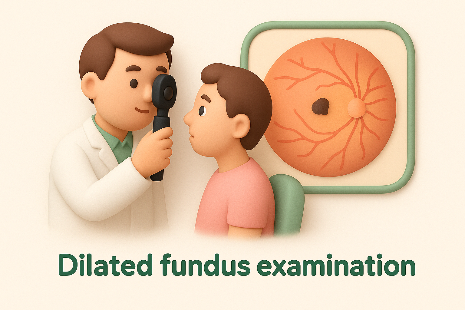
Dilation remains essential because it provides the clearest view of structures at the back of your eye.
Advanced imaging when needed
Some cases require additional tests for a complete assessment. Optical coherence tomography (OCT) produces detailed cross-sectional images of your retina, revealing subtle changes invisible during standard examination. Your specialist might order a B-scan ultrasound if bleeding or cloudiness obscures the view through your pupil. This ultrasound technique maps your eye’s internal structures using sound waves, detecting tears or detachment even when direct visualisation proves impossible. Your eye care professional documents all findings with photographs or diagrams, establishing a baseline for monitoring any changes during follow-up appointments.
Treatment and prevention options
Your treatment path depends entirely on what’s causing your small black spot in vision and how severely it affects your daily activities. Most people with benign floaters need no intervention beyond routine monitoring, while retinal tears or detachments demand urgent surgical repair. Between these extremes lie options like laser treatment for persistent floaters that interfere with your quality of life. Prevention focuses on protecting your eyes from injury and managing systemic health conditions that damage retinal blood vessels.
When observation alone is enough
Doctors recommend watchful waiting when your black spots result from a stable posterior vitreous detachment without warning signs. Your floaters pose no threat to your vision and typically become less noticeable as your brain adapts to filter them out. Schedule follow-up examinations at intervals your specialist suggests, usually every few months initially, then annually once stability is confirmed. Between appointments, monitor for any sudden increases in floaters, new flashes, or vision changes that signal complications. This conservative approach works well because most benign floaters fade from your conscious awareness within weeks to months without any treatment.
Laser treatment for bothersome floaters
Laser vitreolysis offers a minimally invasive solution when floaters significantly disrupt your central vision or interfere with reading and driving. Your ophthalmologist uses a specialised laser to target and break up the collagen clumps causing your floaters, fragmenting them into pieces too small to cast noticeable shadows. The procedure happens in the consulting room under local anaesthetic drops, taking 15 to 30 minutes per session. You return home the same day with minimal restrictions. Success depends on floater location (they must sit at least 2 millimetres from your retina) and your eye’s anatomy. Most patients notice marked improvement though complete elimination isn’t always possible.
Laser vitreolysis provides relief without the risks associated with more invasive surgery.
Surgical options for serious problems
Retinal tears and detachments require prompt surgical intervention to preserve your sight. Your surgeon might perform a vitrectomy, removing the vitreous gel along with any floaters or blood and replacing it with saline solution. Alternatively, a scleral buckle involves placing a silicone band around your eye’s exterior to relieve traction and close tears. For established detachments, specialists inject gas or silicone oil to hold your retina in position while healing occurs. These procedures boast success rates around 90 per cent for reattachment, though recovery involves specific head positioning and follow-up visits. Dense floaters from vitreous haemorrhage also respond to vitrectomy when they persistently cloud your vision.
Preventing future eye complications
Protect your eyes by wearing sunglasses with UV protection whenever you’re outdoors, reducing cumulative damage from sunlight exposure. Control chronic conditions like diabetes and hypertension through medication, diet, and regular monitoring with your general practitioner. Maintain a healthy lifestyle with regular exercise and a diet rich in leafy greens and omega-3 fatty acids. Avoid smoking, which accelerates vitreous changes and retinal damage. Schedule routine dilated eye examinations according to your risk level: annually if you’re over 50, have high myopia, or manage diabetes. Report any new visual symptoms promptly rather than waiting for your next scheduled appointment.

Protecting your vision
Your eyes give you essential warning signs when something changes inside them. A small black spot in vision that appears gradually usually signals harmless age-related floaters, but sudden showers of spots accompanied by flashes or shadows demand immediate attention. The difference between routine monitoring and emergency care depends on how your symptoms develop and what accompanies them. Acting quickly when you notice warning signs preserves your sight and prevents permanent damage from retinal tears or detachments.
Schedule regular dilated eye examinations based on your age and risk factors. Control chronic conditions like diabetes and hypertension that damage retinal blood vessels. Protect your eyes from UV exposure with quality sunglasses, avoid smoking, and maintain a healthy diet rich in leafy greens. Keep a simple diary of any visual changes, noting when they started and what they look like. This information helps your eye specialist assess whether problems are developing.
If you’ve noticed new floaters or experience any concerning visual symptoms, contact Bright Sight Eye Specialists for expert assessment and personalised care from our experienced team.