


When shingles in eye develops, the stakes are high. This painful condition, caused by the varicella-zoster virus reactivating years after a chickenpox infection, can threaten your vision if left untreated. The blisters and inflammation that shingles causes on the skin become far more serious when they affect the delicate structures of your eye.
At Bright Sight Eye Specialists, Dr Rahul Dubey and Dr Capucine Odouard regularly treat patients in Randwick and Sydney’s Eastern Suburbs who present with shingles-related eye complications. Early recognition and prompt treatment are essential to protect long-term vision and reduce the risk of lasting damage.
This article explains the symptoms that should prompt an urgent visit to an eye specialist, outlines the treatment options currently available, and sets realistic expectations for recovery time. Whether you’ve noticed a rash spreading towards your eye or you’re already experiencing eye pain and sensitivity, you’ll find the practical information you need to take the right next steps.
Why shingles in the eye needs urgent care
Your eye contains delicate tissues that can sustain permanent damage when exposed to the varicella-zoster virus. Unlike shingles on your chest or back, which primarily causes discomfort and scarring, shingles affecting the eye structures can lead to vision loss that persists long after the acute infection resolves. The virus triggers inflammation throughout multiple parts of your eye, and this inflammatory response can continue for months or even years if initial treatment is delayed.
The ophthalmic division of the trigeminal nerve, which provides sensation to your forehead, upper eyelid, and the surface of your eye, becomes the pathway for viral spread. When shingles in eye develops, the virus doesn’t simply cause a rash on the skin near your eye. It travels along the nerve fibres and can infect the cornea, iris, and other internal eye structures. This internal involvement creates complications that require specialist intervention rather than standard shingles treatment from a general practitioner.
Early treatment within 72 hours significantly reduces your risk of serious complications and long-term vision problems.
The virus attacks multiple eye structures
The cornea, which serves as your eye’s clear front window, becomes one of the first casualties when shingles affects the eye. Viral particles invade the corneal tissue, causing dendrites (tree-like patterns of infection) that can be seen under magnification. These dendrites damage the delicate corneal cells and leave areas of scarring that blur your vision permanently if left untreated.
Beyond the cornea, the virus frequently triggers uveitis (inflammation of the middle layer of your eye) and can cause elevated intraocular pressure similar to glaucoma. The inflammation affects your iris, ciliary body, and the blood vessels inside your eye. Some patients develop scleritis, where the white outer coat of the eye becomes inflamed and painful. Each of these complications requires different medications and monitoring strategies to prevent lasting damage.
Vision loss can become permanent
When shingles damages your optic nerve or retina, you face the possibility of irreversible vision loss in the affected eye. The inflammation can disrupt blood flow to the retina or cause direct damage to the nerve fibres that transmit visual signals to your brain. Unlike corneal scarring, which can sometimes be treated with corneal transplants, optic nerve damage cannot be reversed with current medical treatments.
Research shows that approximately 10 to 25 percent of patients who develop eye involvement from shingles will experience some degree of permanent visual impairment. The severity ranges from mild blurring to complete vision loss in the affected eye. Your risk increases substantially if treatment is delayed beyond the first few days of symptom onset or if you have underlying health conditions that weaken your immune system.
The 72-hour treatment window matters
Starting antiviral medication within the first 72 hours of rash development dramatically reduces your risk of complications. The medication works by stopping viral replication, which limits the extent of tissue damage and inflammation. After three days, the virus has already caused significant damage to nerve and eye tissues, making treatment less effective at preventing complications.
You should contact an eye specialist immediately if you develop a rash on one side of your forehead or near your eye, particularly if it extends to the tip of your nose (Hutchinson’s sign). This specific pattern indicates a high probability of eye involvement. Similarly, any eye redness, pain, or light sensitivity that accompanies a shingles rash requires same-day assessment by an ophthalmologist who can examine the internal structures of your eye and prescribe appropriate treatment before irreversible damage occurs.
Symptoms and early warning signs
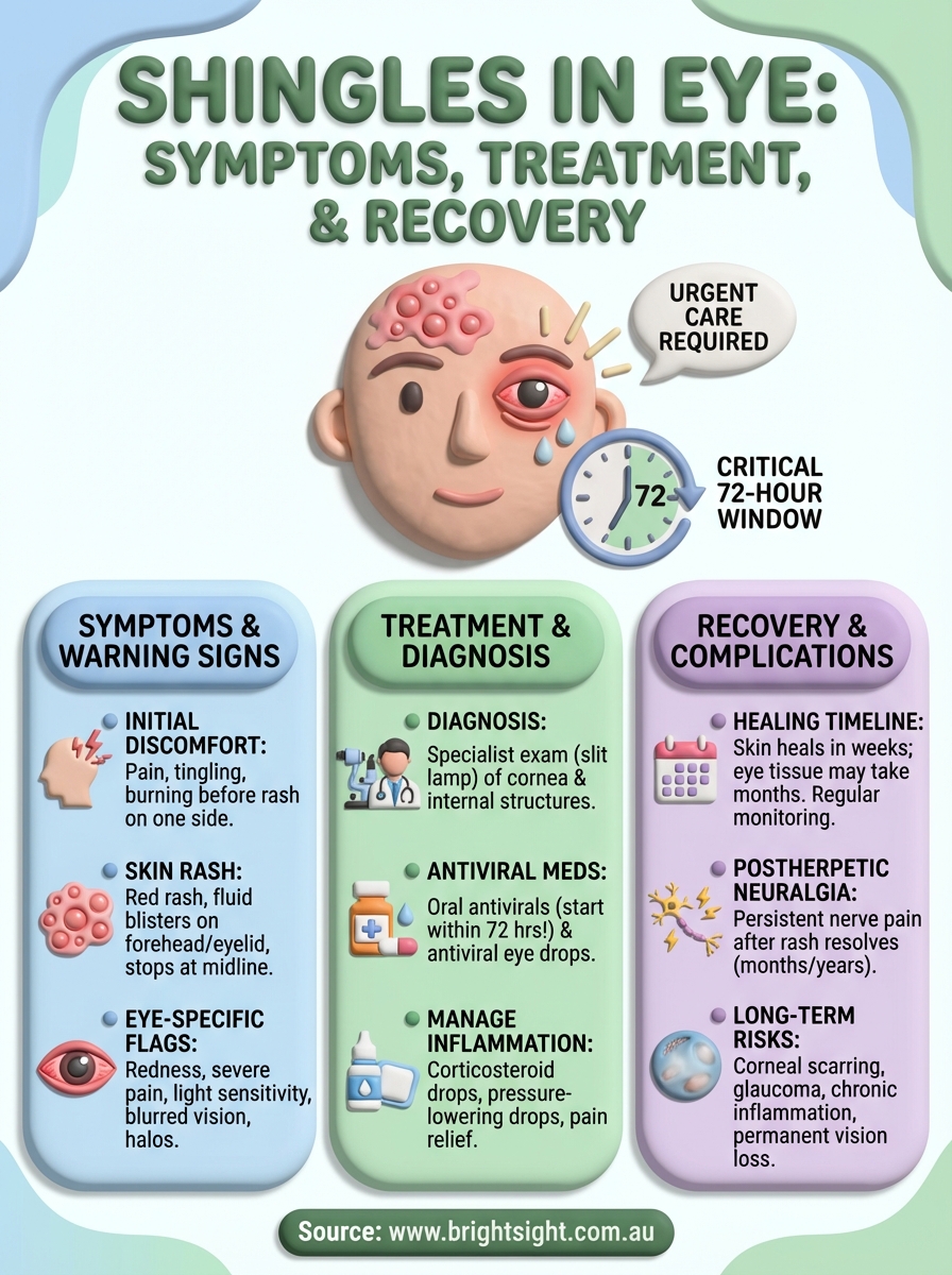
The symptoms of shingles in eye typically begin with pain, tingling, or burning on one side of your forehead or around your eye. This discomfort precedes the visible rash by several days, and many patients initially mistake it for a tension headache or sinus problem. You might also experience fever, fatigue, or sensitivity to touch in the affected area before any blisters appear.
The initial skin symptoms
Your first visible sign will be a red rash that emerges on one side of your forehead, upper eyelid, or the area surrounding your eye. The rash follows the path of the trigeminal nerve and stops precisely at the midline of your face, never crossing to the other side. Within 24 to 48 hours, the rash develops into fluid-filled blisters that cluster together in a distinctive band-like pattern.
Pay particular attention if blisters appear on the tip of your nose (Hutchinson’s sign). This specific location indicates that the nasociliary branch of the nerve is affected, which means the virus has likely spread to internal eye structures. The blisters eventually crust over after several days, but the underlying viral infection continues to pose risks to your eye even after the skin lesions begin healing.
Eye-specific warning signs
Your affected eye may become red and irritated as the virus attacks the eye tissues. You’ll likely notice increased tearing and a discharge that causes your eyelids to stick together, particularly after sleeping. Light sensitivity develops in most cases, making it uncomfortable to be in bright environments or look at screens.
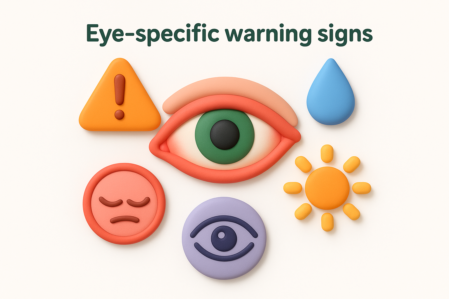
If you experience blurred vision or see halos around lights, contact an eye specialist immediately, as these symptoms indicate corneal involvement.
Vision changes signal more serious complications. Blurred or hazy vision suggests that your cornea has sustained viral damage, while seeing halos around lights or experiencing severe eye pain indicates possible elevated intraocular pressure. Some patients describe feeling like there’s something stuck in their eye (a foreign body sensation) that doesn’t improve with blinking or flushing.
Drooping of your upper eyelid can occur when the virus affects the nerve controlling eyelid movement. You might also notice that your pupil appears irregular in shape or responds abnormally to light, which indicates inflammation of your iris. These symptoms require immediate assessment by an ophthalmologist who can examine the internal structures and begin treatment before permanent damage occurs.
How doctors diagnose shingles in the eye
Your doctor will base the diagnosis of shingles in eye primarily on the characteristic appearance of the rash combined with your symptoms. The distinctive one-sided distribution of blisters on your forehead or around your eye, along with eye redness and pain, provides strong clinical evidence. An ophthalmologist will conduct a thorough examination of both your external skin and internal eye structures to assess the extent of viral involvement.
Diagnosis typically occurs during an urgent consultation when you present with symptoms. Your specialist will ask about your medical history, including whether you’ve had chickenpox in the past and if you have any conditions that weaken your immune system. They’ll document when your symptoms started, as this timeline affects treatment decisions and helps predict your prognosis.
Physical examination of your eye
Your ophthalmologist uses a slit lamp microscope to examine your eye in detail. This specialised equipment allows them to view your cornea, iris, lens, and other internal structures with high magnification. The doctor looks for specific signs like dendritic ulcers (branching patterns on your cornea), inflammation in the anterior chamber, and changes in your intraocular pressure.
During the examination, your specialist will apply fluorescein dye to your eye’s surface. This orange dye glows under blue light and reveals areas where your cornea has sustained damage from the virus. The distinctive tree-like patterns created by viral dendrites become clearly visible, confirming corneal involvement. Your doctor will also check your pupil reactions, measure your eye pressure, and assess whether inflammation has affected your iris or deeper structures.
A comprehensive slit lamp examination can identify viral damage before it causes permanent vision loss, making early specialist assessment critical.
Laboratory tests and additional investigations
Most cases don’t require laboratory confirmation because the clinical presentation provides sufficient evidence for diagnosis. However, your doctor may order polymerase chain reaction (PCR) testing if your presentation is unusual or if you have a compromised immune system. This test analyses samples from your eye or skin lesions to detect viral DNA, confirming the presence of varicella-zoster virus.
Blood tests can help identify underlying conditions that might complicate your treatment or recovery. Your specialist may check your immune function, particularly if you’re under 50 years old (when shingles is less common) or if your symptoms are severe. In rare cases where diagnosis remains uncertain, your doctor might refer you for additional imaging or specialist consultations to rule out other conditions that can mimic shingles symptoms.
How shingles in the eye is treated
Treatment for shingles in eye combines antiviral medications with therapies that address inflammation and specific complications. Your ophthalmologist will prescribe treatments based on which parts of your eye are affected and how severe the infection has become. The goal is to stop viral replication, reduce inflammation, and prevent long-term damage to your vision.
Antiviral medications form the foundation
Your specialist will prescribe oral antiviral drugs such as aciclovir, valaciclovir, or famciclovir within the first 72 hours of rash development. These medications work by preventing the varicella-zoster virus from multiplying, which limits tissue damage and shortens the duration of your symptoms. You’ll typically take the medication for seven to ten days, depending on the severity of your infection and your overall health status.

Starting antiviral treatment within 72 hours reduces your risk of vision-threatening complications by up to 50 percent compared to delayed treatment.
Patients with compromised immune systems may require intravenous antiviral medication administered in hospital. Your doctor will also prescribe antiviral eye drops or ointment if your cornea shows signs of infection. These topical medications deliver concentrated antiviral action directly to the affected eye tissues, complementing the oral medication.
Corticosteroid eye drops reduce inflammation
Your ophthalmologist will prescribe topical corticosteroids to control inflammation in your eye structures. These drops reduce swelling and prevent scarring that could permanently affect your vision. However, your specialist will monitor you closely, as steroids can slow viral clearance and potentially worsen certain types of corneal involvement if used too early in the infection.
Treatment typically begins with frequent dosing (hourly or every two hours during waking hours) that gradually tapers as inflammation resolves. Your doctor might also prescribe dilating drops to reduce pain and prevent complications from inflammation in your iris. The combination of anti-inflammatory and dilating drops addresses both discomfort and the risk of adhesions forming between your iris and lens.
Additional medications target specific complications
Doctors prescribe pressure-lowering drops if your intraocular pressure increases beyond safe levels. Elevated pressure can damage your optic nerve, so your ophthalmologist will monitor this carefully and adjust medications as needed. Some patients require multiple pressure-lowering medications to maintain safe levels during the acute infection phase.
Pain management becomes essential for many patients experiencing severe discomfort from nerve inflammation. Your specialist may recommend oral pain relievers or prescribe stronger medications if standard over-the-counter options prove insufficient. Cold compresses applied to your closed eyelid can provide additional relief, though you should never apply ice directly to your skin or eye.
Recovery time and possible complications
Recovery from shingles in eye varies significantly depending on how quickly you received treatment and which eye structures sustained damage. Most patients experience improvement in acute symptoms within two to four weeks of starting antiviral medication, though complete healing of your eye tissues may take several months. Your ophthalmologist will schedule regular follow-up appointments to monitor your progress and adjust treatment as complications develop or resolve.
Expected timeline for healing
Your skin rash typically crusts over and begins healing within seven to ten days of appearing, though residual redness and discomfort may persist for weeks longer. The corneal dendrites caused by viral infection generally resolve within two to three weeks with appropriate antiviral treatment, but corneal scarring can remain permanently if the infection was severe or treatment was delayed.
Inflammation inside your eye responds more slowly to treatment than surface symptoms. You might need to continue using corticosteroid eye drops for several months as your specialist gradually tapers the dosage. Most patients achieve stable vision within three to six months, though some experience ongoing fluctuations in their sight as inflammation resolves and scar tissue matures.
Recovery depends on prompt treatment, with patients starting antivirals within 72 hours typically experiencing faster healing and fewer long-term complications.
Long-term complications you might face
Postherpetic neuralgia affects approximately 10 to 15 percent of patients who develop shingles in the eye region. This condition causes persistent nerve pain that continues for months or years after the infection resolves, sometimes requiring ongoing pain management with medications designed to treat neuropathic pain.
Chronic eye problems develop in some patients despite appropriate initial treatment. Persistent inflammation (chronic uveitis) can require long-term steroid drops or other immunosuppressive medications to control. Your cornea may develop irregular scarring that distorts your vision, potentially requiring a corneal transplant in severe cases. Some patients develop glaucoma from prolonged inflammation or steroid use, necessitating pressure-lowering drops or surgical intervention. Regular monitoring by your ophthalmologist allows early detection and treatment of these complications before they cause additional vision loss.
What to do next
If you notice a rash developing on one side of your forehead or experience any combination of eye pain, redness, or vision changes, seek specialist assessment immediately. The 72-hour window for starting treatment determines whether you’ll recover fully or face ongoing complications. Don’t wait to see if symptoms improve on their own, as shingles in eye requires urgent medical intervention to protect your sight.
Your next step is to contact Bright Sight Eye Specialists for same-day assessment if you’re experiencing symptoms. Dr Rahul Dubey and Dr Capucine Odouard have extensive experience diagnosing and treating viral eye infections at their Randwick practice. Both specialists use advanced diagnostic equipment to assess the extent of viral damage and can begin appropriate antiviral treatment immediately.
Patients throughout Sydney’s Eastern Suburbs receive comprehensive care that addresses both immediate symptoms and long-term complications, ensuring the best possible outcomes for your vision.