


You notice a sudden shower of tiny dots drifting across your vision, maybe a flicker of light in the corner of your eye. It’s unsettling, and your first instinct is to find out what’s going on. In most cases, these symptoms point to a separated vitreous, a condition where the gel inside your eye pulls away from the retina. It’s one of the most common age-related eye changes, and while it’s usually harmless, it can occasionally signal something more serious like a retinal tear or detachment.
Knowing the difference matters. A straightforward vitreous separation typically needs nothing more than monitoring, but a retinal tear left unchecked can threaten your sight. The symptoms can overlap, which is exactly why getting a prompt, thorough examination is so important when new floaters or flashes appear.
At Bright Sight Eye Specialists in Randwick, Dr. Rahul Dubey and Dr. Capucine Odouard regularly assess and manage vitreous and retinal conditions, from routine posterior vitreous detachments through to complex retinal surgery. This article explains what a separated vitreous actually is, what causes it, what symptoms to watch for, and when you should seek urgent care.
Why a separated vitreous matters
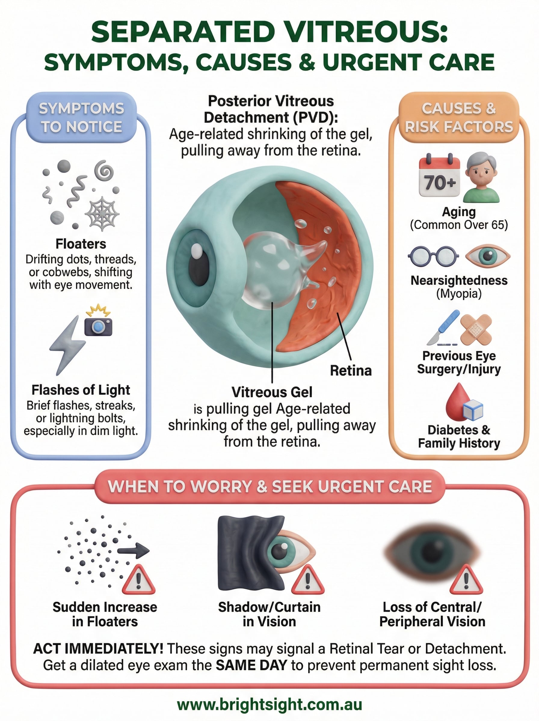
The vitreous is a clear, jelly-like substance that fills the back portion of your eye, pressing gently against your retina. When you are young, this gel stays firmly attached. As you age, it gradually liquefies and shrinks away from the retinal surface in a process called posterior vitreous detachment (PVD). This is what most people mean when they refer to a separated vitreous. For the majority of people, this process causes nothing more than some temporary visual annoyance.
What happens inside your eye
Attached to your retina at multiple points, the vitreous connects most firmly near the optic nerve at the back of your eye. As the gel pulls away from these attachment points, it tugs on the retina and produces the classic symptoms of floaters and flashes. In most cases, the vitreous detaches cleanly and completely, and no lasting damage occurs. Your brain gradually learns to ignore the new floaters, and vision settles over weeks to months.

The problem arises when the vitreous does not detach cleanly. In some people, strands of gel remain firmly adhered to the retina as the rest pulls away. This traction can generate enough force to tear retinal tissue, which is a serious development that demands prompt attention.
The link between vitreous separation and retinal tears
A retinal tear on its own is not automatically a vision emergency, but it can quickly become one. Fluid from inside your eye can seep through the tear and lift the retina away from the underlying tissue, causing a retinal detachment. Once detachment occurs, the retinal cells begin losing their blood supply, and permanent vision loss can follow rapidly without surgical intervention.
Research indicates that around 10 to 15% of people who present with a sudden onset of floaters and flashes have a retinal tear identified at their initial examination.
This is why eye specialists treat new floaters and flashes seriously, even when your vitreous separation turns out to be straightforward. The risk of a serious complication is real, and missing a retinal tear carries consequences that are difficult to reverse.
Symptoms you may notice
The symptoms of a separated vitreous tend to appear suddenly and without warning. Most people notice something unusual in their vision during an ordinary activity, such as reading or looking at a bright sky. Understanding exactly what to look for helps you recognise when to act without delay.
Floaters
When the vitreous gel pulls away from the retina, it leaves behind clumps of collagen fibres that cast shadows on your retina. You see these shadows as floaters, which may appear as dots, threads, cobwebs, or a ring-shaped shadow drifting across your visual field. Floaters tend to shift as your eye moves and then drift slowly when you stop.
Floaters are often most noticeable when you look at a plain, bright background such as a white wall or a clear sky.
Flashes of light
As the vitreous tugs on the retina during separation, it stimulates retinal cells mechanically, producing brief flashes or streaks of light. These flashes typically appear in your peripheral vision and can resemble a camera flash or a lightning bolt. They often become more apparent in dim lighting and may persist for several weeks before settling.
Changes that need urgent attention
Certain symptoms suggest something more serious is occurring. If you notice a sudden increase in floaters, a shower of dark spots, a shadow or curtain creeping across your vision, or any reduction in your central or peripheral sight, you should seek an emergency eye examination the same day rather than wait for a routine appointment.
Causes and risk factors
Most cases of separated vitreous occur simply because the eye ages. Understanding what drives this process, and what makes some people more vulnerable than others, helps you recognise whether you fall into a higher-risk group.
Age-related changes to the vitreous
The vitreous gel is roughly 98% water when you are born, but it also contains collagen fibres and hyaluronic acid that hold its structure together. Over time, these components break down. The gel liquefies from the inside out, forming pockets of fluid that gradually undermine its attachment to the retina. By the time most people reach their 70s or 80s, a posterior vitreous detachment has already occurred in one or both eyes.
The majority of people over 65 have experienced at least one posterior vitreous detachment, often without realising it.
Conditions that increase your risk
Age alone is the strongest driver, but several other factors accelerate vitreous ageing or increase the mechanical forces placed on the retina during separation. You are at higher risk if any of the following apply:
- Short-sightedness (myopia): Longer eyeballs stretch the vitreous and retina, making early separation more likely.
- Previous eye surgery or injury: Cataract surgery, in particular, is associated with a higher rate of PVD in the months following the procedure.
- Diabetes: Diabetic changes to the vitreous and retina increase both the likelihood of separation and the chance of complications.
- Family history: A strong family history of retinal tears or detachment raises your personal risk considerably.
When to worry and seek urgent care
Most people who experience a separated vitreous go through the process without complications. However, certain symptoms act as warning signs that something more dangerous may be happening inside your eye, and these require same-day assessment from an eye specialist rather than a wait-and-see approach.
Symptoms that signal an emergency
A small increase in pre-existing floaters often falls within the normal range of vitreous separation. What demands immediate attention is a sudden large increase in floaters, particularly if they appear as a shower of dark spots or specks filling your visual field. A curtain, shadow, or veil creeping in from the edge of your vision is another critical warning sign that your retina may have detached.
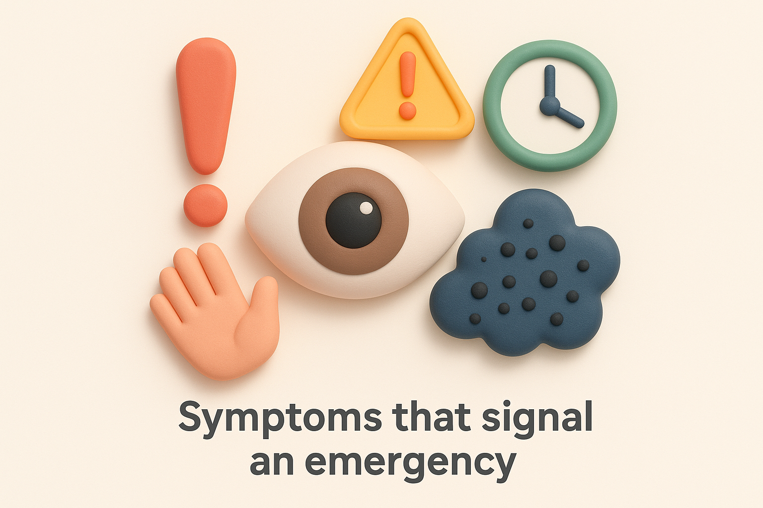
If you notice a shadow spreading across any part of your vision, treat it as an emergency and seek care the same day.
Flashes that persist or intensify over several weeks are also worth reporting promptly, as ongoing traction between the vitreous and retina raises the risk of a tear forming.
Acting within the right timeframe
Retinal tears caught early can often be treated with laser therapy before fluid seeps underneath the retina. Once detachment develops, you need more complex surgical intervention, and the outcome depends heavily on how quickly treatment begins.
Do not wait to see whether symptoms resolve on their own when your sight is potentially at risk. Prompt assessment by a retinal specialist gives you the best chance of preserving your full vision.
Diagnosis and treatment options
When you come in for assessment of a separated vitreous, your specialist will perform a dilated eye examination to inspect both the vitreous gel and your retina thoroughly. This examination remains the most reliable way to confirm whether your vitreous separation has caused any retinal complications.
Getting a dilated examination within 24 to 48 hours of new symptoms gives your specialist the best chance of catching a retinal tear early.
How your eye specialist examines you
Your specialist will use dilating eye drops to widen your pupil, then examine the retina using a slit lamp and an indirect ophthalmoscope. This allows a detailed view of your peripheral retina, where tears most commonly occur. If floaters are dense or a haemorrhage blocks the view, an ultrasound scan of the eye provides the additional information needed.
Beyond the physical examination, optical coherence tomography (OCT) produces cross-sectional images of the retina to detect any subtle traction or swelling that standard examination alone may not reveal.
Treatment: when is it needed?
Most cases of posterior vitreous detachment require no active treatment. Your specialist will advise you to monitor your symptoms and return promptly if new floaters, increased flashes, or any shadow in your vision develops. When a complication is found, treatment depends on what has occurred:
- Laser photocoagulation: Seals a retinal tear before detachment can develop
- Vitrectomy: Removes the vitreous gel to treat retinal detachment or significant floaters
- Scleral buckle: A band placed around the eye to support detached retinal tissue back into position
Next steps for your eyes
A separated vitreous is a natural part of ageing, and for most people it passes without causing lasting harm. What matters is that you take new symptoms seriously and do not put off getting your eyes examined when floaters or flashes appear suddenly. The difference between a routine vitreous detachment and a developing retinal tear often comes down to timing, and your specialist cannot make that distinction without a proper dilated examination.
At Bright Sight Eye Specialists in Randwick, Dr. Rahul Dubey and Dr. Capucine Odouard offer thorough retinal and vitreous assessments for patients across Sydney’s Eastern Suburbs and beyond. Both specialists bring extensive surgical experience to complex retinal conditions, and both are committed to giving you clear, honest information about what is happening in your eyes. If you have noticed new floaters, flashes, or any change to your vision, book an appointment with our team today.