


Tiny black dots drifting across your vision are shadows cast on the retina by small particles floating inside your eye. These specks appear to move when you shift your gaze because they’re suspended in the vitreous humour, the clear gel filling your eyeball. Most people describe them as spots, threads or cobwebs that seem to dart away when you try to look at them directly. While these floaters are usually harmless, they can sometimes signal a more serious problem that needs prompt attention.
This article explains what causes those moving dots in your sight, from normal age related changes to urgent retinal issues. You’ll learn which symptoms suggest a benign floater that can be monitored versus warning signs that require same day assessment. We’ll cover who faces the highest risk, how eye specialists diagnose the underlying cause, and what treatment options are available. By the end, you’ll know exactly when to watch and wait, and when to act quickly to protect your vision.
Why tiny black dots in vision matter
Understanding why you’re seeing tiny black dots in your vision matters because it helps you distinguish between normal age related changes and genuine emergencies. Most floaters are benign, resulting from natural shifts in your vitreous gel as you grow older. However, certain patterns of dots can indicate retinal tears, detachment or bleeding inside your eye that demands immediate specialist care. Missing these warning signs can lead to permanent vision loss, so knowing when to act protects your sight for the long term.
Impact on your quality of life
Daily activities become frustrating when floaters drift across your central vision repeatedly. You might struggle to read small text, notice reduced contrast when looking at screens, or feel distracted during tasks that require sustained focus. Some people find bright backgrounds make the dots more obvious, which can affect outdoor activities or driving on sunny days. While your brain often adapts over time and filters out harmless specks, severe floaters can genuinely impair your ability to work, read or enjoy hobbies without constant visual interruption.
Even harmless floaters can reduce your comfort and confidence in everyday tasks, making early assessment worthwhile.
When dots signal serious eye conditions
Sudden increases in the number of black specks, especially when paired with flashes of light or a shadow across your vision, point to potential retinal damage. These symptoms suggest that the vitreous is tugging on your retina or that a tear has formed, allowing fluid to seep underneath. Acting quickly in these scenarios can mean the difference between a simple office procedure and complex surgery that requires weeks of recovery. Recognising the urgency ensures you preserve your vision and avoid complications that might otherwise be preventable with prompt treatment.
How to respond to tiny black dots in vision
Your response to seeing tiny black dots in vision depends on how suddenly they appear and what other symptoms accompany them. Stay calm and assess the situation rather than panicking, because most cases don’t require emergency treatment. Start by noting when you first noticed the dots, how many you can see, and whether they’re increasing in number. Check if you’re experiencing flashes of light, shadows across your visual field or any eye pain. These observations will help your eye care professional determine urgency and guide the appropriate next steps for your situation.
Immediate steps when you first notice dots
Stop what you’re doing and observe the pattern of your floaters for a few minutes. Look at a plain, bright surface like a white wall or clear sky to see the dots more clearly. Count roughly how many specks you can detect and note their shape, whether they’re stationary or drifting. Write down the time and date you first noticed them, along with any activities you were doing beforehand. This information becomes valuable during your consultation, helping your specialist understand whether your symptoms suggest a benign age-related change or something requiring urgent investigation.

When to schedule a routine eye exam
Book a standard appointment within a few days if you’ve noticed one or two floaters that appear stable and aren’t accompanied by flashes or shadows. Your optometrist or ophthalmologist will dilate your pupils to examine your retina and vitreous thoroughly. This non-urgent scenario typically applies when you’re over 50, have had floaters before, and the current ones aren’t dramatically different from previous experiences. Regular monitoring ensures any gradual changes get documented, providing a baseline for future comparisons if your symptoms evolve.
Emergency situations requiring same-day care
Contact an eye specialist immediately if you experience a sudden shower of new floaters, flashes of light in your peripheral vision, or a dark curtain sweeping across your sight. These symptoms suggest potential retinal tears or detachment that need assessment within hours, not days. Don’t wait overnight if you also notice reduced vision, eye pain or a significant shadow blocking part of your visual field.
Retinal emergencies caught early often require less invasive treatment and have better outcomes than those left untreated for even 24 hours.
Call your ophthalmologist’s emergency line or visit your nearest hospital eye department if your regular clinic is closed.
Common harmless causes and floaters explained
Most cases of seeing tiny black dots in vision stem from natural changes in your eye’s structure that don’t threaten your sight. Posterior vitreous detachment ranks as the leading benign cause, occurring when the gel inside your eye shrinks and pulls away from the retina. Age-related collagen clumping also produces floaters as the vitreous humour becomes less uniform over time. These harmless particles drift across your vision because they float freely in the gel, casting shadows that move with your eye movements. Understanding these common causes helps you recognise when your symptoms fit normal ageing patterns rather than urgent medical problems.
Age-related vitreous changes
Your vitreous humour starts life as a firm, clear gel, but decades of exposure to light and metabolic processes gradually break down its structure. By your 50s and 60s, the vitreous becomes more liquid in some areas while remaining gel-like in others. Collagen fibres that once formed an invisible mesh begin clumping together, creating visible strands and specks that cast shadows on your retina. This process happens to nearly everyone who lives long enough, making it one of the most common reasons people notice floaters. The dots typically appear more obvious against bright backgrounds like blue skies or white walls because the contrast makes the shadows stand out.
Posterior vitreous detachment (PVD)
Posterior vitreous detachment occurs when the vitreous gel shrinks away from the retina’s surface, creating a separation between the two structures. This detachment releases tiny fragments of collagen and cellular debris into the vitreous cavity, which float around and appear as black dots or cobwebs. PVD affects roughly 75% of people over 65 and usually causes no permanent damage to your vision. You might notice a sudden increase in floaters or occasional flashes of light as the vitreous tugs on the retina during separation, but these symptoms typically settle within weeks once the detachment completes.
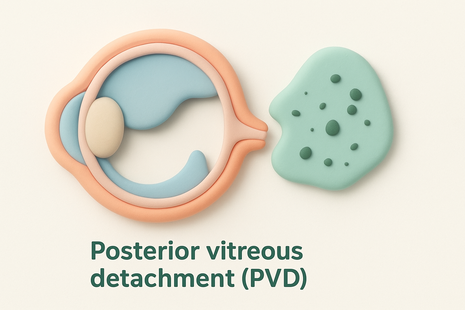
PVD is a natural part of ageing that rarely requires treatment, though you should still have it examined to rule out complications.
Why your brain adapts to floaters
Your visual system has a remarkable ability to filter out static or repetitive distractions that don’t signal danger. Over several weeks to months, your brain learns to suppress the signals from harmless floaters through a process called neural adaptation. This doesn’t make the physical particles disappear, but it reduces how much you consciously notice them during daily activities. Most people find their floaters become far less bothersome after this adaptation period, even though the dots remain visible if they deliberately look for them. This natural adjustment explains why long-term floaters often cause less distress than new ones that suddenly appear.
Serious causes and warning signs
While most floaters result from harmless vitreous changes, several sight-threatening conditions can produce tiny black dots that demand immediate medical attention. Retinal tears, detachment and bleeding inside the eye all create similar visual disturbances but require urgent specialist care to prevent permanent vision loss. Recognising the difference between benign age-related floaters and these serious causes relies on identifying specific accompanying symptoms and understanding how quickly your dots appeared. Your response time in seeking treatment directly affects your chances of preserving clear sight, making awareness of these warning signs crucial.
Retinal tears and traction
A retinal tear occurs when the vitreous gel pulls forcefully enough to create a full-thickness break in your retina’s delicate tissue. This traction often happens during posterior vitreous detachment when the gel remains firmly attached to certain spots on your retina. You might notice sudden flashes of light in your peripheral vision alongside new floaters because the mechanical tugging stimulates your retina’s photoreceptors. These flashes typically appear as brief, flickering lights or lightning-like streaks that persist even in complete darkness. Tears create an entry point for fluid to seep underneath your retina, risking progression to detachment within hours or days if left untreated.
Retinal detachment
Retinal detachment happens when fluid accumulates beneath your retina, separating it from the underlying layer that supplies oxygen and nutrients to its cells. The detached portion stops functioning properly, creating a shadow or curtain effect that advances across your visual field as more retina lifts away. You’ll typically notice this as a dark area blocking your peripheral or central vision, depending on where the detachment begins. Unlike floaters that drift and move, this shadow remains fixed in position and gradually expands if you don’t receive emergency treatment. Surgical intervention within 24 to 48 hours offers the best chance of reattaching your retina and recovering useful vision in the affected area.
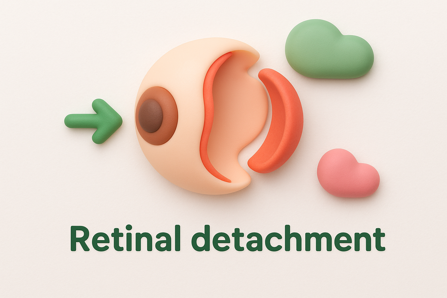
Any sudden curtain-like shadow sweeping across your sight requires same-day ophthalmology assessment to prevent irreversible damage.
Vitreous haemorrhage
Bleeding into your vitreous cavity produces numerous dark specks that can appear suddenly as tiny black dots scattered throughout your vision. This haemorrhage usually stems from damaged blood vessels in conditions like diabetic retinopathy, retinal tears or trauma to your eye. Fresh blood cells floating in the vitreous cast shadows that look denser and more numerous than typical age-related floaters. You might describe the appearance as looking through a red haze or seeing hundreds of small spots drifting simultaneously. Severe bleeding can significantly blur your vision or create a dark veil that obscures large portions of your visual field, requiring urgent assessment to identify and treat the underlying cause.
Critical warning signs
Several specific symptoms signal that seeing tiny black dots in vision stems from a serious condition rather than harmless floaters. Seek immediate care if you experience a sudden shower of dozens of new floaters appearing within minutes or hours. Contact an eye specialist the same day if you notice flashing lights that persist or repeat frequently, especially in your peripheral vision. A dark shadow or curtain advancing across any part of your sight demands emergency attention regardless of whether you have eye pain. Loss of peripheral vision, sudden blurriness that doesn’t clear with blinking, or a combination of floaters plus flashes always warrants urgent professional examination. Recording the exact time symptoms began and any activities that preceded them helps your specialist assess urgency and plan appropriate intervention.
Who is most at risk
Certain groups face higher chances of seeing tiny black dots in vision due to factors that stress or alter the vitreous humour and retina. Age ranks as the single strongest predictor, but your eye anatomy, medical history and underlying health conditions all influence your likelihood of developing floaters or serious complications. Understanding your personal risk profile helps you know whether you need annual screening, more frequent monitoring or heightened awareness of sudden symptom changes. Risk doesn’t guarantee problems, but it signals when you should stay more vigilant about new visual disturbances.
Age and natural deterioration
Your risk of floaters climbs steadily after age 50 as the vitreous gel inside your eye undergoes irreversible structural breakdown. By your 60s and 70s, posterior vitreous detachment becomes almost universal, releasing collagen fragments that appear as drifting specks. Older adults also face higher rates of retinal tears and detachment because decades of vitreous traction weaken the retina’s attachment points. Regular dilated eye exams become essential once you reach middle age, even if you’ve never had vision problems before.
High myopia and elongated eyes
People with severe short-sightedness have eyeballs stretched longer than normal, which thins both the retina and vitreous humour throughout their lives. This anatomical difference means you’re more prone to early onset floaters, often noticing them in your 30s or 40s rather than later decades. The stretched retina tears more easily under vitreous traction, raising your risk of detachment several-fold compared to people with normal or far-sighted vision.
High myopia multiplies your chances of serious retinal complications, making annual specialist reviews crucial regardless of symptoms.
Previous eye surgery or trauma
Any procedure or injury that disturbs your eye’s internal structures increases your vulnerability to floaters and retinal problems. Cataract surgery and vitrectomy accelerate vitreous liquefaction, while blunt trauma can create immediate tears or scarring that manifests years later. Inform your eye specialist about past surgical history or significant eye injuries during every examination.
Systemic health conditions
Diabetes and high blood pressure damage the tiny blood vessels supplying your retina, leading to bleeding, swelling and abnormal new vessel growth. These vascular changes produce floaters through haemorrhage or inflammatory debris in your vitreous. Maintaining tight control of blood sugar and blood pressure reduces your risk substantially, though regular retinal screening remains necessary even with good management.
How eye specialists diagnose black dots
Eye specialists follow a systematic approach to determine whether you’re seeing tiny black dots in vision due to harmless floaters or something requiring urgent treatment. Your assessment begins with detailed questioning about when symptoms started, how they’ve progressed, and any accompanying signs like flashes or shadows. The specialist will then perform a thorough dilated eye examination to view your retina and vitreous directly, looking for tears, detachment, bleeding or other structural changes. Advanced imaging may supplement this examination when the view is obscured or when subtle changes need confirmation. This comprehensive process ensures accurate diagnosis while ruling out sight-threatening conditions that demand immediate intervention.
Patient history and symptom timeline
Your specialist needs precise details about when you first noticed the black dots and how they’ve behaved since onset. They’ll ask whether you saw a sudden shower of specks or gradual appearance of one or two floaters over weeks. Questions about flashing lights, curtain-like shadows, eye trauma, previous surgery and systemic conditions like diabetes help narrow the possible causes. Documenting whether symptoms appeared after specific activities, during certain lighting conditions or in particular parts of your visual field guides the physical examination that follows. Bringing notes about symptom timing and any changes you’ve observed saves appointment time and ensures you don’t forget critical details under pressure.
Dilated pupil examination
Dilation drops temporarily widen your pupils, allowing your specialist to examine the entire retina and vitreous gel through specialised lenses and ophthalmoscopes. The drops take 15 to 20 minutes to work fully and may blur your near vision for several hours afterwards. During examination, your specialist looks for posterior vitreous detachment, retinal tears, haemorrhage and signs of traction where the vitreous remains attached to your retina. They’ll inspect the peripheral retina carefully because tears often occur in these outer areas first. This examination remains the gold standard for diagnosing floater causes because it reveals structural changes that imaging alone might miss.
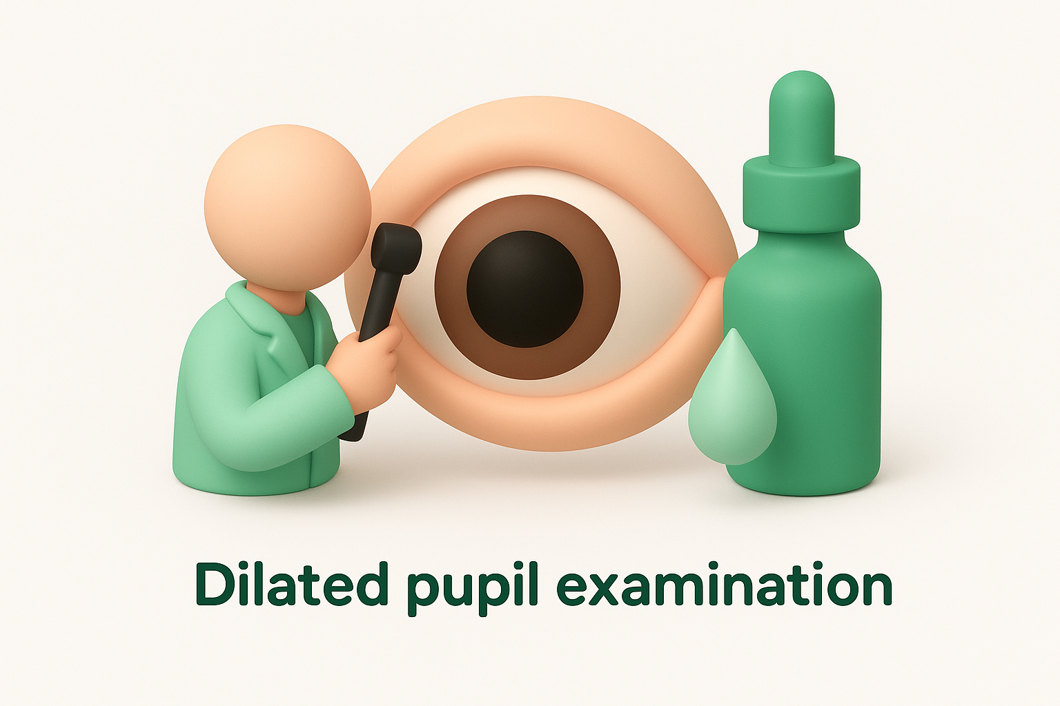
Dilated examination provides the clearest view of retinal health and catches most serious conditions before they progress to sight loss.
Advanced imaging when needed
Optical coherence tomography (OCT) produces high-resolution cross-sectional images of your retina, showing layers in detail that direct viewing cannot achieve. Your specialist orders OCT when they suspect macular involvement, subtle detachment or vitreoretinal traction that needs precise measurement before treatment planning. B-scan ultrasound becomes essential if bleeding or dense floaters prevent clear visual examination of your retina. These imaging techniques complement rather than replace dilated examination, offering additional information that confirms diagnoses and guides surgical decisions when intervention becomes necessary.
Treatment options and what to expect
Treatment for seeing tiny black dots in vision varies widely depending on what’s causing your symptoms and how much they affect your daily life. Harmless floaters from age-related vitreous changes rarely need active intervention beyond regular monitoring, while retinal tears or detachment demand urgent surgical repair to prevent permanent vision loss. Your eye specialist tailors recommendations to your specific diagnosis, considering factors like floater density, location, your visual demands at work or home, and overall eye health. Understanding each treatment option helps you make informed decisions and know what recovery involves if intervention becomes necessary.
Observation and monitoring
Your specialist recommends watchful waiting when examination confirms your black dots stem from benign posterior vitreous detachment without retinal tears or bleeding. This approach involves scheduled follow-up appointments at intervals of several weeks to months, allowing your ophthalmologist to track whether symptoms remain stable or progress. During this period, your brain gradually adapts to the floaters through neural filtering, making them less noticeable even though they physically remain in your vitreous. You’ll receive clear instructions about warning signs that warrant immediate reassessment, such as sudden increases in floater number, new flashes or shadows across your vision.
Most people find their floaters become significantly less bothersome after three to six months without any treatment. Your specialist documents the baseline appearance and position of your floaters during initial examination, creating a reference point for future comparisons if your symptoms change later.
Laser vitreolysis for persistent floaters
Laser vitreolysis offers a minimally invasive outpatient option when floaters persistently interfere with your reading, driving or other essential activities despite months of observation. Your specialist uses a specialised YAG laser to target and break up the collagen clumps or strands casting shadows on your retina. The procedure takes 15 to 30 minutes per eye and requires only topical anaesthetic drops, causing minimal discomfort during treatment.
Laser vitreolysis works best for large, well-defined floaters positioned safely away from your retina and lens.
Recovery involves temporary blurred vision for a few hours and possible mild light sensitivity for a day or two. You might notice immediate improvement in floater density, though some people need two or three treatment sessions spaced weeks apart to achieve satisfactory results. The laser doesn’t eliminate every floater, but it typically reduces the most bothersome ones enough to restore comfortable vision for daily tasks.
Vitrectomy surgery
Vitrectomy becomes necessary when severe floaters significantly impair your vision or when complications like vitreous haemorrhage, retinal tears or detachment require direct surgical access to your eye’s interior. Your surgeon makes tiny incisions to remove the vitreous gel entirely, replacing it with a clear saline solution that maintains your eye’s shape without containing debris. This procedure happens under local or general anaesthetic and takes one to two hours, depending on complexity.
Postoperative recovery requires you to avoid strenuous activity for several weeks and attend frequent follow-up appointments to monitor healing. Your vision improves gradually over weeks to months, though you face increased risk of cataract formation within one to two years after vitrectomy. Infection, retinal detachment and elevated eye pressure represent uncommon but serious complications that your surgical team monitors closely throughout recovery.
Emergency repair for tears and detachment
Retinal tears caught early receive immediate laser photocoagulation or cryotherapy to seal the break before fluid accumulates underneath. These office-based procedures create controlled scarring around the tear, bonding your retina firmly to prevent detachment. If detachment has already begun, you need urgent vitrectomy, scleral buckle or pneumatic retinopexy within 24 to 48 hours to reattach your retina and restore blood flow to affected areas. Success rates exceed 90% when treatment happens promptly, preserving useful vision in most cases.

Next steps for your eye health
Taking action when you notice seeing tiny black dots in vision protects your sight and gives you peace of mind about what’s happening inside your eyes. Schedule a dilated eye examination if you’ve recently developed floaters, even when they seem mild or infrequent. Your optometrist or ophthalmologist will determine whether your symptoms stem from harmless age-related changes or require urgent attention. Keep a brief record of when your dots appeared, how many you can see, and any accompanying flashes or shadows to share during your appointment.
Maintaining regular eye health check-ups becomes increasingly important after age 50, when vitreous changes accelerate and retinal complications grow more common. Monitor your vision daily by looking at plain backgrounds like walls or clear skies, noting any sudden increases in floater number or new visual disturbances. If you experience warning signs such as flashes, curtains or showers of new specks, contact an eye specialist immediately rather than waiting for your next scheduled visit. Bright Sight Eye Specialists provides comprehensive retinal assessments and treatment for floaters and related conditions throughout Sydney’s Eastern Suburbs.