


You’re reading a book or looking at the sky when you notice them, tiny specks, threads, or cobweb-like shapes drifting across your vision. Seeing floaters in eyes is an experience most people have at some point, and for many, it’s simply a normal part of ageing. However, these visual disturbances can sometimes signal something more serious happening inside your eye.
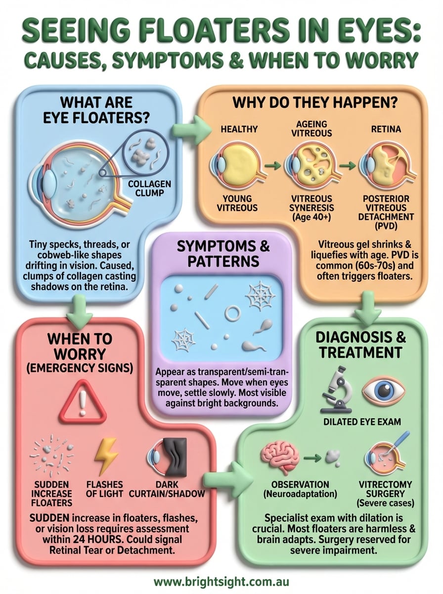
Eye floaters are caused by changes in the vitreous, the gel-like substance that fills the inside of your eyeball. As we get older, this gel naturally shrinks and becomes more liquid, causing tiny fibres to clump together and cast shadows on the retina. While this process is usually harmless, a sudden increase in floaters, especially when accompanied by flashes of light or vision loss, can indicate conditions like retinal detachment that require urgent attention.
At Bright Sight Eye Specialists, Dr Rahul Dubey and our team regularly help patients in Randwick and Sydney’s Eastern Suburbs understand and manage eye floaters. With expertise in retinal conditions and advanced surgical treatments, we can determine whether your floaters are a benign nuisance or a symptom requiring treatment. This article explains what causes floaters, the symptoms to watch for, and when you should seek professional care.
Why you see floaters in your eyes
The floaters you notice drifting across your field of vision originate from changes inside your eyeball, specifically in the vitreous humour. This clear, gel-like substance makes up about 80% of your eye’s volume and sits between the lens at the front and the retina at the back. Understanding what happens to this gel explains why seeing floaters in eyes becomes more common as you age.
The vitreous gel and its natural changes
Your vitreous starts out firm and uniform when you’re young, maintaining a consistent jelly-like texture that helps your eyeball keep its shape. However, from around age 40 onwards, this gel begins to liquefy and shrink through a process called vitreous syneresis. The once-smooth gel develops pockets of liquid, and the remaining gel becomes less stable.

As this liquefaction continues, the vitreous eventually pulls away from the retina in what specialists call a posterior vitreous detachment (PVD). This separation is a normal part of ageing and happens to most people by their sixties or seventies. The detachment itself often triggers a noticeable increase in floaters, though it doesn’t always cause problems.
While vitreous detachment is common and usually harmless, it’s the most frequent time when serious complications can occur.
What creates the shadows you see
The floaters themselves are tiny clumps of collagen fibres that form when the vitreous gel breaks down. These fibres stick together instead of remaining evenly distributed throughout the gel. When light enters your eye, these clumps cast shadows on your retina, which your brain interprets as the shapes you see floating in your vision. The shadows move as your eye moves because the floaters are suspended in the liquid vitreous and shift with your eye’s motion.
How to tell harmless floaters from emergencies
Most floaters develop gradually and pose no threat to your vision, but certain warning signs require immediate medical attention. The key difference lies in how quickly the floaters appear and what other symptoms accompany them. While seeing floaters in eyes typically results from normal ageing, sudden changes can indicate serious retinal problems that need urgent treatment.
Signs that indicate an emergency
You need to see an eye specialist urgently if you experience a sudden shower of new floaters appearing within hours or days, rather than noticing them gradually over months or years. This rapid onset often signals that the vitreous has torn or pulled on your retina during detachment. The situation becomes particularly concerning when floaters appear alongside flashes of light (like camera flashes in your peripheral vision) or a dark curtain or shadow blocking part of your visual field.
Any combination of new floaters with flashes or vision loss requires assessment within 24 hours to rule out retinal tears or detachment.
Characteristics of harmless floaters
Benign floaters typically remain stable in number and don’t worsen over time. You’ve probably had them for years, and while they may become more noticeable against bright backgrounds like the sky, they don’t multiply suddenly or change dramatically. These floaters move when your eyes move and settle when you stop looking around, behaving predictably and consistently without affecting your overall vision quality.
Symptoms and patterns that help you identify floaters
The way floaters appear and behave in your vision follows recognisable patterns that help you understand whether you’re seeing floaters in eyes or experiencing a different visual disturbance. These characteristics remain consistent across most people, though the specific shapes and sizes vary based on your individual eye anatomy and the type of vitreous changes you’re experiencing.
Common visual descriptions
Floaters typically appear as transparent or semi-transparent shapes that drift across your field of vision. You might describe them as dots, circles, lines, cobwebs, or threads that move when you try to look at them directly. Some people see shapes that resemble small insects or tadpoles, while others notice circular rings or irregular blobs. These shapes become most visible against uniform, bright backgrounds like white walls, clear blue skies, or computer screens.
The shapes you see correspond to the actual structure of collagen clumps floating in your vitreous gel.
Movement patterns and behaviour
Your floaters move in predictable ways that distinguish them from other visual symptoms. They drift slowly when your eyes are still and swing into view when you move your gaze quickly, continuing to float for a second or two after your eyes stop moving. This delayed settling motion happens because the floaters suspend in liquid and follow the flow of your vitreous humour rather than staying fixed in one position.
How eye specialists diagnose new floaters
When you visit an eye specialist about seeing floaters in eyes, the examination focuses on examining your retina and assessing the vitreous gel. The diagnostic process goes beyond simply confirming you have floaters; it aims to identify any retinal tears, holes, or detachment that might be causing them. This thorough evaluation helps distinguish between harmless age-related changes and conditions requiring urgent treatment.
What happens during your examination
Your specialist begins with dilating eye drops that widen your pupils, allowing a complete view of your retina and vitreous. After the drops take effect (usually 15 to 20 minutes), the doctor uses a slit lamp microscope combined with special lenses to examine the inside of your eye. This equipment provides a magnified, three-dimensional view of your vitreous and retina, revealing any clumps, tears, or areas where the vitreous has pulled away from the retinal surface.
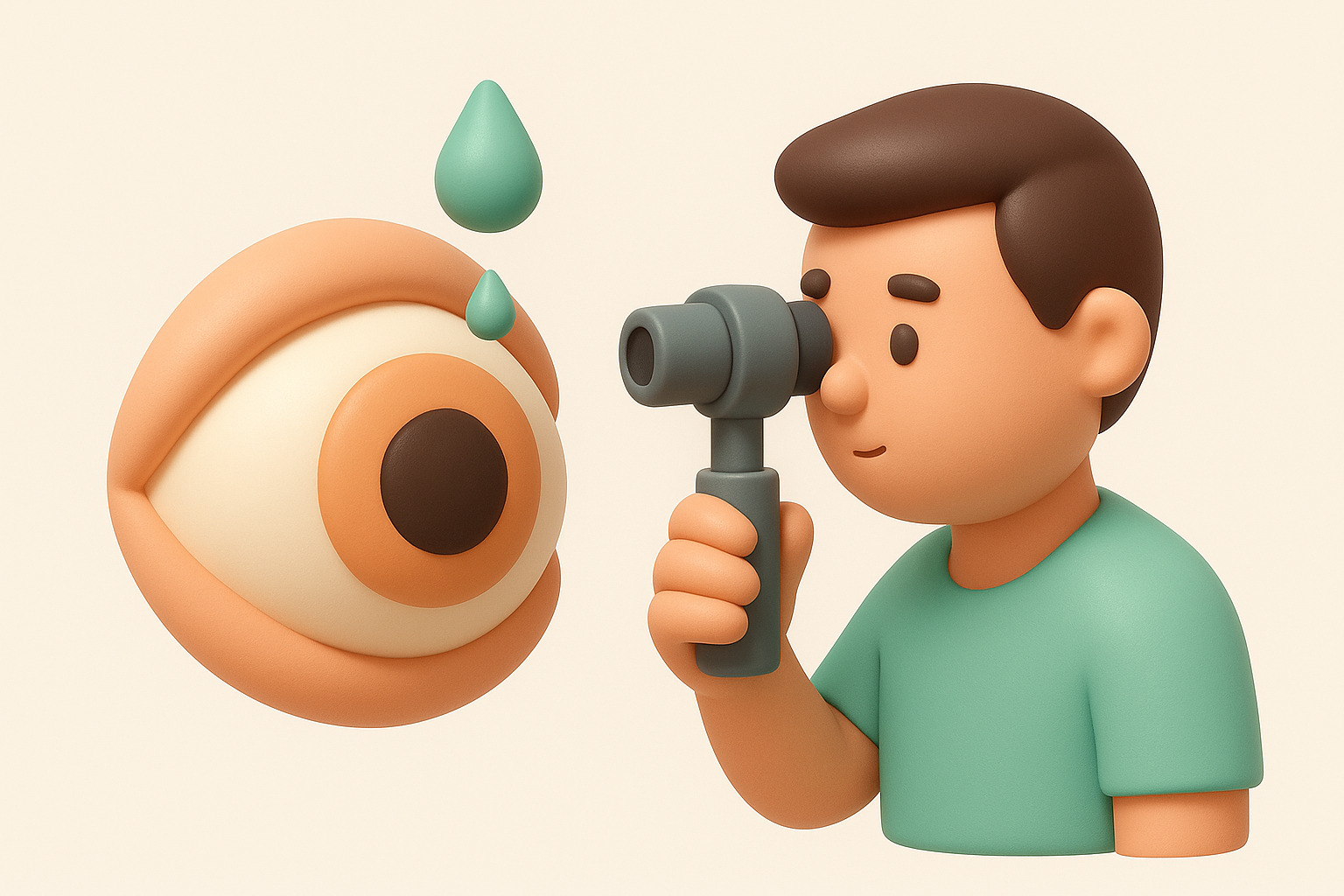
A dilated examination remains the most reliable method for detecting retinal tears that could lead to vision-threatening complications.
Additional imaging when needed
Some cases require optical coherence tomography (OCT), a scanning device that creates detailed cross-sectional images of your retina. This technology proves particularly useful when your specialist needs to assess structural changes in the macula or evaluate areas that standard examination cannot fully visualise due to dense floaters or other obstructions.
Treatment options and what actually helps
The approach to treating floaters depends entirely on how severely they affect your vision and whether they indicate underlying problems. Most people who report seeing floaters in eyes don’t require any medical intervention, as the brain typically adapts to their presence over weeks or months. However, when floaters result from retinal tears or cause significant visual disruption, several treatment options exist.
When no treatment is necessary
Your eye specialist will recommend observation rather than treatment if your floaters stem from normal vitreous changes without retinal complications. The shadows become less noticeable as your brain learns to ignore them, a process called neuroadaptation that happens gradually over time. You’ll find that moving your eyes to shift the floaters out of your central vision provides temporary relief, though this strategy works best for occasional annoyance rather than constant disruption.
Most people find their floaters become significantly less bothersome within three to six months, even when the floaters themselves remain present.
Surgical interventions for problematic floaters
Vitrectomy surgery removes the vitreous gel containing the floaters and replaces it with a clear saline solution. This procedure achieves complete floater removal but carries risks including cataracts, retinal detachment, and infection. Specialists typically reserve this option for severe cases where floaters substantially impair your daily activities and quality of life.
What to do next
Seeing floaters in eyes affects most people at some point, and understanding the difference between normal age-related changes and warning signs of retinal problems helps you make informed decisions about your eye health. You now know that gradual floaters rarely cause concern, while sudden increases accompanied by flashes or vision loss require urgent specialist assessment.
If you’ve developed new floaters or notice any worrying symptoms, don’t wait to see whether they improve on their own. Dr Rahul Dubey and Dr Capucine Odouard at Bright Sight Eye Specialists provide comprehensive retinal examinations using advanced diagnostic technology to assess your condition accurately. Our team serves patients throughout Sydney’s Eastern Suburbs, offering transparent advice about whether your floaters need treatment or simply monitoring.
Contact our Randwick practice to arrange a thorough eye examination. Early detection of retinal tears or detachment makes treatment significantly more straightforward and preserves your vision more effectively than delayed intervention.