


You’re reading, driving, or simply looking at a clear blue sky when you notice them, tiny dark shapes drifting lazily across your vision. Seeing black specks is remarkably common, and while the experience can feel unsettling, understanding what’s happening inside your eye can put your mind at ease.
These floating specks, medically known as floaters, are typically harmless clumps of cells or protein within the vitreous gel that fills your eye. However, in certain cases, they can signal something more serious, such as a retinal tear or detachment, that requires prompt attention. Knowing the difference between everyday floaters and genuine warning signs is essential for protecting your long-term vision.
At Bright Sight Eye Specialists, Dr Rahul Dubey and our team regularly help patients across Sydney’s Eastern Suburbs understand and manage floaters, providing expert assessment and treatment when necessary. This article explains the causes behind those black specks in your vision, outlines when you should seek professional care, and covers the treatment options available to restore your comfort and confidence in your eyesight.
Why seeing black specks matters
Your vision connects you to the world around you, from recognising your grandchildren’s faces to safely navigating busy roads in Randwick or Bondi. When seeing black specks interrupts these daily moments, it’s natural to wonder whether you should brush it off or take action. The answer matters because your eyes don’t regenerate like skin or liver tissue, and damage to the retina or vitreous can become permanent if you miss the window for treatment.
Your quality of life depends on clear vision
Most people tolerate occasional floaters without much disruption, but when black specks multiply or interfere with reading, driving, or screen work, your ability to function comfortably declines. You might find yourself tilting your head to avoid certain angles, squinting more often, or feeling frustrated during activities you once enjoyed. These adjustments seem minor at first, yet over weeks or months they chip away at your independence and confidence.
The longer you delay addressing troublesome floaters, the more they can affect your daily routines and overall wellbeing.
Professional assessment helps you understand whether your symptoms will likely improve on their own or require intervention, giving you clarity about your path forward rather than leaving you in limbo.
Early detection prevents irreversible damage
The critical reason to take black specks seriously lies in what they might signal beyond simple floaters. A sudden increase in floaters, especially when paired with flashes of light or a shadow in your peripheral vision, can indicate a retinal tear or detachment. Your retina has no pain receptors, so you won’t feel damage occurring, and delays of even days or weeks can mean the difference between a straightforward laser procedure and complex surgery with uncertain outcomes.
Conditions like posterior vitreous detachment (PVD) are common and usually harmless, but they create the conditions for retinal tears in roughly 10 to 15 per cent of cases. Catching a tear before it progresses to full detachment allows your specialist to seal it with laser or cryotherapy, preserving your sight and avoiding more invasive treatment. Waiting too long removes these simpler options and increases the risk of permanent vision loss.
Peace of mind has genuine value
Living with uncertainty about your eye health creates unnecessary stress that affects your mental wellbeing and ability to focus on what matters. You might worry silently about whether those specks will worsen, search symptoms online late at night, or avoid activities you love because you’re anxious about your vision. Professional diagnosis replaces this anxiety with facts, giving you either reassurance that your floaters are benign or a clear treatment plan to address the underlying issue.
Understanding your situation also helps you make informed decisions about your lifestyle and future. You’ll know whether you need regular monitoring, should adjust certain habits, or can carry on with confidence that your eyes are healthy and stable.
What black specks usually are
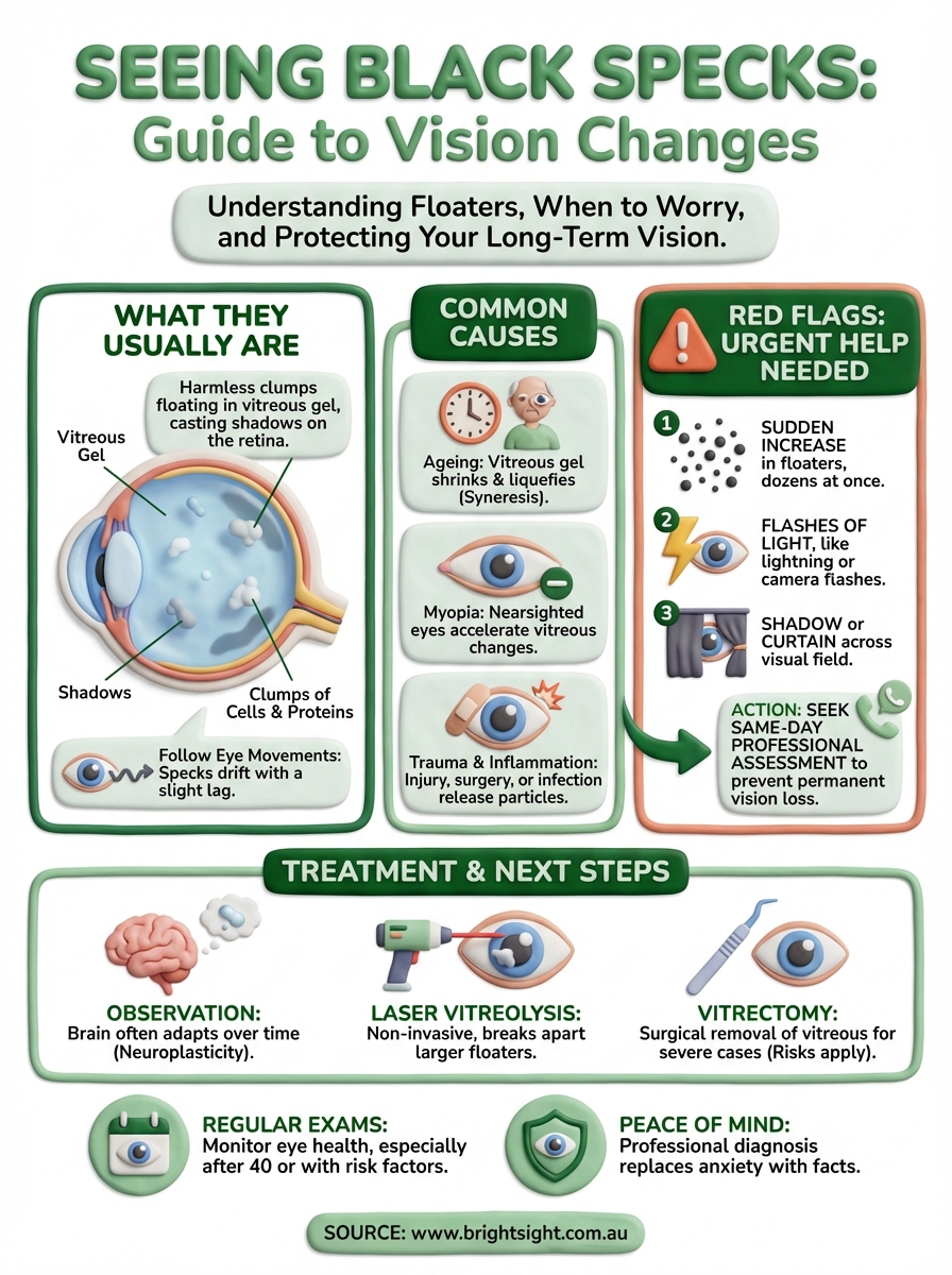
The black specks you notice drifting across your vision are almost always eye floaters, which consist of tiny clumps of cells or protein fibres floating inside the vitreous gel that fills your eyeball. This gel-like substance makes up roughly 80 per cent of your eye’s volume and helps maintain its spherical shape while keeping the retina properly positioned against the back wall. When fragments within the vitreous cast shadows on your retina, you perceive them as dark shapes or specks that move as your eyes move.
The vitreous gel and its role
Your vitreous starts life as a clear, smooth gel with a consistency similar to egg white, but changes naturally as you age. The gel begins to liquefy and shrink over time, typically from your 40s onwards, causing some of the collagen fibres within it to clump together rather than remaining evenly distributed. These clumps create the shadows on your retina that you interpret as floating specks, strings, or cobweb-like shapes in your field of vision.

Most people develop floaters gradually without noticing them until they become more pronounced, though some experience a sudden appearance when the vitreous pulls away from the retina in what specialists call posterior vitreous detachment. This process happens to nearly everyone eventually, usually between ages 50 and 75, and represents a normal part of ageing rather than disease.
What you actually see
Floaters take various forms depending on their size, shape, and position within your vitreous. You might notice small dots, longer thread-like strands, ring-shaped circles, or cloudy patches that drift slowly across your vision. They become most obvious against bright, uniform backgrounds like a white wall, blue sky, or computer screen, where the contrast makes them stand out more clearly.
The specks appear to move when you shift your gaze because they float freely within the vitreous gel, drifting slightly before settling back down.
When seeing black specks this way, you’ll find they don’t stay fixed in one spot like a smudge on your glasses would. Instead, they follow your eye movements with a slight lag and continue drifting even after your eyes stop moving, which confirms their location inside your eye rather than on its surface.
Common causes of black specks in vision
Several factors trigger the appearance of floaters, and understanding these causes helps you determine whether your symptoms need professional attention or simply reflect normal changes in your eyes. Most cases of seeing black specks stem from age-related processes that affect nearly everyone, though certain conditions and lifestyle factors can accelerate or worsen the issue.
Age-related vitreous changes
Your vitreous undergoes natural deterioration as you get older, with the gel becoming more liquid and less uniform in consistency. This process, called syneresis, causes collagen fibres within the vitreous to clump together and cast shadows on your retina. By age 80, roughly two-thirds of people experience some degree of vitreous liquefaction, making floaters an extremely common occurrence among older adults.
Posterior vitreous detachment happens when the vitreous gel pulls away from the retina completely, often occurring between ages 50 and 75. You might notice a sudden increase in floaters when this separation occurs, along with occasional flashes of light as the vitreous tugs on the retina before releasing. While PVD itself rarely causes harm, the pulling action creates a small risk of retinal tears during the detachment process.
The separation of your vitreous from the retina represents a normal milestone in eye ageing rather than a disease requiring treatment.
Myopia and structural factors
Near-sightedness makes you more susceptible to floaters because myopic eyes tend to be longer than average, which stretches the vitreous gel thinner and accelerates its breakdown. People with moderate to high myopia often develop floaters earlier in life, sometimes noticing them in their 30s or 40s rather than later decades. The structural difference in your eye shape creates mechanical stress that promotes earlier vitreous changes.
Eye trauma and inflammation
Physical injury to your eye, whether from sports, accidents, or surgical procedures, can disturb the vitreous and cause new floaters to appear suddenly. The trauma releases cells and protein into the vitreous cavity that create shadows on your retina. Similarly, inflammation inside your eye from conditions like uveitis or infections introduces inflammatory cells that float in the vitreous, producing the black specks you perceive in your vision.
Certain medications, particularly those injected into the eye to treat retinal conditions, can also introduce small bubbles or particles that temporarily increase floater symptoms before the body absorbs them over weeks or months.
Red flags and when to seek urgent help
While most floaters represent harmless age-related changes, certain warning signs indicate serious retinal problems that require immediate professional assessment. Learning to recognise these red flags helps you distinguish between symptoms you can monitor at home and those demanding urgent medical attention to prevent permanent vision loss.
Sudden onset symptoms requiring same-day care
You need to contact an eye specialist the same day if you experience a sudden increase in floaters, particularly when dozens appear at once rather than one or two developing gradually. This rapid onset often signals posterior vitreous detachment or, more seriously, a retinal tear where the vitreous has pulled hard enough to damage your retina. Accompanying flashes of light that resemble lightning bolts or camera flashes, especially in your peripheral vision, strengthen the urgency because they indicate ongoing traction on your retina.
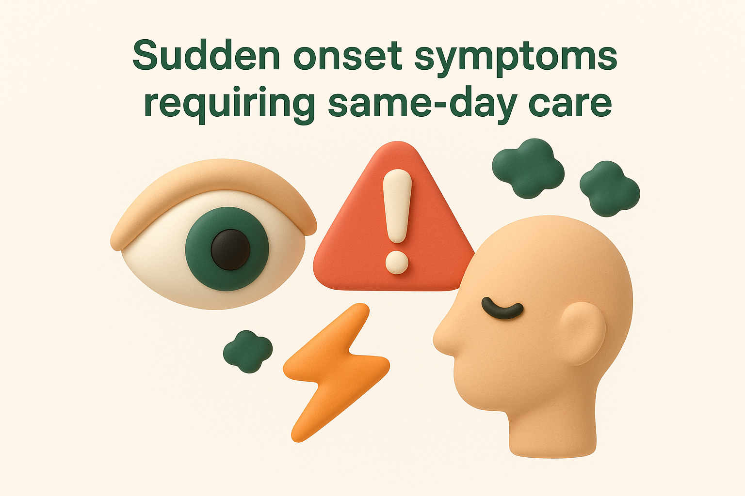
A shadow or curtain moving across your field of vision represents an absolute emergency, as this symptom suggests your retina has already detached and is lifting away from the back of your eye. Every hour counts when detachment occurs, because the longer your retina remains separated from its blood supply, the higher your risk of permanent vision loss in the affected area.
When seeing black specks arrives suddenly with flashes or shadows, you’re experiencing symptoms that require professional examination within hours, not days.
Other warning signs needing prompt attention
Notice whether your floaters appear alongside reduced vision in one eye, as this combination suggests something beyond simple vitreous changes. Bleeding inside your eye from diabetic retinopathy, torn blood vessels, or other conditions creates floaters while simultaneously blocking light from reaching your retina properly. You might also notice that straight lines appear wavy or distorted, which points to potential macular involvement rather than just vitreous issues.
Changes in your peripheral vision, such as dark spots or missing patches that don’t move when you shift your gaze, differ from typical floaters and warrant investigation. These fixed blind spots might indicate retinal damage or other serious conditions affecting your visual field.
When to schedule routine appointments
Gradual increases in floaters over months or years without accompanying symptoms typically don’t require emergency care, though you should mention them at your next routine eye examination. Similarly, existing floaters that remain stable in number and appearance rarely signal danger, but documenting them helps your specialist track any future changes that might need attention.
How eye specialists diagnose the cause
Determining what’s causing your floaters requires a systematic evaluation that goes beyond simply looking at your eyes. When you visit Bright Sight Eye Specialists or any retinal practice, your specialist combines your medical history with specialised examination techniques and, when necessary, advanced imaging to identify whether you’re experiencing benign floaters or something requiring treatment. This thorough approach ensures nothing serious gets missed while avoiding unnecessary tests or procedures.
Initial assessment and your symptom history
Your specialist begins by asking detailed questions about when you first noticed the floaters, whether they appeared suddenly or gradually, and if you’ve experienced flashes, shadows, or vision loss. This history provides crucial clues about the underlying cause, as sudden onset with flashes points toward posterior vitreous detachment or retinal tears, while gradual development suggests age-related changes. Your eye health background matters too, including any history of myopia, eye surgery, trauma, diabetes, or inflammatory conditions that increase your risk of retinal complications.
Documenting your general health helps identify systemic conditions that might affect your eyes, such as high blood pressure that can cause retinal bleeding or autoimmune disorders linked to eye inflammation. This conversation typically takes five to ten minutes but provides your specialist with essential context for interpreting the physical findings.
Detailed examination with dilated pupils
Your specialist performs a comprehensive eye examination using dilating drops that widen your pupils, allowing clear visualisation of your vitreous and retina. The drops take 15 to 20 minutes to work fully and blur your near vision temporarily, but they’re essential for seeing the peripheral retina where tears and detachments often begin. Once dilated, your specialist uses a slit lamp with special lenses to examine your vitreous for floaters, blood, or inflammatory cells.
A dilated examination remains the gold standard for diagnosing the cause of floaters because it provides direct visualisation of your entire retina and vitreous.
Direct ophthalmoscopy or indirect ophthalmoscopy techniques allow your specialist to scan your retinal surface systematically, checking for tears, holes, detachments, or other abnormalities that might explain your symptoms when seeing black specks. The examination typically takes ten to fifteen minutes and causes no discomfort beyond bright lights shining in your eyes.
Advanced imaging for complex cases
Most floater assessments don’t require imaging beyond the clinical examination, but your specialist might order optical coherence tomography (OCT) scans if they suspect macular involvement or need detailed cross-sectional views of your retina. OCT works like an ultrasound but uses light waves instead of sound, creating high-resolution images that reveal subtle structural changes invisible during routine examination. Ultrasound scanning becomes useful when blood or inflammation prevents clear visualisation of your retina, helping your specialist map any detachments or abnormalities through the opaque media.
Treatment options for floaters and retinal issues
Treatment choices for seeing black specks depend entirely on whether your floaters stem from harmless vitreous changes or more serious retinal problems requiring intervention. Your specialist tailors their approach to your specific situation, weighing the severity of your symptoms against the risks and benefits of each option. Most people with uncomplicated floaters never need treatment beyond reassurance, while those with retinal tears or detachments require prompt procedures to prevent permanent vision loss.
When observation is the best approach
If your examination reveals benign floaters without retinal complications, your specialist typically recommends watchful waiting rather than active treatment. Your brain gradually adapts to floaters over weeks or months, making them less noticeable even though they remain physically present. This neurological adaptation, called neuroplasticity, helps most patients regain their quality of life without intervention, though the process requires patience and realistic expectations about outcomes.
Your brain’s ability to filter out unchanging visual stimuli means floaters often become less bothersome over time, even without medical treatment.
Regular monitoring appointments every six to twelve months help track whether your floaters remain stable or develop patterns suggesting emerging complications that warrant treatment.
Laser vitreolysis for persistent floaters
Laser vitreolysis offers a non-invasive option for patients whose floaters significantly impair their daily function despite conservative management. Your specialist uses a specialised YAG laser to target and break apart the larger floater clumps, vaporising them into tiny fragments that become less visible. The procedure takes place in the clinic without anaesthesia beyond numbing drops, lasting fifteen to thirty minutes per session.
Success rates vary depending on floater type and location, with some patients experiencing substantial improvement while others gain only modest benefit. Your specialist carefully assesses whether your floaters suit this approach, as the laser works best for larger, well-defined floaters positioned away from your retina and lens.
Vitrectomy for severe cases
Vitrectomy removes the vitreous gel entirely, replacing it with saline solution that eliminates floaters permanently. Surgeons reserve this option for patients with severe floater symptoms that interfere dramatically with their work and daily activities, as the procedure carries risks including cataract formation, retinal detachment, and infection. The surgery requires operating theatre facilities and takes one to two hours under local or general anaesthesia.
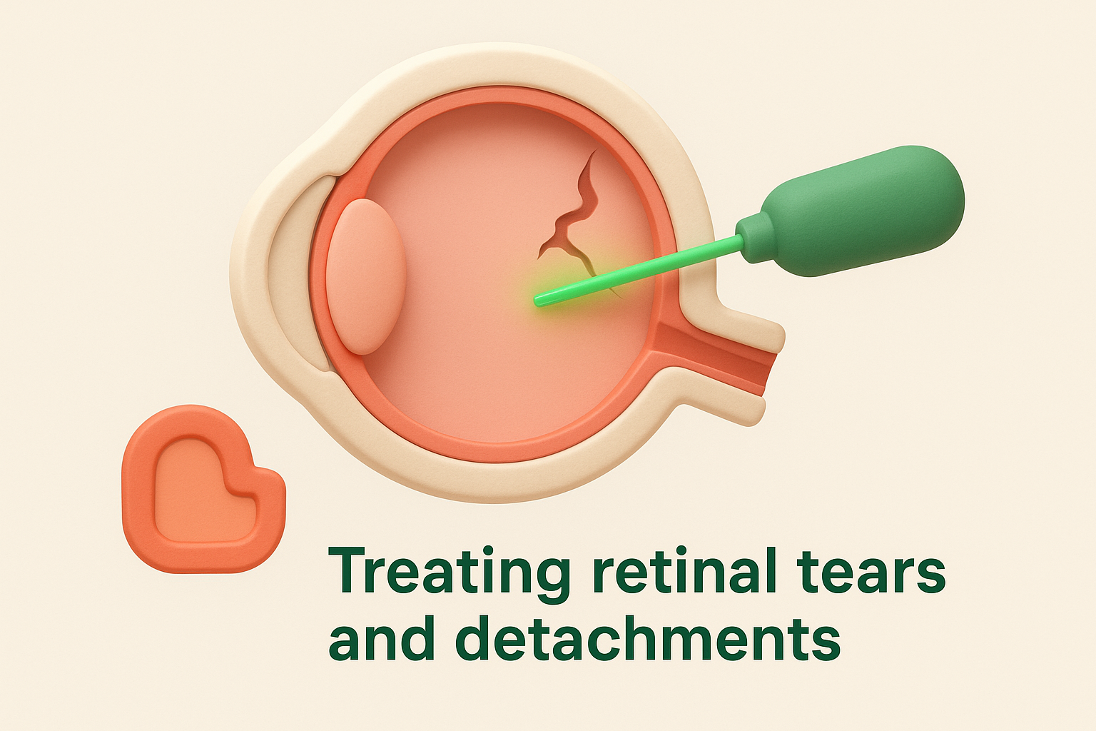
Treating retinal tears and detachments
Retinal tears need immediate sealing with laser photocoagulation or cryotherapy, which creates controlled scarring that welds your retina back in place. These procedures take minutes in the clinic and prevent progression to full detachment. Established retinal detachments require surgical repair through vitrectomy, scleral buckling, or pneumatic retinopexy, with the specific technique depending on the detachment’s location and severity.
What you can do now to protect your sight
Taking proactive steps to maintain your eye health reduces your risk of complications that might cause seeing black specks or more serious vision problems. While you can’t prevent all age-related changes in your vitreous, you can control several factors that influence your overall eye health and catch potential issues before they progress to permanent damage. These practical measures require minimal effort but deliver substantial benefits over your lifetime.
Schedule regular comprehensive eye examinations
Book a dilated eye examination every one to two years if you’re over 40, or annually if you have risk factors like high myopia, diabetes, or a family history of retinal disease. These check-ups allow your specialist to detect early signs of retinal tears, detachments, or other conditions before they cause noticeable symptoms. Don’t wait until you experience problems to visit an eye specialist, as many serious conditions develop silently in their early stages when treatment works best.
Regular monitoring creates a baseline record of your eye health, making it easier for your specialist to spot subtle changes that might indicate emerging problems.
Your specialist can also update your prescription, assess your intraocular pressure for glaucoma risk, and check for cataracts or macular degeneration that commonly develop alongside floaters in older adults.
Protect your eyes from injury and strain
Wear appropriate safety glasses during activities that risk eye injury, including sports, yard work, or using power tools. Even minor trauma can disturb your vitreous or damage your retina, creating floaters or more serious complications. Consider UV-blocking sunglasses when outdoors, as ultraviolet exposure contributes to various eye conditions over time, though direct links to floaters remain unclear.
Taking regular breaks during screen work reduces eye strain and fatigue, though these habits won’t prevent floaters from developing.
Manage systemic health conditions
Control your blood sugar levels if you have diabetes, as unmanaged diabetes causes retinal bleeding and other complications that increase floaters and threaten your vision. Similarly, maintaining healthy blood pressure protects your retinal blood vessels from damage. Smoking cessation improves circulation to your eyes and reduces inflammation throughout your body, supporting better long-term eye health. These systemic factors influence whether minor issues like floaters progress to serious vision-threatening conditions requiring urgent intervention.
Next steps for clearer vision
Understanding the cause behind seeing black specks empowers you to respond appropriately rather than ignoring symptoms that might need attention. Most floaters represent harmless age-related changes that your brain learns to filter out over time, but sudden increases in floaters, flashes of light, or vision loss require same-day assessment to protect your sight from permanent damage. The difference between these scenarios often comes down to timing, making professional evaluation your safest choice when uncertainty exists.
Taking action starts with a comprehensive eye examination that identifies whether your symptoms stem from benign vitreous changes or serious retinal complications requiring treatment. If you’re experiencing concerning floaters or simply want peace of mind about your eye health, book a consultation at Bright Sight Eye Specialists in Sydney’s Eastern Suburbs. Dr Rahul Dubey and our team provide expert assessment and personalised treatment plans that address your specific situation, helping you maintain the clear, comfortable vision you need for life’s important moments.