


Your retina is a thin layer of light-sensitive tissue at the back of your eye, and when something goes wrong with it, your vision can deteriorate quickly. Retina eye surgery refers to a group of surgical procedures designed to repair damage to this critical structure, from tears and detachments to conditions like macular holes and epiretinal membranes. If you or someone you care about is facing one of these diagnoses, understanding your surgical options is a practical first step toward protecting your sight.
The good news is that modern retinal surgery has come a long way. Procedures are more precise, recovery times are shorter, and outcomes are significantly better than they were even a decade ago. But there’s still a lot to take in, different procedure types, varying recovery timelines, potential risks, and the question of what it will actually cost with or without private health insurance.
At Bright Sight Eye Specialists in Randwick, Dr Rahul Dubey performs advanced retinal and macular surgery for patients across Sydney’s Eastern Suburbs and beyond. His training at Moorfields Eye Hospital in London, one of the world’s leading eye hospitals, gives him deep expertise in treating complex retinal conditions. This article walks you through the main types of retina eye surgery, what each procedure involves, what recovery looks like, and the costs you can expect so you can approach your next appointment with confidence.
Why retina eye surgery matters
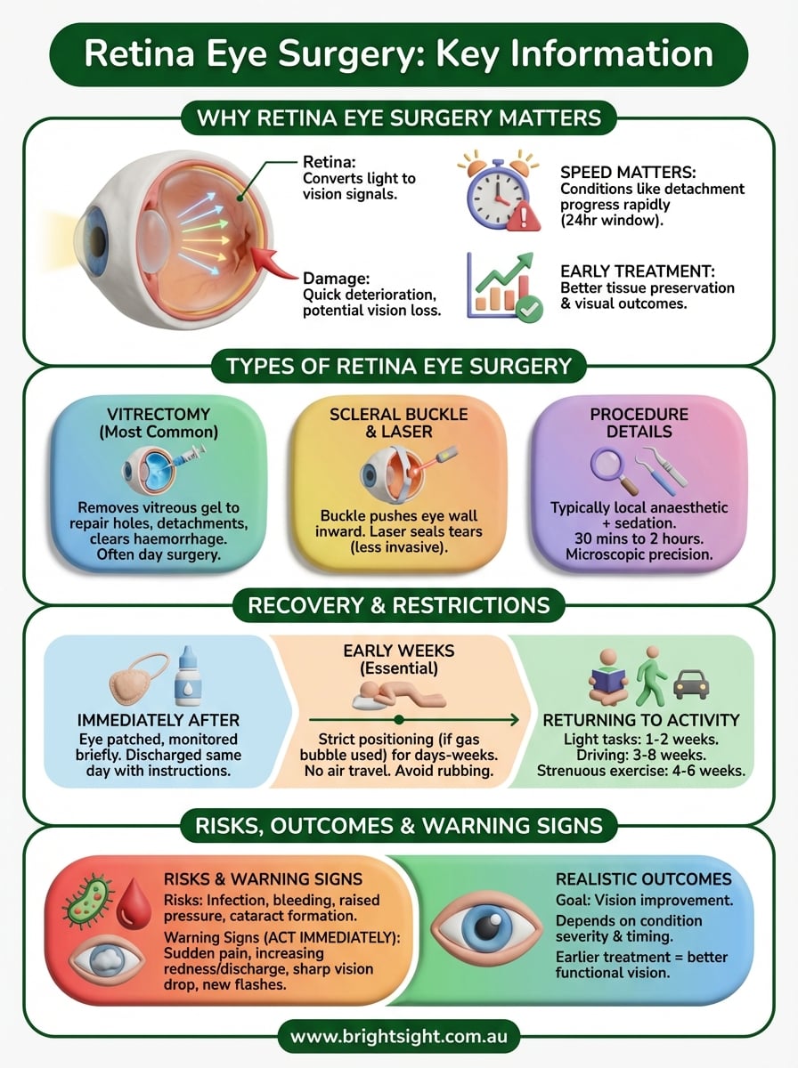
The retina does the work of converting light into electrical signals that your brain reads as vision. When it’s damaged, your central or peripheral vision deteriorates, and in serious cases, you can lose your sight permanently. What makes retinal conditions particularly concerning is how quickly some of them progress. A small retinal tear, if left untreated, can develop into a full detachment within days.
Retinal detachment is considered a medical emergency, and delaying treatment by even 24 hours can determine whether your central vision is saved.
Conditions that make surgery necessary
Several eye conditions require retina eye surgery to stop or reverse vision loss. Retinal detachment occurs when the retina lifts away from the tissue that supplies it with oxygen and nutrients, and without prompt surgical repair, the detached area loses function. Macular holes and epiretinal membranes are conditions that distort or block your central vision, the part you rely on for reading, driving, and recognising faces. Vitreous floaters, when dense or persistent, can also block enough of your visual field to warrant surgical removal.
Diabetic retinopathy and retinal vein occlusion can cause bleeding inside the eye or abnormal blood vessel growth, both of which may require surgical intervention to prevent lasting damage. These conditions often develop gradually, which is why regular eye checks matter so much for people with diabetes or high blood pressure.
Why early treatment produces better results
Timing matters a great deal with retinal conditions. The sooner surgery is performed, the more retinal tissue can be preserved, and the better your final visual outcome is likely to be. In macular hole surgery, for example, holes caught early have a significantly higher closure rate and a stronger chance of meaningful vision recovery. Waiting until a condition has worsened often means your surgeon has less to work with, and your recovery will take longer.
Types of retina eye surgery
Not all retinal conditions require the same surgical approach. Your surgeon will recommend a procedure based on the specific condition, its severity, and how much of your retina is affected. Understanding the main procedure types helps you know what you are agreeing to and what to expect.
Vitrectomy
Vitrectomy is the most common form of retina eye surgery and involves removing the vitreous gel from inside your eye to access the retina directly. Your surgeon uses this approach to repair macular holes, peel epiretinal membranes, treat retinal detachments, and clear vitreous haemorrhage caused by diabetic eye disease. Once the vitreous is removed, the eye is filled with a gas bubble or silicone oil to hold the retina in place while it heals.

In many cases, vitrectomy is performed as day surgery under local anaesthetic, meaning you go home the same day.
Scleral buckle and laser treatment
A scleral buckle is a procedure where a flexible band is placed around the outside of the eye to gently push the wall of the eye inward against a detached retina. It is often used alongside vitrectomy for complex detachments. Laser photocoagulation is a less invasive option used to seal retinal tears before they progress to full detachment, and it is typically completed in an outpatient setting without any incisions.
What happens before, during and after surgery
Knowing what to expect at each stage of retina eye surgery makes the experience far less daunting. Your surgeon will guide you through the process, but having a clear picture beforehand helps you prepare practically and mentally for what lies ahead.
Before your procedure
In the weeks leading up to surgery, you will attend a pre-operative assessment where your surgeon examines your retina in detail, reviews your medical history, and confirms the surgical plan. You may need to stop certain medications, particularly blood thinners, and arrange for someone to drive you home. Fasting is required for several hours before the procedure if you are having sedation or general anaesthesia.
During the operation
Most retinal procedures are performed under local anaesthetic with sedation, so you are comfortable and still without being fully unconscious. The surgery typically takes between 30 minutes and two hours depending on complexity. Your surgeon works through tiny incisions using microscopic instruments and a high-magnification operating system to repair the retina with precision.
For most patients, retina eye surgery is completed as day surgery, meaning no overnight hospital stay is required.
Immediately after surgery
Once the procedure is finished, your eye is patched and protected while the anaesthetic wears off. Staff monitor you briefly before discharge and provide clear instructions on eye drops, positioning requirements, and what symptoms to watch for at home.
Recovery time and activity restrictions
Recovery from retina eye surgery varies based on the procedure you had and the condition being treated. Most patients notice gradual improvement over several weeks, though your vision may appear blurry or distorted initially, particularly if a gas bubble was placed inside the eye during surgery.
Your vision may look worse before it improves during the first one to two weeks after surgery.
Positioning requirements and early restrictions
If your surgeon used a gas bubble during your vitrectomy, you will need to hold a specific head position for several days to two weeks. Strict compliance with positioning is essential for the bubble to press correctly against the retina and support healing. Key restrictions during this phase include:
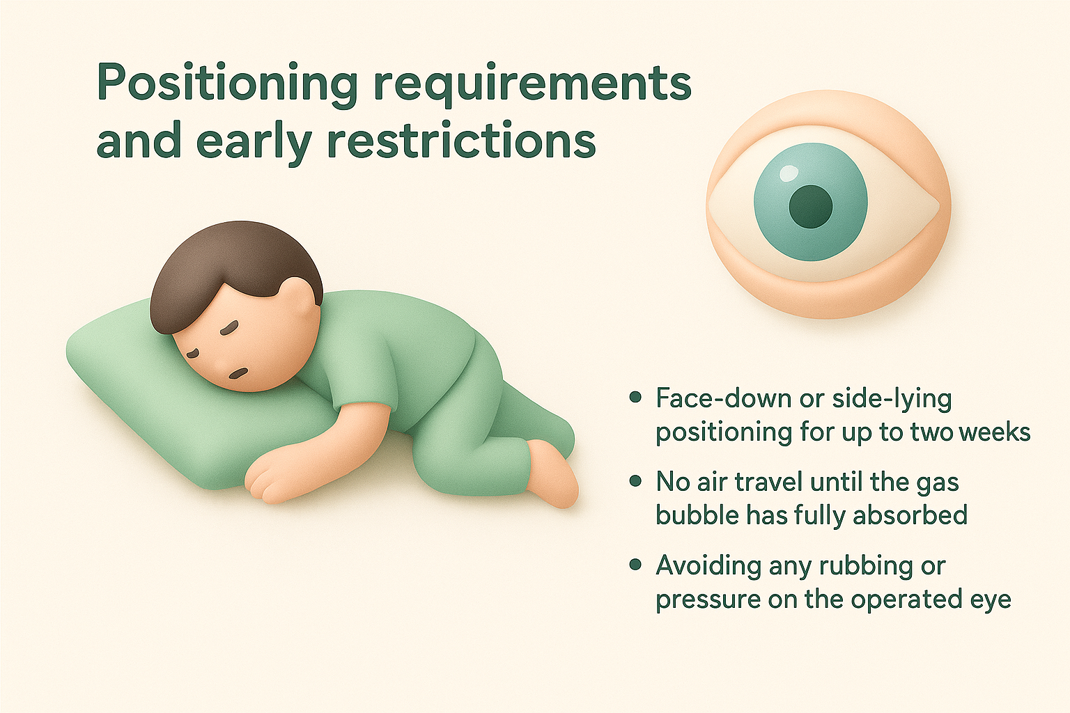
- Face-down or side-lying positioning for up to two weeks
- No air travel until the gas bubble has fully absorbed
- Avoiding any rubbing or pressure on the operated eye
Returning to everyday activities
Light daily tasks such as reading or short walks are generally possible within one to two weeks. Your surgeon will confirm when your vision meets the standard required for driving, which typically takes three to eight weeks depending on your recovery progress.
Strenuous exercise, heavy lifting, and swimming should be avoided for four to six weeks. Most people return to full activity step by step, following the specific guidance their surgeon provides at each follow-up appointment.
Risks, outcomes and warning signs
Retina eye surgery carries a strong track record of success, but like any surgical procedure, it comes with risks you should understand before giving consent. Most complications are rare and manageable, and your surgeon will discuss your individual risk profile in detail during your pre-operative consultation.
Risks and realistic outcomes
Infection, bleeding, and raised eye pressure are among the more common risks associated with vitrectomy and other retinal procedures. Cataract formation after surgery is also well-documented, and some patients require cataract surgery within two years of a vitrectomy. Vision improvement is the goal, but outcomes depend heavily on how long the condition was present before surgery and how much retinal damage had already occurred.
The earlier a retinal detachment or macular hole is treated, the more likely you are to recover functional vision.
Warning signs to act on immediately
Following surgery, your eye will feel sensitive and mildly uncomfortable for the first week or two. Certain symptoms, however, indicate something more serious is happening and require prompt attention. Contact your surgeon without delay if you experience any of the following:
- A sudden increase in pain that paracetamol does not relieve
- Increasing redness, discharge, or swelling around the eye
- A sharp drop in vision or new flashes of light
- Severe headache alongside eye pain
Do not wait for your scheduled follow-up if these symptoms appear.
Next steps
Retinal conditions respond best to treatment when they are caught and addressed promptly. If you have been referred to a specialist, are experiencing new visual symptoms, or want a clearer picture of your options before your next appointment, taking action now rather than later gives you the best chance of protecting your sight. Retina eye surgery has strong outcomes when performed by an experienced surgeon with access to advanced equipment, and the decision to proceed becomes much easier once you understand exactly what is involved.
At Bright Sight Eye Specialists in Randwick, Dr Rahul Dubey and Dr Capucine Odouard see patients from across Sydney’s Eastern Suburbs and beyond, offering thorough assessments and clear treatment plans tailored to your situation. Whether you are facing a diagnosis or simply have questions, getting the right advice early makes a real difference. Book a consultation with our team to take the next step toward better eye health.