


A torn or detached retina can threaten your sight within hours. The good news: modern surgical techniques can repair retina damage effectively, and in many cases, preserve or restore functional vision when treatment happens promptly.
But the term “retinal repair” covers a range of procedures, from in-office laser treatments to complex surgeries performed under anaesthesia. Understanding which approach applies to your situation, and what recovery actually looks like, helps you make confident, informed decisions about your care. Knowing what to expect removes much of the anxiety that comes with an urgent or unexpected diagnosis, and puts you in a stronger position to act quickly when it matters most.
At Bright Sight Eye Specialists in Randwick, Dr Rahul Dubey performs retinal and macular surgery using advanced techniques developed through his training at Moorfields Eye Hospital in London. Our team treats conditions including retinal detachment, macular holes, epiretinal membranes, and vitreous haemorrhage, with transparent pricing and a patient-first approach throughout the process.
This article walks you through the main types of retinal repair, how each procedure works, what influences your surgeon’s recommendation, and what to realistically expect during recovery. Whether you’re facing a new diagnosis or researching on behalf of someone you care about, you’ll find clear, practical information to guide your next steps.
What repair retina means
The retina is a thin layer of light-sensitive tissue that lines the back wall of your eye. When light enters your eye, the retina converts it into electrical signals that travel through the optic nerve to your brain, producing the images you see. When a doctor says they need to repair retina damage, they mean any procedure that restores the retina to its correct position or corrects structural problems that prevent this process from working properly.
How the retina works
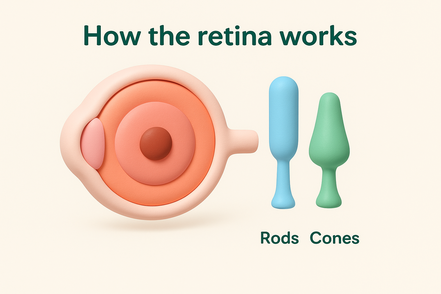
Your retina contains two types of photoreceptor cells: rods and cones. Rods handle low-light and peripheral vision, while cones manage colour perception and fine detail, particularly in the central retinal zone called the macula. Together, these cells form a remarkably sensitive system that processes everything from bright outdoor light to the dim glow of a lamp across the room.
Because the retina depends on a constant supply of oxygen and nutrients from the blood vessels beneath it, anything that separates the retina from that underlying layer starts to damage those cells almost immediately. This is why timing plays such a critical role in outcomes: the longer the retina sits without adequate blood supply, the more cells are lost, and the harder it becomes to recover useful vision even after a successful repair.
What can damage the retina
Several conditions compromise retinal integrity to the point where treatment becomes necessary. The most common include:
- Retinal tears and detachments: A break in the retina allows vitreous fluid to seep underneath it, separating it from the blood supply it relies on.
- Macular holes: A small opening forms in the centre of the macula, directly affecting your ability to see fine detail and straight-ahead images clearly.
- Epiretinal membranes: Scar tissue forms on the retinal surface and pulls on the underlying tissue, causing distortion or blurring.
- Vitreous haemorrhage: Bleeding into the vitreous gel blocks light from reaching the retina and can obscure the retina from your surgeon’s view.
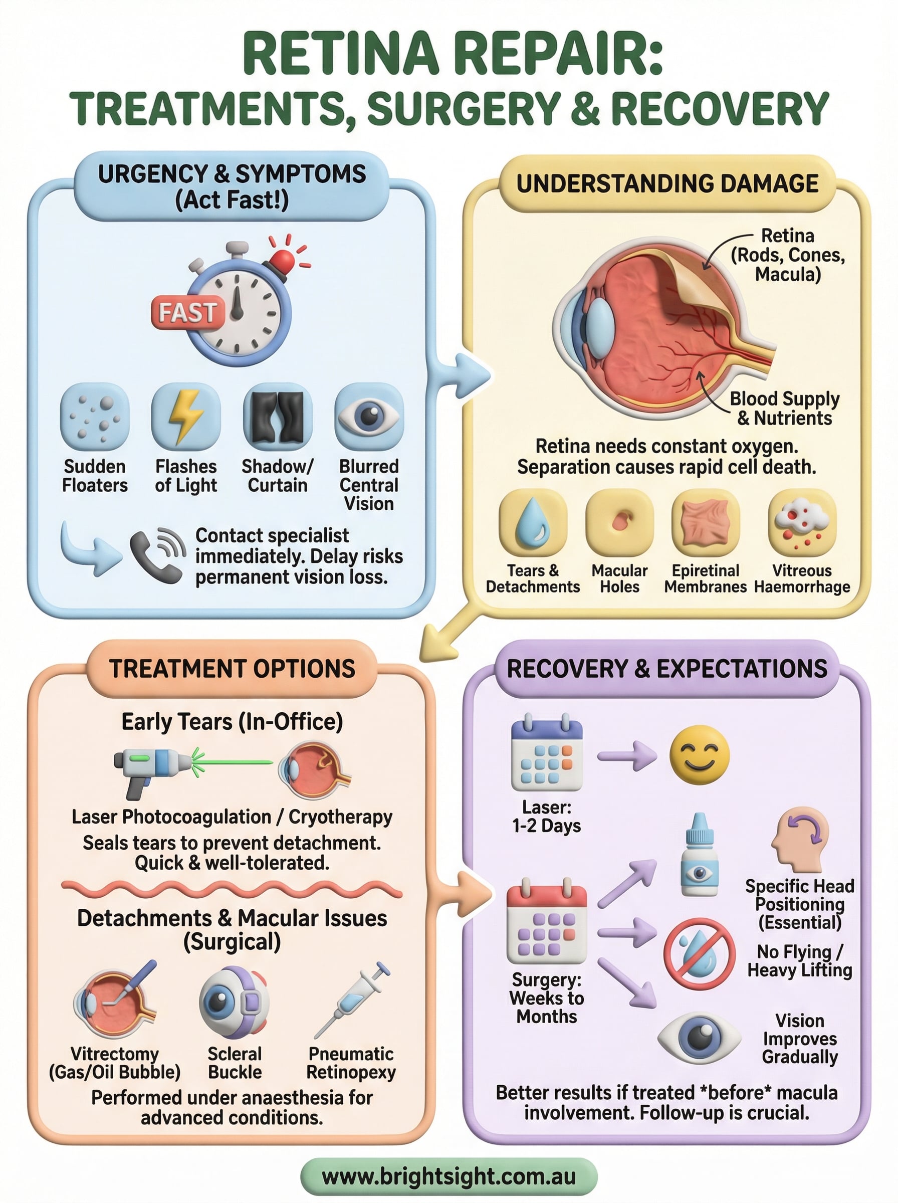
A retinal tear caught before it becomes a full detachment can often be treated with a quick in-office laser procedure, while a full detachment requires surgery under anaesthesia.
Each of these conditions calls for a different treatment approach, and the degree of damage at the time of diagnosis directly shapes what recovery looks like. Understanding which condition applies to your situation, and how far it has progressed, is where every meaningful conversation with your eye specialist begins.
Why you must treat retina problems fast
Retinal tissue does not regenerate. Once the cells that line the back of your eye die from lack of oxygen, no procedure can bring them back. This is why speed matters more with retinal conditions than with almost any other eye problem. The difference between acting within hours and waiting a few days can mean the difference between recovering useful vision and losing it permanently.
What happens when you delay
When your retina detaches or a tear allows fluid to accumulate underneath it, the photoreceptor cells in the affected area begin to deteriorate rapidly. Central vision is particularly vulnerable because the macula sits in the most oxygen-dependent zone of the retina. If a detachment reaches the macula before surgery takes place, outcomes drop significantly, even after a technically successful repair.
A retinal detachment that spares the macula can often achieve near-normal visual recovery after surgery, while one that involves the macula frequently leaves permanent central vision loss.
Symptoms you should not ignore
Certain warning signs point directly to retinal damage and warrant same-day contact with an eye specialist. Do not wait to see if these resolve on their own:
- A sudden increase in floaters, especially if they appear in a shower or cluster
- Flashes of light in your peripheral vision, particularly in a dark room
- A shadow, curtain, or dark area spreading across your visual field
- Sudden blurred or distorted central vision
When you notice any of these symptoms, contact your eye specialist immediately. Prompt assessment allows your surgeon to repair retina damage at the earliest opportunity, giving you the strongest possible chance of a good outcome.
How eye specialists repair the retina
The method your surgeon uses to repair retina damage depends on the type of condition, how far it has progressed, and which part of the retina is affected. There is no single procedure that fits every situation, so your specialist will match the technique to your specific diagnosis after a thorough examination.
Laser and cryotherapy for tears
When your surgeon identifies a retinal tear before it becomes a full detachment, laser photocoagulation is usually the first-line treatment. During this in-office procedure, a focused laser beam creates tiny burns around the tear, forming scar tissue that seals the retina to the underlying tissue and prevents fluid from getting underneath. Cryotherapy uses a cold probe to achieve the same result when laser access is more difficult. Both are quick, well-tolerated procedures that typically take less than 30 minutes and require no incisions.
Treating a retinal tear with laser before fluid accumulates beneath it can prevent a detachment entirely, making it one of the most effective interventions in eye care.
Surgical options for detachments and macular conditions
More advanced conditions require surgery performed under local or general anaesthesia. Your surgeon will recommend one of the following approaches based on your diagnosis:
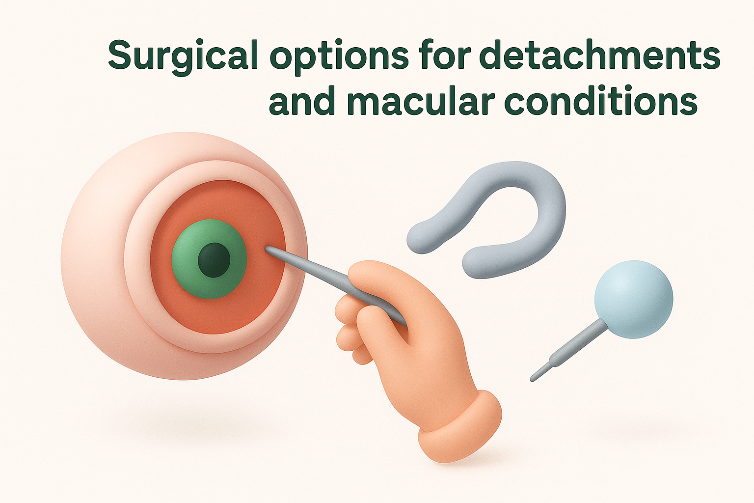
- Vitrectomy: The vitreous gel is removed and replaced with gas, air, or silicone oil to hold the retina in place while it heals. This treats detachments, macular holes, and epiretinal membranes.
- Scleral buckle: A silicone band is placed around the outside of the eye to gently push the wall inward and close the break.
- Pneumatic retinopexy: A gas bubble is injected into the eye to press the retina back against the wall in select cases.
Recovery after retina repair surgery
Recovery time after you repair retina damage varies depending on which procedure you had and how advanced your condition was before surgery. Laser treatment for a tear usually causes minimal disruption, with most people returning to normal activity within a day or two. Surgical procedures like vitrectomy or scleral buckle require a longer, more structured recovery period that can span several weeks to months.
What to expect in the first few weeks
Your surgeon will prescribe anti-inflammatory and antibiotic eye drops to reduce swelling and prevent infection after surgery. Vision often appears blurry, distorted, or partially obscured in the early weeks, particularly if your surgeon placed a gas bubble inside the eye. This is normal. The bubble gradually absorbs on its own, and your vision typically improves progressively as the retina heals and inflammation settles.
Do not fly in an aeroplane while a gas bubble remains in your eye, as changes in cabin pressure can cause a dangerous rise in eye pressure.
Position requirements and activity limits
If your surgeon used a gas bubble during vitrectomy, you will likely need to maintain a specific head position for several days after the procedure. This positioning keeps the bubble in contact with the repair site and is critical to a successful outcome. Your surgical team will give you clear instructions on which position applies to your situation.
During recovery, you should avoid strenuous exercise, heavy lifting, and rubbing the eye until your surgeon clears you. Most people can return to light daily activities within one to two weeks, but full visual recovery often takes three to six months depending on whether the macula was involved.
Risks, results, and common questions
No surgical procedure is without risk, and retinal surgery is no exception. Understanding what can go wrong, and how likely those complications actually are, helps you weigh your options clearly and have a more productive conversation with your surgeon before you proceed.
What are the main risks of retinal surgery
Your surgeon will discuss your individual risk profile during your consultation, but the most common concerns across retinal procedures include:
- Raised eye pressure in the days following surgery, which your surgical team monitors closely
- Cataract formation accelerating after vitrectomy, particularly in patients over 50
- Re-detachment of the retina, which may require a second procedure
- Infection inside the eye (endophthalmitis), which is rare but serious if it occurs
Most complications are manageable when detected early, which is why attending every scheduled follow-up appointment after surgery is not optional.
What results can you realistically expect
Results depend heavily on whether the macula was involved before surgery and how quickly treatment began. When a surgeon can repair retina damage before the macula detaches, the majority of patients regain vision that is functional for daily tasks such as driving, reading, and recognising faces. When the macula was already affected at the time of surgery, some central vision loss often persists even after a technically successful repair.
Frequently asked questions
Can the retina detach again after surgery? Yes, re-detachment affects a small percentage of cases and typically requires further intervention. Will you need glasses after retinal surgery? Your prescription may shift after vitrectomy, especially if a cataract develops, so your optometrist will reassess your vision once healing is complete.
What to do now
If you’ve read this far, you likely have a specific concern about your retinal health, either for yourself or someone close to you. The core message from this article is clear: retinal conditions do not improve on their own, and waiting even a short time can significantly affect what is possible. Whether you are experiencing new floaters, flashes of light, or a confirmed diagnosis, the right next step is to speak with a specialist who can assess and repair retina damage before it progresses further.
At Bright Sight Eye Specialists in Randwick, Dr Rahul Dubey provides comprehensive retinal and macular surgery with the depth of experience that comes from his training at Moorfields Eye Hospital in London. Transparent pricing and a patient-first approach are part of every consultation. Book an appointment with our team to get a clear picture of your situation and understand your treatment options.