


Posterior vitreous detachment (PVD) happens when the gel inside your eye pulls away from the retina at the back. Your eye contains a clear, jelly-like substance called the vitreous that fills the space between your lens and retina. As you age, this gel becomes more liquid and gradually shrinks. Eventually it can no longer fill the entire space, so it separates from the retinal surface. This natural process typically occurs after age 60 and affects most people at some point in their lives.
This article explains what you need to know about PVD and your eye health. You’ll learn how to recognise the warning signs like floaters and flashes of light. We’ll cover what causes this condition and who faces higher risk. You’ll understand how doctors diagnose PVD during an eye examination and what treatment options exist. Most importantly, you’ll know when to seek immediate care. While PVD itself rarely threatens your vision, it can sometimes lead to serious complications like retinal tears or detachment that do require urgent attention.
Why PVD in the eye matters
Understanding PVD in the eye helps you protect your vision and respond appropriately when symptoms appear. This condition affects approximately 75% of people over age 65, making it one of the most common age-related changes in the eye. While the separation itself rarely causes permanent vision loss, your awareness matters because PVD can trigger serious complications in about 10 to 15% of cases. These complications include retinal tears, retinal detachment, and vitreous haemorrhage, all of which require immediate treatment to preserve your sight.
The impact on quality of life
Your daily activities can be disrupted when floaters from PVD appear in your central vision. Reading becomes more difficult when dark spots obscure the text. Driving may feel unsafe when floaters distract you, particularly in bright sunlight or when looking at light-coloured surfaces. Many people experience anxiety and frustration during the first few weeks of PVD, especially if they haven’t been warned about what to expect.
The emotional burden of sudden vision changes often goes unrecognised, yet it significantly affects how people cope with the condition.
Professional reassurance and accurate information help reduce this distress. Knowing what’s happening inside your eye and when to worry makes the experience far less frightening.
How to recognise and respond to PVD
Recognising PVD in your eye starts with understanding its characteristic symptoms and knowing how your body signals potential complications. The two main symptoms are floaters and flashes of light, which typically appear suddenly over a few hours or days. Most people notice floaters first, describing them as spots, cobwebs, or threads drifting across their vision. These symptoms may feel alarming when they first occur, but learning to identify normal PVD versus warning signs helps you respond appropriately and seek care when necessary.
The telltale signs of PVD
Floaters appear as dark shapes that move when you shift your gaze and seem to float away when you try to look directly at them. You might see small dots, curved lines, or a ring-shaped shadow called a Weiss ring. These become most noticeable against plain backgrounds like a white wall, blue sky, or computer screen. Flashes of light typically appear at the side edges of your vision, particularly in dim lighting or when you move your eyes quickly. The flashes last only a fraction of a second and often look like brief streaks or arcs of light.

Your symptoms will likely change over the first few weeks. Floaters may appear more prominent initially, then gradually settle towards the bottom of your vision as gravity pulls the condensed vitreous gel downward. Most people notice fewer flashes within four to six weeks as the vitreous completes its separation from the retina.
When to seek urgent care
Contact an eye specialist immediately if you notice a sudden shower of new floaters, like someone threw pepper across your vision. This suggests bleeding inside the eye or a retinal tear. A curtain or shadow spreading across your vision indicates possible retinal detachment, which requires surgery within hours to days to prevent permanent vision loss.
Sudden vision changes always warrant same-day assessment, even if you’ve been told your PVD is "normal" and to expect floaters.
Ring your ophthalmologist straightaway if you see persistent flashes that increase in frequency or a sudden decrease in vision. These symptoms don’t necessarily mean disaster, but only an examination can rule out complications that threaten your sight.
What causes PVD and who is at risk
Your vitreous gel changes naturally as you age, leading to PVD in the eye through a process that affects nearly everyone eventually. The gel consists of 99% water held together by collagen fibres, which gradually break down over time. This breakdown causes the gel to liquefy and contract, creating pockets of liquid within the previously uniform substance. Eventually, the gel collapses on itself and separates from the retina, completing the PVD process. This separation typically occurs without warning and progresses over several weeks.

The natural aging process behind PVD
Age represents the single most important factor in developing PVD. The condition rarely occurs before age 40 and becomes increasingly common throughout your 50s and 60s. By age 65, three out of four people will have experienced PVD in at least one eye. Your second eye typically develops PVD within one to two years after the first, though the timeline varies considerably between individuals.
Your body’s natural aging process, not lifestyle choices or poor health, drives most cases of PVD.
Risk factors that increase your chances
Myopia (short-sightedness) accelerates PVD development, often causing it to occur 10 to 15 years earlier than in people with normal vision. Your longer eyeball shape stretches the vitreous and speeds up its liquefaction. Recent cataract surgery increases your risk because the procedure disturbs the vitreous and can trigger earlier separation. Eye injuries that cause inflammation inside your eye may also prompt premature PVD. Additionally, diabetic eye disease can lead to abnormal vitreous changes that increase separation risk at younger ages.
How PVD is diagnosed in clinic
Your eye specialist diagnoses PVD in the eye through a thorough dilated examination that allows clear visualisation of your vitreous and retina. The appointment typically takes 45 to 60 minutes, including time for the dilating drops to work. Your ophthalmologist places drops in your eyes to widen your pupils, which may cause temporary blurring and light sensitivity for three to four hours afterward. You shouldn’t drive yourself home after this examination because your vision remains impaired whilst the drops wear off.
The examination process
The specialist examines your eye using a slit lamp combined with special lenses that provide a magnified view of your vitreous cavity and retinal surface. They look for signs of vitreous separation, including the classic Weiss ring (a circular opacity that represents the detached vitreous around your optic nerve). Your doctor specifically checks for retinal tears, holes, or areas of traction where the vitreous may be pulling on your retina. This examination reveals whether your PVD has caused any complications requiring immediate treatment.
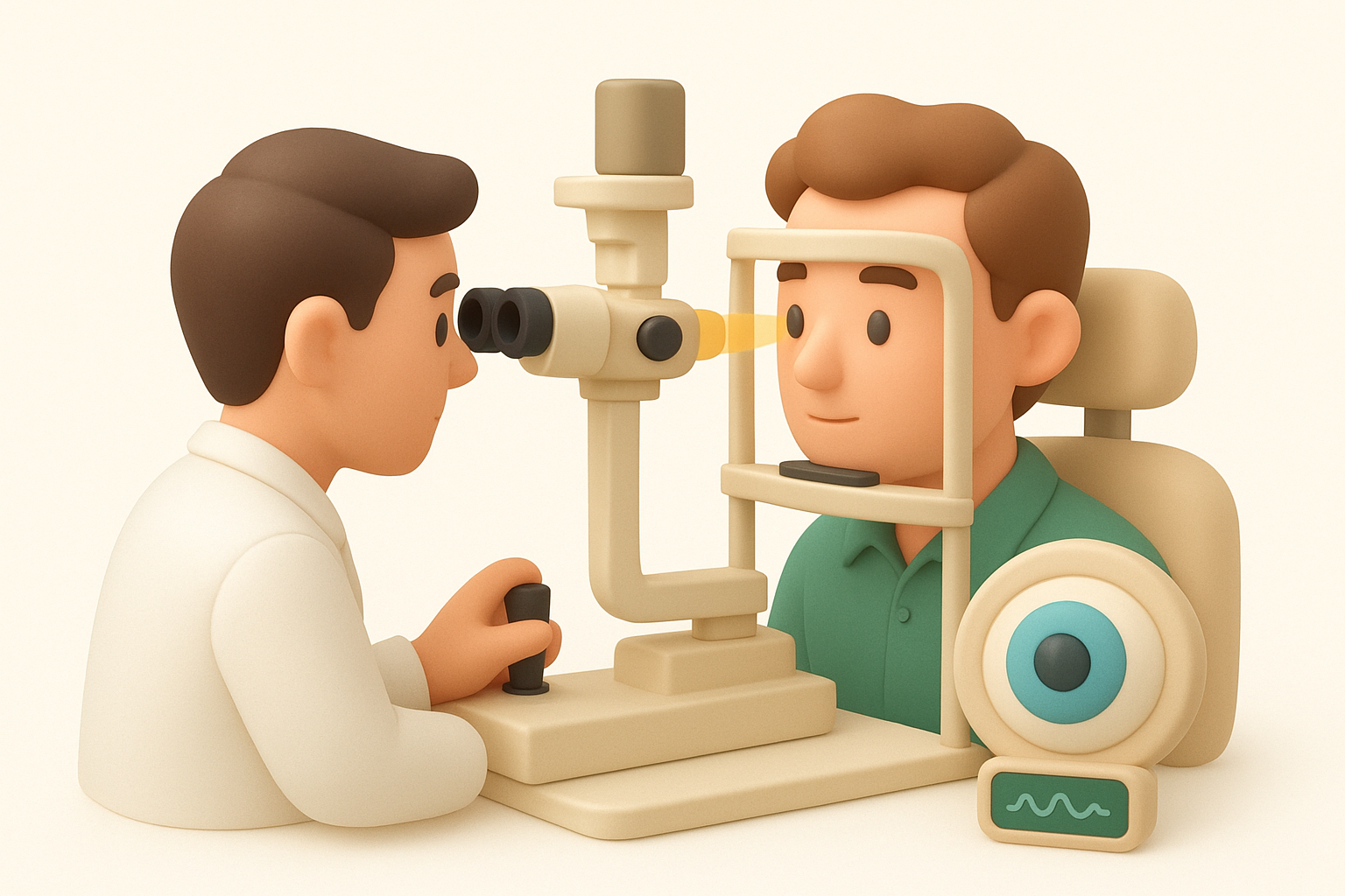
A dilated examination remains the gold standard for diagnosing PVD and ruling out sight-threatening complications.
Additional tests like optical coherence tomography (OCT) or ultrasound may be necessary if your vitreous appears too cloudy for direct visualisation. OCT creates detailed cross-sectional images of your retina and can detect subtle macular changes. Ultrasound proves particularly useful when bleeding obscures the view of your retina.
Treatment options, recovery and daily life
Most cases of PVD in the eye require no medical treatment because the condition resolves naturally over time. Your ophthalmologist will monitor you through follow-up examinations at 4 to 6 weeks and again at 3 months after initial diagnosis. These appointments check for delayed complications like retinal tears that can develop even when your first examination showed no problems. Your specialist treats any complications directly rather than treating the PVD itself.
Living with floaters day-to-day
Floaters typically become less bothersome within 3 to 6 months as your brain learns to ignore them and they settle below your central vision. Moving your eyes in circles can shift a floater temporarily when it blocks something you’re trying to read. Wearing sunglasses outdoors reduces the contrast that makes floaters more visible against bright backgrounds. Adjusting screen brightness and using dark mode settings helps when working on computers or mobile devices.

Your brain’s remarkable ability to filter out unchanging stimuli means most floaters fade from your awareness naturally over time.
When treatment becomes necessary
Vitrectomy surgery removes persistent floaters that significantly impair your quality of life after 6 to 12 months. Surgeons remove the vitreous gel and replace it with saline solution during this procedure. However, surgery carries risks including cataract formation, retinal tears, and infection, so specialists reserve it for severe cases only. Your ophthalmologist will discuss whether surgery’s potential benefits outweigh these risks based on your specific symptoms and lifestyle impact. Treatment for complications like retinal tears involves laser therapy or surgical repair, typically performed urgently to prevent retinal detachment and preserve your vision.

Next steps for your eye health
Schedule a comprehensive eye examination if you notice new floaters, flashes, or any sudden changes in your vision. Early detection of complications from PVD in the eye makes treatment simpler and more effective. Your regular optometrist can perform initial screening, though you’ll need referral to a retinal specialist if any concerning signs appear. Don’t wait weeks hoping symptoms will improve on their own, particularly if you notice multiple new floaters or a shadow in your peripheral vision.
Maintaining routine eye check-ups becomes increasingly important after age 50, even without symptoms. Annual examinations detect not only PVD but other age-related conditions like glaucoma and macular degeneration. Contact Bright Sight Eye Specialists to arrange assessment with experienced retinal surgeons who understand the full spectrum of vitreous and retinal conditions. Protecting your sight starts with staying informed and seeking prompt care when your eyes signal changes.