


A small crescent of blood at the edge of your optic nerve head might seem insignificant on a routine eye exam report, but an optic disk hemorrhage is one of the most important clinical signs your eye specialist can detect. These tiny bleeds, often invisible to the person who has them, are strongly associated with glaucoma progression, and catching them early can change the course of treatment.
If your optometrist or GP has flagged a disc haemorrhage, or if you’ve come across the term while researching your own eye health, you likely have questions. What causes it? Does it mean you have glaucoma? And what should you do next? These are exactly the kinds of concerns that deserve clear, straightforward answers rather than vague reassurance. Understanding what’s happening at the optic nerve gives you a genuine advantage in protecting your long-term vision.
At Bright Sight Eye Specialists in Randwick, Dr Rahul Dubey and Dr Capucine Odouard assess and manage conditions affecting the optic nerve, retina and macula every day. Our team believes that well-informed patients make better decisions about their eye health, which is why we’ve put together this detailed guide. Below, you’ll find a thorough explanation of optic disk haemorrhages: what they are, why they matter, their connection to glaucoma, and the steps you and your eye care team should take if one is found.
What an optic disc haemorrhage is
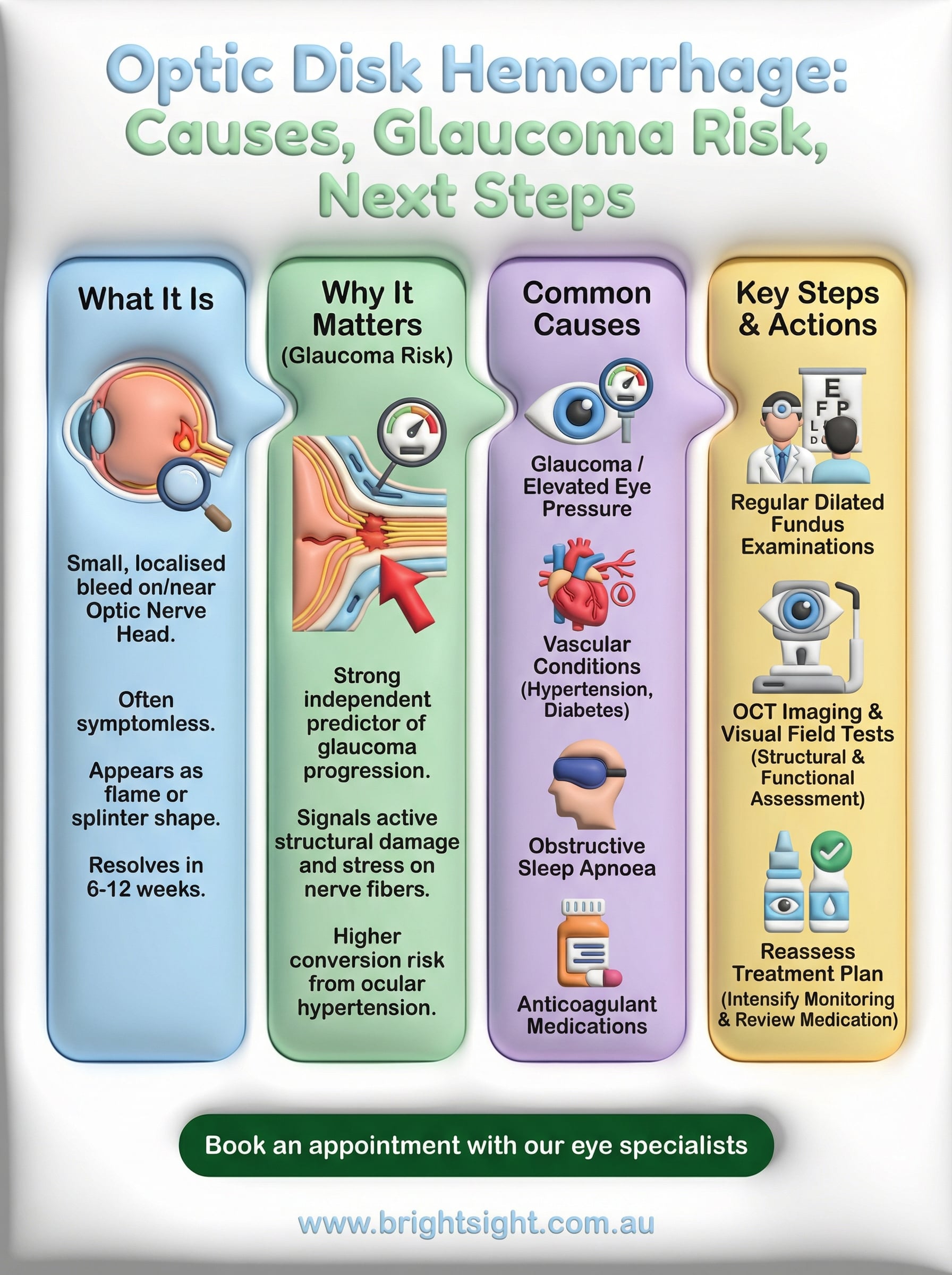
An optic disc haemorrhage is a small, localised bleed that occurs on or immediately adjacent to the optic nerve head, which is the point where your optic nerve exits the back of the eye. You cannot feel it, and it causes no pain or sudden change in your vision. Despite its small size, it carries significant clinical weight because it often signals that something is going wrong with the pressure or blood supply supporting the optic nerve fibres.
An optic disk hemorrhage is rarely visible to the person who has one, yet it can be one of the earliest detectable signs of glaucomatous damage to the optic nerve.
This haemorrhage is distinct from a retinal bleed. It sits specifically at the optic nerve head, most often in the upper or lower portions of the disc margin, which ophthalmologists refer to as the superior or inferior poles. Its presence is a finding on examination rather than a symptom you would notice yourself, which is exactly why regular, thorough eye assessments matter so much as you get older.
The anatomy behind the bleed
Your optic nerve head contains a dense network of small blood vessels that supply the nerve fibres responsible for transmitting visual information to your brain. When one of these tiny vessels ruptures, blood seeps into the nerve fibre layer immediately surrounding the disc. This produces the characteristic flame-shaped or splinter-like appearance that your eye specialist looks for during a dilated fundus examination.
Structurally, the nerve fibre layer at this location is particularly vulnerable to mechanical stress and fluctuations in blood flow. Raised intraocular pressure, impaired vascular autoregulation, or subtle structural changes within the disc itself can all contribute to vessel rupture at this point. Understanding this anatomy helps explain why clinicians pay such close attention to any bleed found at the optic nerve, regardless of how small it appears.
What it looks like on examination
When your eye specialist examines your optic disc, an optic disc haemorrhage appears as a thin, red or dark-red streak that crosses the edge of the disc margin, typically radiating outward into the surrounding retina. Most haemorrhages are small, no wider than one or two nerve fibre bundles, and they resolve on their own within six to twelve weeks without any treatment directed at the bleed itself.

Because they resolve and can recur, spotting them requires careful and consistent examination over time. Your specialist may observe:
- A fresh, bright red streak in the nerve fibre layer at the disc margin
- A fading, darker haemorrhage that is partially absorbed
- Subtle notching or thinning of the nerve fibre layer where a previous bleed has resolved
Photograph-based disc documentation is essential for tracking these changes, since a single clinic visit can easily miss a haemorrhage that cleared between appointments. Serial retinal imaging gives your care team a far more complete picture of what has been happening at the optic nerve over months and years, rather than just a snapshot from one point in time.
Why it matters for glaucoma risk
Finding an optic disk hemorrhage during a routine eye examination is a significant clinical event, not an incidental observation to note and move past. Research consistently shows that a disc haemorrhage is one of the strongest independent predictors of glaucoma development in people with elevated eye pressure, and a reliable marker of active structural damage in those already living with the disease. Put simply, a haemorrhage at the optic nerve substantially raises the probability that your optic nerve fibres are under damaging stress right now.
The link between disc haemorrhage and glaucoma progression
Large clinical trials, including the Ocular Hypertension Treatment Study, have confirmed that patients who develop a disc haemorrhage face a significantly higher risk of converting from ocular hypertension to established glaucoma than those who do not. In people already diagnosed with glaucoma, a new haemorrhage often signals that current treatment is not adequately controlling the disease, making it a clear prompt to review intraocular pressure targets and medication plans. Your specialist will not simply observe and wait; the finding calls for direct action.
A disc haemorrhage in a glaucoma patient is one of the clearest signals that pressure control needs to be re-evaluated and treatment intensified.
Why the location of the bleed matters
Haemorrhages occurring at the inferior or superior poles of the optic disc carry particular concern because these regions correspond to the areas of the visual field most commonly affected first by glaucomatous damage. A bleed in either zone suggests that the nerve fibre bundles responsible for your peripheral and paracentral vision are under stress, even if your visual field test results appear normal at this stage.
Your risk profile amplifies this concern further. If you have a thin cornea, a large optic cup-to-disc ratio, or a confirmed family history of glaucoma, a disc haemorrhage carries even greater clinical weight as a warning sign that closer monitoring and potentially earlier treatment are warranted without further delay.
Common causes and look-alikes
Not every optic disk hemorrhage points to glaucoma, and your eye specialist will consider several possible explanations before drawing conclusions. Understanding the range of causes helps you appreciate why a thorough clinical assessment matters far more than a single observation in isolation.
What causes a disc haemorrhage
The most common cause of a disc haemorrhage is glaucoma or elevated intraocular pressure, which creates mechanical strain on the small vessels at the nerve head. However, other systemic and local factors can also rupture these vessels. Vascular conditions including hypertension, diabetes, and obstructive sleep apnoea all reduce the reliability of blood flow to the optic nerve, making vessel walls more prone to breaking under normal or mildly raised pressure.
Disc haemorrhages in patients with well-controlled intraocular pressure still warrant investigation, as vascular disease and structural disc vulnerability may be driving the bleed independently of glaucoma.
Additional causes your specialist will consider include:
- Posterior vitreous detachment, where the vitreous gel pulling away from the retina drags on blood vessels near the disc
- Optic disc drusen, which are calcium deposits within the nerve head that can distort local vasculature
- Optic neuritis or other inflammatory conditions affecting the nerve directly
- Anticoagulant medications, which reduce the blood’s ability to clot and increase bleeding risk throughout the body, including at the optic nerve
Conditions that can look like a disc haemorrhage
Several findings can resemble a genuine disc haemorrhage on casual examination, which is why careful interpretation by an experienced ophthalmologist is essential. A preretinal haemorrhage from a retinal vessel can sit close to the disc and appear similar, but its origin and clinical significance differ substantially from a true nerve-head bleed.
Flame haemorrhages from a branch retinal vein occlusion can also extend near the disc margin, making them easy to confuse with a primary disc haemorrhage during a brief examination. Your specialist will trace the haemorrhage back to its precise anatomical source, using retinal imaging and vascular assessment to distinguish between these conditions and determine which one is actually driving the finding.
How doctors confirm it
Confirming an optic disk hemorrhage requires direct clinical examination combined with imaging tools that give your specialist an objective, reproducible record of what is happening at the optic nerve. No single test is sufficient on its own; the diagnosis comes from combining findings across several assessment methods to build an accurate picture of both the bleed itself and any associated structural damage to the surrounding nerve fibre layer.
Dilated fundus examination
Your eye specialist begins with a dilated fundus examination, which involves widening your pupil with eye drops and then examining the optic nerve head using a slit-lamp biomicroscope paired with a condensing lens. This gives a magnified, stereoscopic view of the disc margin, allowing the clinician to identify the characteristic splinter or flame-shaped bleed and assess its precise location relative to the disc poles, particularly at the superior and inferior zones where glaucomatous damage tends to appear first.
Because haemorrhages resolve within six to twelve weeks and can recur at different sites, fundus photography at each appointment is standard practice. A photograph taken today becomes the reference point for your next visit, making it far easier to detect new bleeds or confirm that a previous one has cleared fully.
Optical coherence tomography and structural assessment
OCT (optical coherence tomography) is now a core part of any thorough optic nerve workup. This non-contact imaging scan measures the thickness of the retinal nerve fibre layer around the optic disc with high precision, revealing whether the haemorrhage sits alongside areas of thinning or structural loss that are not yet visible on fundus examination alone.
OCT can detect nerve fibre layer thinning before a visual field defect becomes measurable, making it one of the most valuable tools your specialist has for catching glaucomatous damage early.
Pairing OCT findings with a formal visual field test allows your specialist to assess whether nerve damage has already translated into functional loss. Together, these tools confirm the clinical significance of the haemorrhage, help determine its most likely cause, and establish a clear baseline for monitoring progression over the months ahead.
What happens next in care and follow-up
When an optic disk hemorrhage is confirmed, your care does not stop at the diagnosis. Your eye specialist will use the finding as a direct prompt to reassess your current treatment plan and establish a more intensive monitoring schedule going forward. The goal is to identify whether the underlying cause is adequately managed and to catch any new bleeds or structural changes before they lead to measurable vision loss.
Reviewing your treatment plan
Your specialist will review your intraocular pressure readings and medication history to determine whether your current regimen is controlling eye pressure to a safe level for your optic nerve. If you are already using pressure-lowering drops and a haemorrhage has still appeared, that is a strong signal that your target pressure needs to be revised downward. In some cases, your specialist may recommend adding a second medication, switching to a different drug class, or discussing laser treatment such as selective laser trabeculoplasty to achieve better pressure reduction.
A disc haemorrhage in a patient already on treatment is one of the clearest prompts to intensify therapy rather than simply continue the existing plan.
Your specialist will also review any systemic risk factors that may be contributing independently of eye pressure, including blood pressure, diabetes management, and any medications that increase bleeding risk. Addressing these factors alongside your eye-specific treatment gives your optic nerve the best possible environment for stability.
Monitoring intervals and long-term follow-up
Following a confirmed haemorrhage, your review intervals will shorten significantly compared to a standard glaucoma check. Most specialists will schedule a repeat dilated fundus examination and OCT scan within two to four months, rather than waiting the six to twelve months typical for stable cases. This closer schedule gives your care team the opportunity to detect a recurrent bleed early and respond quickly.
Long-term follow-up remains essential because haemorrhages can recur at the same or different disc locations, sometimes without any change in symptoms or visual field results. Keeping your scheduled appointments, even when your vision feels unchanged, is how you and your specialist stay ahead of any progression before it becomes permanent.
When to seek urgent help
Most optic disk hemorrhage findings are managed through planned appointments rather than emergency visits, but certain symptoms alongside a known disc haemorrhage demand immediate attention. Your eye specialist will have discussed a monitoring plan with you, and in the majority of cases that plan unfolds over weeks and months without urgency. However, your eye is capable of developing separate, serious conditions that can coincide with a disc haemorrhage finding, and recognising the difference between routine monitoring and a genuine emergency protects your vision.
Knowing which symptoms require same-day assessment means you act quickly when it counts and avoid unnecessary anxiety at every minor change.
Symptoms that require immediate assessment
Some visual changes indicate a condition that cannot safely wait for a scheduled appointment. If you experience a sudden loss of vision in one or both eyes, partial or complete, you need same-day assessment at an emergency eye clinic or hospital eye unit. Similarly, a new curtain, shadow, or veil across part of your visual field that appears suddenly suggests a retinal detachment or vascular event, neither of which responds well to delayed treatment.
Other symptoms that warrant urgent review include:
- A rapid increase in floaters or flashes of light, particularly if combined with any visual field change
- Sudden blurring or distortion of your central vision that was not present the day before
- Eye pain accompanied by redness and nausea, which can indicate a sudden rise in intraocular pressure
- Any vision change that develops within hours rather than gradually over days
When your scheduled plan still applies
If your vision is stable and unchanged between appointments and you simply notice that an existing haemorrhage has faded or shifted slightly in appearance, that does not require emergency contact. Your scheduled review appointment exists precisely to assess these incremental changes in a controlled setting. Contacting your specialist to describe the change and confirm whether it alters your review timeline is always reasonable, but clear and stable vision without new symptoms means your monitoring plan remains appropriate and there is no need to present urgently.
Key takeaways and next step
An optic disk hemorrhage is a small but clinically important finding that warrants careful attention, not dismissal. It signals mechanical or vascular stress at the optic nerve, carries a strong association with glaucoma development and progression, and can recur without warning. Early detection through regular dilated eye examinations, OCT imaging, and visual field testing gives you and your specialist the clearest possible picture of your nerve health before permanent damage sets in.
Your most important action is straightforward: do not wait and hope the finding resolves on its own. A confirmed haemorrhage at the optic nerve deserves expert review, a reassessed treatment plan, and a tighter monitoring schedule tailored to your specific risk profile. Whether you have been referred by an optometrist or you are managing an existing glaucoma diagnosis, specialist input makes a direct difference to your long-term visual outcome.
Book an appointment with our eye specialists to discuss your optic nerve health today.