


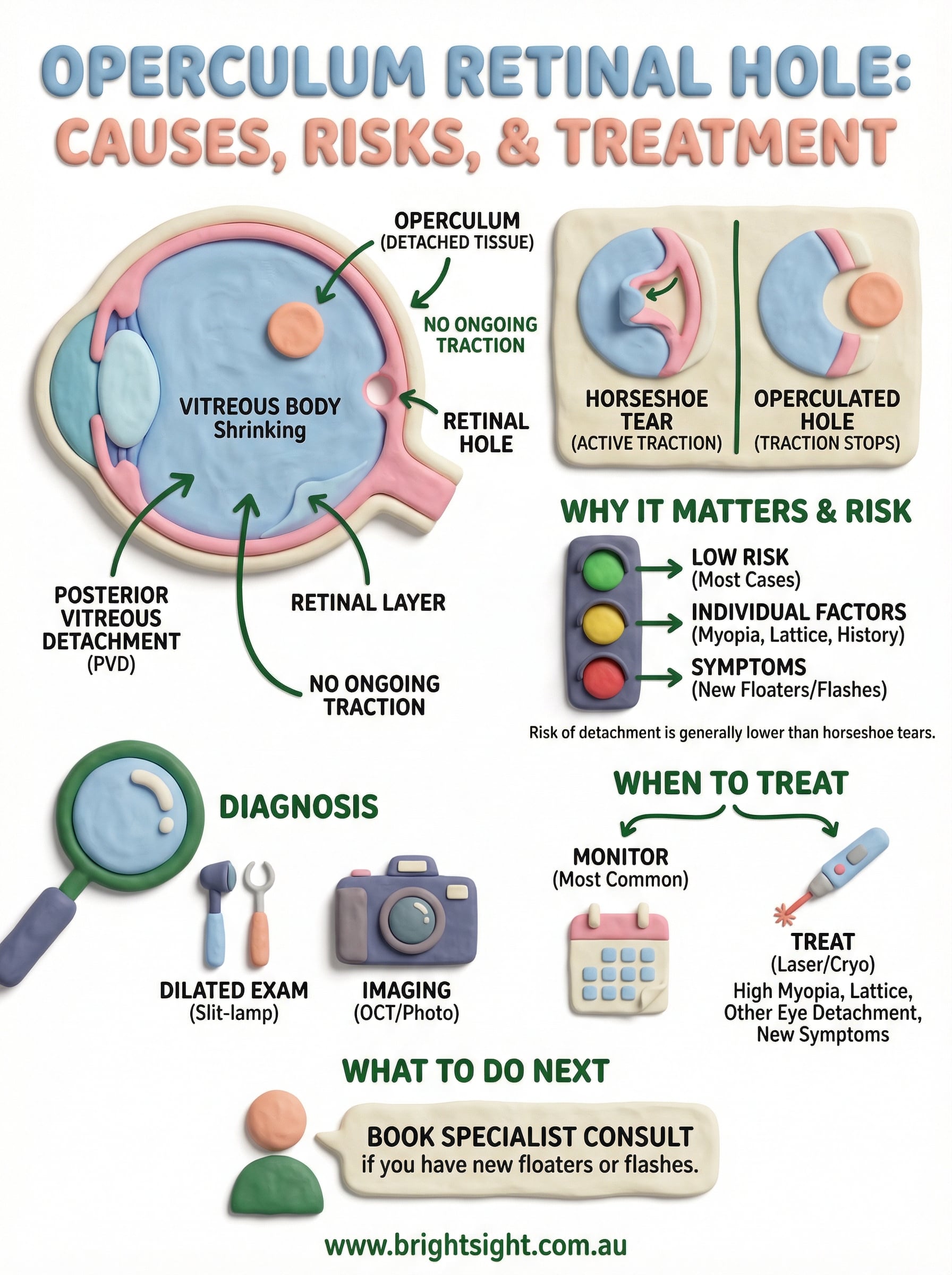
An operculum retinal hole forms when a small, round piece of retinal tissue pulls away completely from the surrounding retina, typically during a posterior vitreous detachment. Unlike horseshoe tears, where the flap stays partially attached and continues to tug on the retina, an operculated hole has no ongoing traction. That distinction matters because it directly affects your risk of retinal detachment and whether you need treatment at all.
If your optometrist or GP has flagged this finding during a routine eye check, you’re likely wondering how serious it is and what happens next. The good news is that most operculated holes are low-risk, but not all cases are straightforward, and some do require closer attention or intervention.
At Bright Sight Eye Specialists in Randwick, our ophthalmologists, including Dr Rahul Dubey and Dr Capucine Odouard, regularly assess and manage retinal holes as part of our comprehensive retinal care. In this article, we’ll explain what causes an operculated hole, how it compares to other retinal breaks, the actual risk it poses, and when treatment becomes necessary.
Why an operculated retinal hole matters
The retina lines the back of your eye and converts light into the electrical signals your brain reads as vision. Any full-thickness break in the retinal tissue creates a potential pathway for fluid to travel underneath, which is exactly how a retinal detachment begins. Understanding why an operculum retinal hole behaves differently from other retinal breaks helps you and your specialist make the right call on management.
The retina’s role in your vision
The retina is roughly 0.5mm thick and covers most of the inner surface of your eye. Photoreceptor cells – the rods and cones tightly packed into the retinal layers – do all the heavy lifting when it comes to detecting light and colour. A full-thickness hole through this layer removes those photoreceptors from that exact spot, and the immediate clinical question becomes whether fluid from the vitreous cavity will move through the opening and lift the surrounding retina away from its blood supply.
With an operculated hole specifically, the freed disc of tissue (the operculum) floats in the vitreous gel just in front of the hole. Because the operculum is completely detached, it cannot exert ongoing traction on the hole’s edges the way an adherent flap does in a horseshoe tear. That single anatomical detail is why ophthalmologists generally treat operculated holes with considerably less urgency than other retinal break types.
The real risk of retinal detachment
An operculated hole carries a substantially lower lifetime risk of retinal detachment than a horseshoe tear – most published clinical data places that risk well below 1% in eyes without other predisposing factors.
Despite those reassuring figures, certain individual characteristics can shift the picture significantly. If the hole sits within an area of lattice degeneration, if you are highly myopic (short-sighted), or if your other eye has already experienced a retinal detachment, those factors will weigh heavily in your ophthalmologist’s assessment. Your age, the size of the hole, and whether you are noticing new floaters or flashes also matter. The baseline risk is low, but your personal profile determines whether watchful waiting is genuinely appropriate or whether a closer look is warranted.
How an operculated retinal hole happens
An operculum retinal hole almost always traces back to the natural ageing of the vitreous gel that fills your eye. Over time, the vitreous shrinks and separates from the retina, a process called posterior vitreous detachment (PVD). In most cases that separation is uneventful, but when there is an unusually firm adhesion between the vitreous and a patch of retinal tissue, the pull can punch out a small, round disc of retina rather than simply peeling away cleanly.
The mechanics of vitreous separation
During a PVD, the vitreous collapses inward and pulls away from the retinal surface. Where adhesion is especially tight, the force concentrates at one point. If the bond is stronger than the retinal tissue itself, the vitreous detaches by tearing out a small plug rather than releasing cleanly. That extracted plug becomes the operculum, leaving a round, full-thickness hole behind. The operculum then floats freely in the vitreous cavity, which is why traction on the hole’s edges stops once the separation is complete.
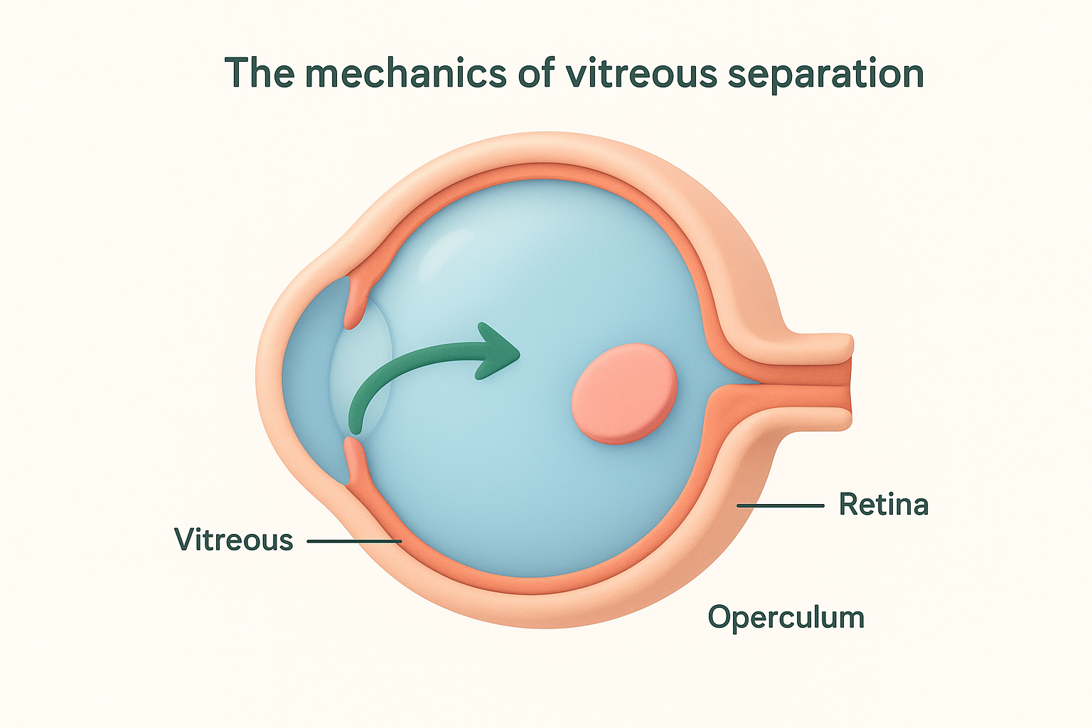
The moment the operculum detaches fully, the mechanical force driving further damage effectively disappears, which is the key reason these holes carry a lower detachment risk than tears where vitreous adhesion persists.
Who is most likely to develop one
Short-sighted individuals are at higher risk because myopic eyes tend to have a thinner retina and altered vitreoretinal adhesion patterns. Age is also a significant factor, as PVD becomes increasingly common after 50, making operculated holes more likely to appear in older adults during routine retinal examinations.
How it differs from other retinal breaks
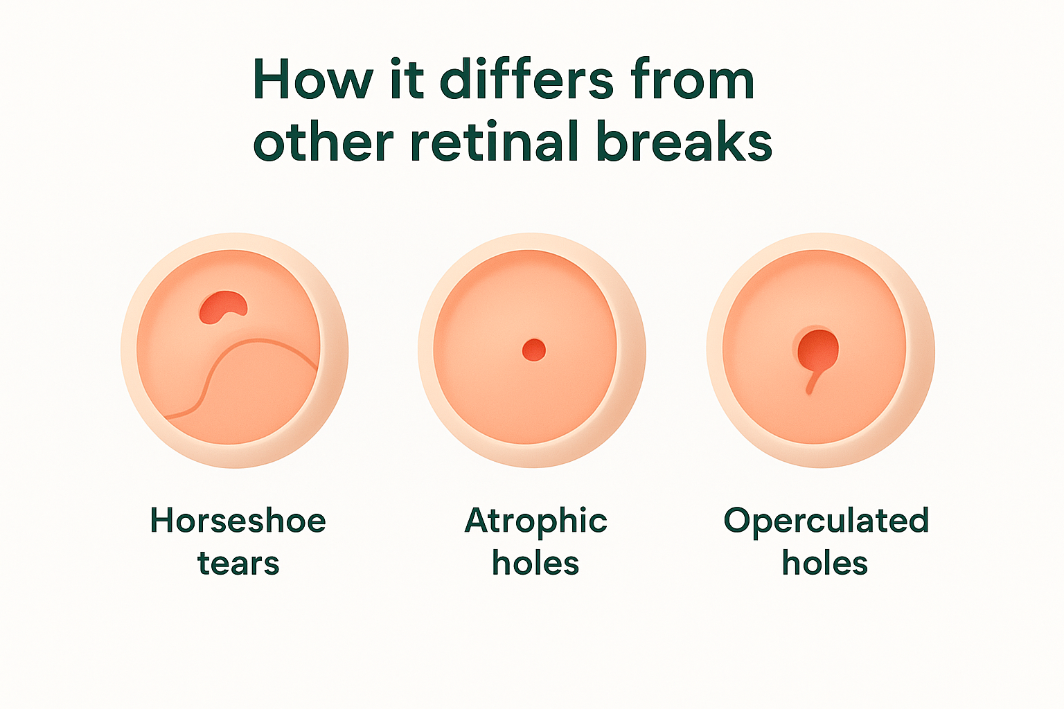
Retinal breaks fall into several distinct categories, and where an operculum retinal hole sits within that classification determines almost everything about your risk level and management pathway. The three main types, horseshoe tears, atrophic holes, and operculated holes, each carry different risks because of the underlying mechanism that created them.
Horseshoe tears
A horseshoe tear forms when the vitreous pulls on the retina but does not fully detach, leaving a flap of retinal tissue with one edge still anchored. That persistent traction continues tugging on the retinal edge, actively driving fluid underneath the retina and making detachment considerably more likely. This is why ophthalmologists typically recommend prompt laser treatment for horseshoe tears rather than watchful waiting.
An operculated hole, by contrast, carries far less urgency because the vitreous has already released completely, removing the mechanical force that drives most retinal detachments.
Atrophic holes
Atrophic holes develop without any vitreous traction at all. They arise from gradual thinning of the retinal tissue itself, typically within areas of lattice degeneration, and tend to affect younger, short-sighted individuals. While they also carry a low detachment risk, their cause and clinical context differ substantially from operculated holes, which result from an acute PVD event rather than slow tissue degeneration.
Knowing which type of break you have is not just a matter of terminology. It shapes every subsequent decision your specialist makes, from how soon you return for a follow-up review to whether treatment is on the table at all.
How doctors diagnose it
Diagnosing an operculum retinal hole requires a thorough examination of the peripheral retina, which sits well outside the central field of view and cannot be assessed without specific techniques and equipment. Your ophthalmologist will typically begin by asking about any recent onset of floaters or flashes, as these symptoms often accompany the posterior vitreous detachment that triggers this type of hole.
Dilated fundus examination
Dilating your pupils with eye drops is the essential first step. Pupil dilation widens the aperture through which your doctor examines the retina, allowing a detailed view of the peripheral areas where operculated holes most commonly occur. Your ophthalmologist will use a slit-lamp biomicroscope combined with a condensing lens, or an indirect ophthalmoscope with scleral indentation, to systematically scan the full retinal surface and identify any breaks, associated lattice degeneration, or signs of subretinal fluid.
Scleral indentation, where gentle pressure is applied to the outer eye wall, brings peripheral retinal structures into clear view and is particularly useful for confirming the presence and extent of a retinal hole.
Imaging and documentation
Once a hole is identified, wide-field retinal photography or optical coherence tomography (OCT) provides a permanent record that allows your specialist to track any changes at future appointments. OCT is especially useful for assessing whether fluid has begun to accumulate beneath the retina around the hole. Accurate documentation at the initial visit establishes a reliable baseline, which is critical for comparing findings at every subsequent review.
When to monitor vs treat
The decision to monitor or treat an operculum retinal hole depends on the specific factors your ophthalmologist weighs at your initial consultation and at every follow-up appointment. Most operculated holes in otherwise healthy eyes require no active treatment, but certain clinical features can shift that recommendation toward intervention.
Cases that suit monitoring
If your hole is small and isolated, and the vitreous has fully separated without any residual traction remaining, watchful waiting is the standard approach. Your specialist will typically schedule a review within six to eight weeks of the initial finding, then extend the review interval if nothing changes. During that monitoring period, you need to report any sudden increase in floaters, new flashes of light, or a shadow creeping into your peripheral vision without delay, as these symptoms can indicate the hole is progressing.
Reporting new visual symptoms promptly between appointments is one of the most important things you can do to protect your vision during a monitoring period.
When treatment becomes appropriate
Laser photocoagulation or cryotherapy seals an operculated hole by creating a controlled scar that bonds the retina firmly to the underlying tissue. Your ophthalmologist is more likely to recommend this approach if specific risk factors apply to your situation:
- High myopia (significant short-sightedness)
- Lattice degeneration near the hole
- A history of retinal detachment in your other eye
- A large hole or evidence of subretinal fluid on imaging
These factors raise your overall detachment risk enough that the small procedural risks of sealing the hole proactively are clearly justified.
What to do next
An operculum retinal hole is rarely an emergency, but it does require a proper specialist assessment to confirm the type of break, rule out any associated risk factors, and establish a clear monitoring or treatment plan. Knowing your personal risk level with certainty is far more useful than relying on general statistics that may not reflect your individual situation.
If you have been told you have a retinal hole, or if you are currently experiencing new floaters, flashes of light, or any change in your peripheral vision, booking a consultation with an experienced ophthalmologist is the right next step. At Bright Sight Eye Specialists in Randwick, Dr Rahul Dubey and Dr Capucine Odouard assess and manage retinal conditions using current diagnostic technology and a clear, straightforward approach to patient care. Book your retinal consultation today and get a complete picture of your eye health.