


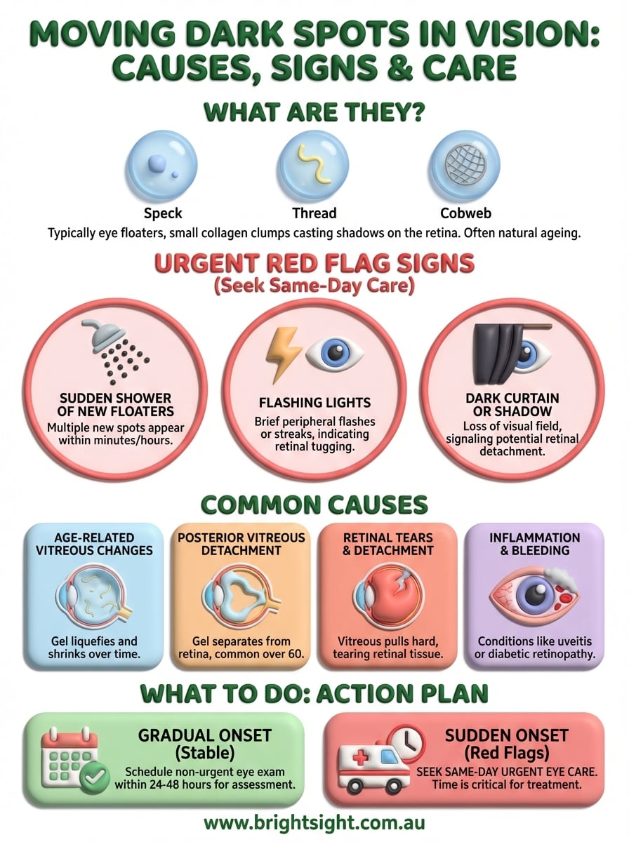
A moving dark spot in your vision is typically an eye floater, a small shadow cast by clumps of collagen fibres floating in the vitreous gel inside your eye. These spots can look like specks, threads, cobwebs or clouds that drift when you move your eyes. While they are often harmless and a natural part of ageing, a sudden appearance of new dark spots requires immediate medical attention.
This article explains what causes moving dark spots, how to tell the difference between routine floaters and serious conditions like retinal tears or detachment, and when you need to see an eye specialist urgently. You’ll learn about the various conditions that can create dark spots in your vision, from age-related vitreous changes to more serious retinal problems. We’ll also cover what to do when you first notice these spots, warning signs that demand immediate care, and how Bright Sight Eye Specialists can assess and treat your concerns with advanced diagnostic technology and personalised treatment options.
Why a moving dark spot needs attention
Your eyes reveal important information about your overall health, and a moving dark spot in vision can be your first warning of serious problems. While most floaters develop gradually and pose no threat, the sudden appearance of new spots indicates that something has changed inside your eye. The vitreous gel that fills your eye can pull away from the retina as you age, and this natural process sometimes leads to complications that require immediate treatment. Understanding which floaters need attention helps you protect your vision and seek care at the right time.
The difference between routine and urgent floaters
You can usually ignore a few stable floaters that have been present for months or years. These long-standing floaters result from normal ageing changes in the vitreous, and your brain learns to filter them out over time. However, a sudden shower of new dark spots or a dramatic increase in the number of floaters signals that the vitreous has pulled away from the retina more forcefully than expected. The quantity and timing matter far more than the appearance of individual floaters when determining urgency.

The timing and quantity of new floaters determine whether you need immediate care or can wait for a scheduled appointment.
What sudden changes signal
Sudden floaters often appear alongside other symptoms that indicate retinal stress or damage. Flashing lights in your peripheral vision suggest the retina is being tugged or stimulated by the separating vitreous. A dark curtain or shadow covering part of your visual field means the retina may have already detached from the back of your eye. These combinations of symptoms require urgent assessment because retinal tears and detachments become more difficult to repair as time passes, and delays can result in permanent vision loss that affects your quality of life.
How to respond when you notice a moving dark spot
Your response to a moving dark spot in vision depends on how suddenly it appeared and what other symptoms accompany it. Stop what you’re doing and take a moment to assess the situation carefully. Cover one eye at a time to determine which eye is affected, and note whether you see the spot in both eyes or just one. Single-eye floaters often indicate a vitreous or retinal issue in that specific eye, while bilateral symptoms may suggest different causes. This initial assessment helps you communicate clearly with your eye specialist and determine the urgency of your situation.
Assess the urgency of your symptoms
You need to contact an eye specialist immediately if you notice multiple new dark spots appearing suddenly, especially if they arrive within minutes or hours rather than days or weeks. Check for accompanying flashing lights by looking at a plain surface like a white wall or clear sky, then turning your head side to side. Peripheral flashes that persist or worsen indicate the vitreous is actively pulling on your retina. Also look for any shadows or curtains blocking parts of your visual field, as these suggest possible retinal detachment requiring emergency treatment within hours.
Contact an eye specialist immediately if new floaters appear alongside flashing lights or vision loss, as these combinations indicate potential retinal damage.
Document what you’re experiencing
Write down exactly when you first noticed the moving dark spot and how many spots you can count. Record the size and shape of each floater, such as whether they look like dots, threads, cobwebs or clouds. Note any changes throughout the day, including whether the spots increase in number or if new symptoms develop. Take photos or videos of your visual field if possible by drawing what you see or using smartphone apps designed to track floaters. This documentation helps your eye specialist understand the progression of your symptoms and makes diagnosis more accurate.
Take practical precautions
Avoid activities that require sharp vision or quick reactions until you’ve had your eyes examined, particularly driving at night when floaters become more noticeable against dark backgrounds. Reduce your risk of further complications by avoiding heavy lifting, straining, or any activities that rapidly increase eye pressure. Rest your eyes regularly and avoid rubbing them, which can worsen existing problems. Schedule an appointment with an eye specialist within 24 to 48 hours for gradual-onset floaters, but seek same-day care for sudden changes. Most specialists reserve urgent slots for acute floater cases because early intervention can prevent permanent vision loss.
Common causes of a moving dark spot
Most moving dark spots in vision result from changes to the vitreous gel that fills your eye, though several conditions can create similar symptoms. The vitreous starts as a firm, clear gel in childhood but gradually liquefies and shrinks as you age, creating pockets of fluid and clumps of collagen fibres. These clumps cast shadows on your retina when light passes through your eye, creating the moving dark spots you see. Understanding the specific cause helps you know whether you need treatment, ongoing monitoring, or simple reassurance that your symptoms are part of normal ageing.
Age-related vitreous changes
Your vitreous begins to change texture after age 40, and most people develop at least a few floaters by their 60s or 70s. The gel liquefies gradually and contracts away from the eye wall, creating strands of condensed collagen that move freely in the remaining fluid. These age-related floaters typically appear slowly over months or years, giving your brain time to adapt and filter them out. You notice them most when looking at bright, plain backgrounds like white walls, blue skies, or computer screens, as the contrast makes the shadows more visible against light backgrounds.
People who are nearsighted experience vitreous changes earlier than those with normal vision, often noticing their first floaters in their 30s or 40s. The elongated shape of a nearsighted eye stretches the vitreous more than normal, accelerating the liquefaction process. You may also develop more floaters if you’ve had eye surgery, eye inflammation, or an eye injury, as these conditions can disturb the vitreous structure and create additional clumps.
Posterior vitreous detachment
Posterior vitreous detachment occurs when the vitreous gel separates completely from the retina at the back of your eye, a process that affects most people over 60. The separation happens naturally as the vitreous shrinks with age, pulling away from the retinal surface where it was previously attached. You typically notice a sudden increase in floaters when this happens, often accompanied by brief flashes of light in your peripheral vision. These flashes occur when the separating vitreous tugs on the retina, mechanically stimulating the light-sensitive cells that interpret any stimulation as light.
Posterior vitreous detachment is a normal ageing process, but it creates a critical window when retinal tears are most likely to occur.
The detachment process usually takes several weeks to complete, during which you may see new floaters appear or existing ones become more prominent. Most people adapt within three to six months as the vitreous settles into its new position and the brain learns to ignore the floaters. However, the separation creates tension on the retina, particularly in areas where the vitreous is more strongly adhered, and this tension can cause the complications discussed below.
Retinal tears and detachment
Retinal tears develop when the separating vitreous pulls hard enough to rip the delicate retinal tissue, creating an opening that allows fluid to seep underneath. You typically notice a sudden shower of dark spots, like someone shaking pepper in your vision, when a retinal tear occurs. The spots represent red blood cells floating in the vitreous after tiny blood vessels tear along with the retina. Without prompt treatment, fluid continues to accumulate under the tear, gradually lifting the retina away from its blood supply and causing a retinal detachment.

Retinal detachment creates additional symptoms beyond the initial floaters, including a shadow or curtain that blocks part of your visual field. The shadow expands over hours or days as more retina detaches, starting in the periphery and potentially spreading to your central vision. You need emergency treatment to reattach the retina and prevent permanent vision loss, which is why understanding these warning signs matters so much.
Eye inflammation and bleeding
Inflammation inside your eye can cause white blood cells to float in the vitreous, creating moving dark spots similar to those from vitreous changes. Posterior uveitis affects the back portion of the eye and may develop from infections, autoimmune diseases like lupus or sarcoidosis, or sometimes for unknown reasons. You usually experience additional symptoms with uveitis, including eye pain, redness, light sensitivity, and blurred vision that distinguish it from simple floaters.
Bleeding into the vitreous creates a different pattern of floaters, often appearing as a dense cloud or haze rather than distinct spots. Diabetic retinopathy causes fragile blood vessels to leak or rupture, filling the vitreous with blood cells that block light and create moving shadows. Retinal vein occlusion, high blood pressure, and eye injuries can also lead to vitreous haemorrhage. The blood typically clears over several weeks to months, though you may need treatment for the underlying condition to prevent future bleeding.
Other contributing conditions
Central serous chorioretinopathy creates fluid buildup under your retina rather than floaters in the vitreous, but it can cause a dark or grey area in your central vision that some people describe as a spot. This condition affects middle-aged adults, particularly men with high stress levels, and usually resolves spontaneously within three to six months. Age-related macular degeneration can cause dark spots in your central vision when abnormal blood vessels leak fluid or blood under the macula, though these spots typically don’t move as true floaters do. You face higher risk of macular degeneration if you’re over 60, smoke, have a family history of the condition, or are Caucasian.
When to seek urgent eye care
Recognising when a moving dark spot in vision requires immediate medical attention can save your sight, as the difference between routine monitoring and emergency care often comes down to specific warning signs. The timing of your response directly affects the treatment options available and your chances of preserving full vision. You should contact an eye specialist the same day if you experience certain combinations of symptoms, even if you feel no pain, as many serious eye conditions remain painless until significant damage has occurred. Acting quickly gives your specialist the best opportunity to intervene before permanent changes develop.
Red flag symptoms that require immediate attention
You need to seek urgent care within hours if you notice a sudden shower of new floaters appearing all at once, particularly if accompanied by flashing lights in your peripheral vision. These flashes look like brief sparks or lightning streaks at the edge of your visual field and indicate the vitreous is actively pulling on your retina. Contact an eye specialist immediately if you see a dark curtain, shadow, or veil blocking any portion of your vision, as this suggests the retina has already detached and requires emergency surgical repair within 24 to 48 hours to prevent permanent vision loss.
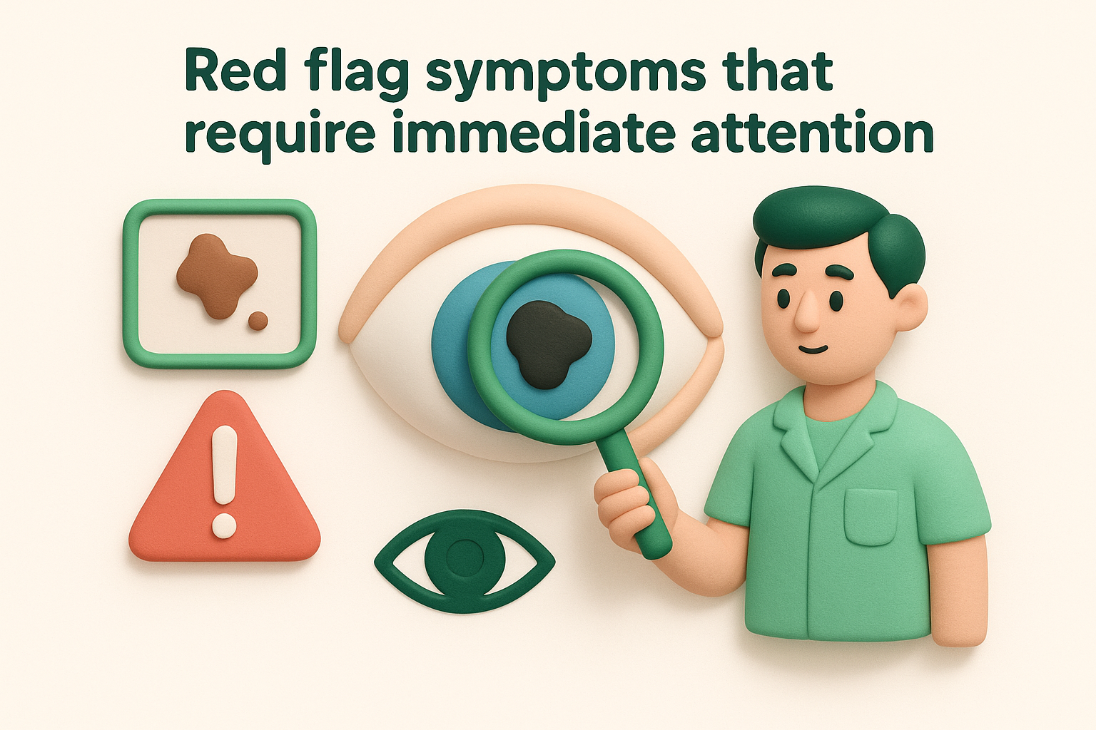
Retinal detachment becomes progressively more difficult to repair as time passes, making immediate care essential for preserving your vision.
Additional urgent symptoms include a significant increase in the number or size of existing floaters over a short period, dense clouds of floaters that obscure your vision, or reddish-tinted floaters suggesting bleeding inside your eye. You should also seek immediate care if you experience sudden vision loss, severe eye pain alongside new floaters, or if you’ve recently sustained an eye injury and now notice new dark spots. These combinations signal conditions that deteriorate rapidly without intervention.
Time windows that matter
The critical window for treating retinal tears spans 24 to 72 hours from when symptoms first appear, as prompt laser treatment or cryotherapy can seal the tear before fluid accumulates underneath. Delays beyond this window increase your risk of progression to full retinal detachment, which requires more extensive surgery and carries a higher risk of permanent vision changes. You have slightly more time with a partial retinal detachment, but you should still receive treatment within a few days to prevent the detachment from spreading to your central vision, which controls your ability to read, drive, and recognise faces.
What happens during an urgent eye examination
Your eye specialist will dilate your pupils with special drops to examine the entire retina, including the far periphery where tears often occur first. The examination takes 30 to 45 minutes once the drops take effect, allowing detailed visualisation of the vitreous, retina, and any areas of concern. Your specialist uses bright lights and specialised lenses to inspect every section of your retina, looking for tears, bleeding, inflammation, or other causes of your symptoms. Most patients receive a definitive diagnosis during this visit, along with a clear treatment plan if intervention is needed, or reassurance and monitoring instructions if no urgent problems are found.
Care at Bright Sight Eye Specialists
Dr Rahul Dubey and his team at Bright Sight Eye Specialists provide comprehensive assessment and treatment for patients experiencing a moving dark spot in vision. The practice uses advanced diagnostic technology to examine your retina in detail, including digital imaging that captures high-resolution photographs of your eye’s interior structures. You receive same-day urgent appointments when symptoms suggest potential retinal problems, ensuring prompt evaluation when timing matters most for protecting your vision.
Advanced diagnostic capabilities
Your examination includes dilated fundoscopy and detailed retinal imaging that allows Dr Dubey to identify retinal tears, vitreous changes, or other causes of your symptoms with precision. The practice’s optical coherence tomography (OCT) scanner creates cross-sectional images of your retina, revealing subtle changes that traditional examination methods might miss. You benefit from clear explanations of your diagnosis, with images and diagrams that help you understand exactly what’s happening inside your eye and why specific treatments are recommended.

Advanced imaging technology at Bright Sight Eye Specialists enables precise diagnosis and personalised treatment planning for complex retinal conditions.
Treatment options available
Dr Dubey provides a full range of treatments for conditions causing moving dark spots, from monitoring stable floaters to performing urgent laser treatment for retinal tears and surgical repair for retinal detachments. The practice offers transparent pricing with most procedures available with no gap or known gap costs for insured patients, eliminating financial uncertainty when you need treatment. Your care includes thorough follow-up to monitor your recovery and ensure optimal outcomes, with accessible communication if new concerns develop after your initial visit.
Next steps for your eye health
Your response to a moving dark spot in vision determines whether you preserve your sight or risk permanent damage. Schedule an eye examination within 24 to 48 hours for gradual-onset floaters, but seek same-day care if you notice sudden changes, flashing lights, or vision loss. Regular eye checks help detect problems early, particularly if you’re over 40, nearsighted, or have diabetes. Contact Bright Sight Eye Specialists for comprehensive assessment and treatment of floaters and retinal conditions, with advanced diagnostic technology and transparent pricing for all procedures.