


You glance at a blank wall or blue sky and there it is again: a tiny dark speck drifting through your vision. You try to focus on it but it slips away, floating just out of reach. That little black spot might seem harmless, yet it can leave you wondering whether something serious is happening inside your eye.
Most of these floating specks are eye floaters, shadows cast by small clumps of gel or cells inside your eye. They form when the clear jelly that fills your eyeball changes with age or other factors. While many floaters are benign and require no treatment, certain patterns signal urgent problems like retinal tears or detachment that can threaten your sight.
This article walks you through what causes that black spot, how to tell if it’s a routine floater or a warning sign, and when you need to see an eye specialist without delay. You’ll learn the diagnostic steps your doctor will follow, available treatments, and simple ways to protect your vision going forward.
Why a little black spot in vision matters
That little black spot in vision isn’t just a visual annoyance. Your eyes send constant signals to your brain about the world around you, and any disruption to this process deserves attention. While many black spots are harmless floaters that your brain learns to ignore, some signal damage to the delicate structures inside your eye. Knowing the difference can protect you from preventable vision loss.
When a harmless floater becomes an emergency
Retinal problems develop quickly once they start. A small tear in your retina can progress to full detachment within hours or days, and detached retinas require emergency surgery to save your sight. If you dismiss that black spot as "just getting older," you might miss the narrow window when simple laser treatment can prevent serious complications. The Royal Australian and New Zealand College of Ophthalmologists reports that prompt treatment of retinal tears achieves success rates above 90 per cent.
Early intervention makes the difference between a quick office procedure and complex surgery with uncertain outcomes.
How visual disturbances affect daily life
Even benign floaters disrupt your daily activities more than you might expect. Reading becomes frustrating when dark specks drift across the page, and driving poses safety concerns when floaters obscure road signs or pedestrians. Your work performance suffers if you spend meetings watching shadows float through presentations. These visual disturbances create constant mental fatigue as your brain works overtime to filter out the interference. Understanding when treatment options exist helps you reclaim clear, comfortable vision rather than simply accepting the nuisance.
How to tell if the black spot is serious
Not every little black spot in vision requires a trip to the emergency department. You need clear criteria to separate routine floaters from sight-threatening emergencies. The key lies in recognising specific warning patterns rather than panicking at every speck. Your symptoms tell a story, and learning to read that story helps you make confident decisions about when to seek urgent care.
Signs of a benign floater
Harmless floaters share predictable characteristics that distinguish them from dangerous conditions. You notice them most against bright backgrounds like white walls or clear skies, and they drift slowly when you move your eyes. These spots have been present for weeks or months without sudden changes in number or appearance. You can see them clearly one moment, then they float out of your direct line of sight the next.
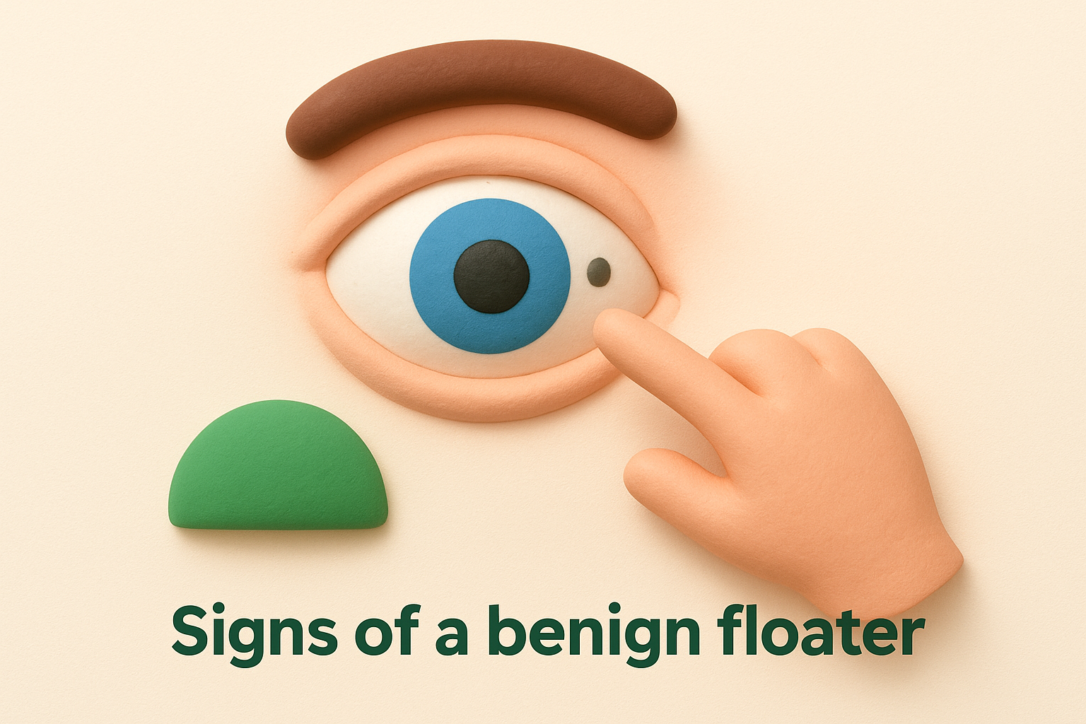
The spots remain stable over time, meaning you don’t see more of them appearing each day. Your peripheral vision stays clear, with no shadows or curtains blocking the edges of your sight. Most importantly, you experience no flashes of light accompanying these floaters. Benign floaters might annoy you, but they don’t interfere with your ability to see details or navigate safely.
Red flags that demand immediate attention
Certain symptoms transform a simple floater into a medical emergency. You see a sudden shower of new spots appearing within hours, often described as hundreds of tiny specks floating across your vision. Flashes of light accompany these floaters, particularly in your peripheral vision, resembling camera flashes or lightning bolts in the corner of your eye.
Any curtain-like shadow spreading across your vision signals potential retinal detachment requiring same-day treatment.
A grey curtain or veil creeps across part of your visual field, blocking your sight like a shade being drawn. Your vision suddenly becomes blurred or distorted in ways you haven’t experienced before. These symptoms develop rapidly, sometimes within minutes to hours, rather than gradually over weeks. You might notice the symptoms worsen when you look up or down, as gravity affects the detaching retina.
The time window for treatment
Speed determines your outcome when serious retinal problems occur. Retinal tears require treatment within 24 to 48 hours to prevent progression to detachment. Once full detachment begins, you have a narrow window of days before permanent vision loss occurs in the affected area. The Queensland Government Health Department emphasises that immediate assessment improves surgical success rates significantly. Contact an eye specialist the same day you notice warning signs, even if symptoms appear outside normal clinic hours.
Common causes of a little black spot in vision
Understanding what creates that little black spot in vision helps you distinguish between normal ageing changes and conditions requiring medical attention. The vitreous humour, a clear gel filling your eyeball, undergoes structural transformations throughout your life. These changes produce shadows on your retina that appear as floating spots. While most causes are benign, some signal underlying eye disease or systemic health problems.
Age-related vitreous changes
Your vitreous humour starts life as a firm, clear gel but gradually liquefies as you age. This natural process begins in your forties and accelerates through your fifties and sixties. The gel contains collagen fibres that maintain its structure, and when these fibres clump together they cast shadows on your retina. You perceive these shadows as black spots, threads, or cobweb-like shapes drifting across your vision.
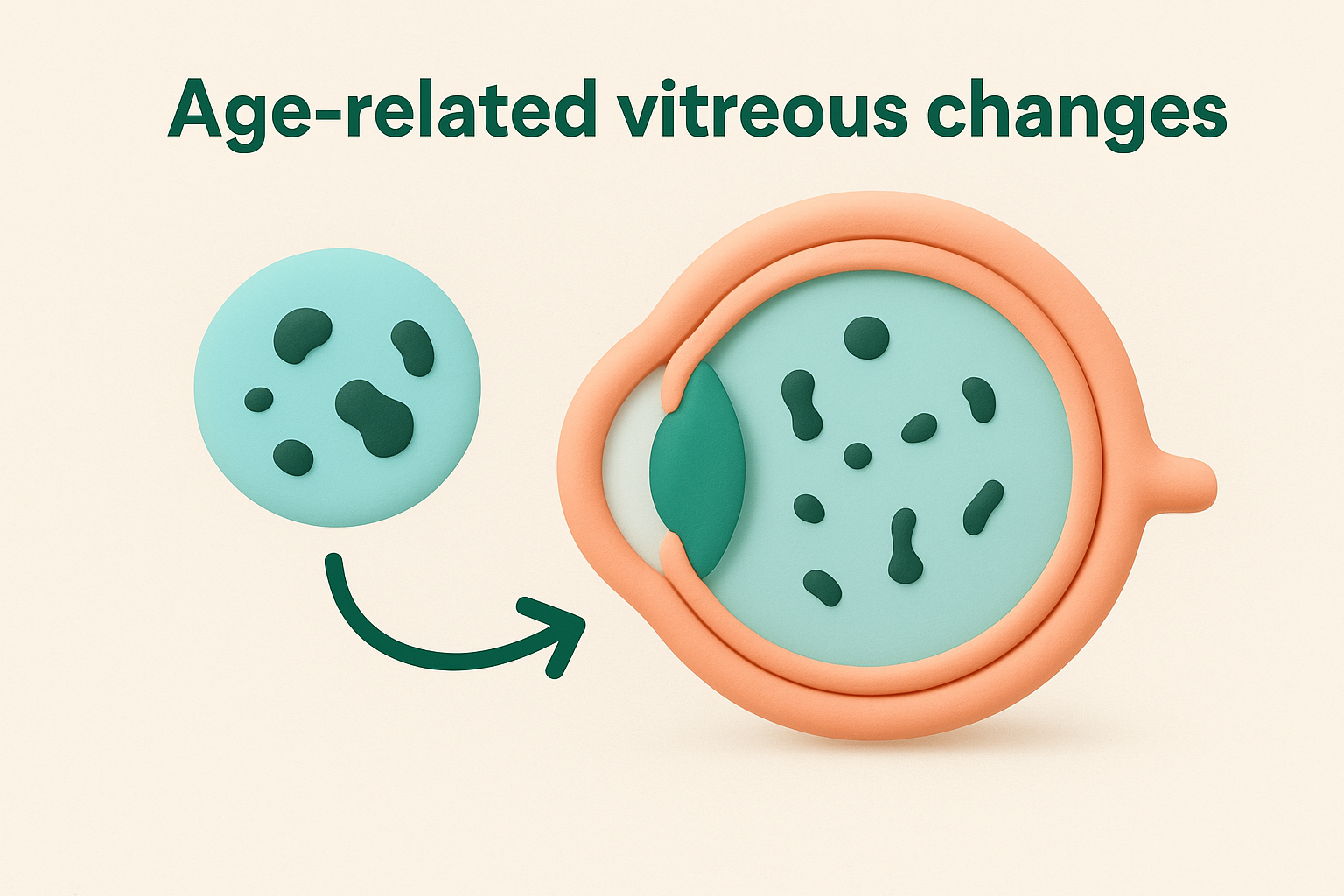
These age-related floaters affect most people eventually. Your brain typically adapts within weeks to months, filtering out the visual noise so the spots become less noticeable. The floaters remain present but your conscious awareness of them diminishes over time. This adaptation process works best when the floaters stay consistent rather than increasing rapidly.
Posterior vitreous detachment
Posterior vitreous detachment (PVD) occurs when your vitreous gel separates from the retina at the back of your eye. This separation is a normal ageing event that happens to most people by age 70. During PVD, the vitreous pulls away from its attachment points, sometimes releasing tiny fragments that become floaters. You might notice a sudden increase in spots or a ring-shaped floater that appears when the gel detaches from around your optic nerve.
PVD itself is harmless, but the tugging motion during separation can tear your retina in vulnerable areas.
Most PVD cases resolve without complications. Your eye specialist monitors the situation to ensure no retinal tears develop during the detachment process. Complete separation usually takes several weeks, after which the risk of complications decreases significantly.
Eye inflammation and bleeding
Inflammation inside your eye (uveitis) releases inflammatory cells into the vitreous that appear as small floating spots. Diabetic retinopathy or high blood pressure can cause tiny blood vessels in your retina to leak or rupture, sending blood cells into the vitreous space. These blood cells create numerous small dark spots that move with your eye movements.
Vitreous haemorrhage from bleeding produces different symptoms than typical floaters. You might see many small red or dark spots appearing suddenly, sometimes described as a cloud of gnats. The blood can make your vision hazy and significantly interfere with your ability to see clearly. This condition requires prompt evaluation to identify and treat the bleeding source.
Impact of systemic diseases
Diabetes affects small blood vessels throughout your body, including those in your retina. Diabetic retinopathy causes these vessels to become fragile and leak, producing floaters alongside other vision changes. High blood pressure similarly damages retinal vessels, increasing your risk of vitreous bleeding and spot formation. Managing these systemic conditions through medication and lifestyle changes reduces your risk of developing vision-threatening floaters.
Warning signs that need urgent care
Recognising the difference between a routine floater and an eye emergency can save your sight. Most people who notice a little black spot in vision experience benign changes that develop gradually over time. However, certain patterns signal serious retinal damage requiring immediate specialist assessment. You need to act within hours, not days, when specific warning signs appear.
A sudden shower of new floaters
You wake up or finish a task and suddenly notice dozens or hundreds of new black spots clouding your vision. This dramatic increase differs completely from the single persistent floater you might have noticed before. Patients often describe this experience as seeing gnats, cobwebs, or pepper scattered across their visual field. The sudden appearance indicates potential vitreous haemorrhage or retinal tearing, both requiring same-day evaluation.
A sudden increase in floaters represents one of the most reliable warning signs of retinal tear or detachment.
This symptom demands action even if you feel no pain. Retinal problems rarely cause discomfort, so painless visual changes can still signal serious conditions. Contact an eye specialist immediately rather than waiting to see if the spots resolve on their own.
Flashing lights or photopsias
Brief flashes of light in your peripheral vision indicate that something is tugging on your retina. These flashes resemble lightning bolts, camera flashes, or sparkles appearing in the corners of your visual field. You notice them most often in dim lighting or when you move your eyes quickly. The flashing occurs because mechanical stimulation of your retina triggers the same nerve signals normally activated by light.
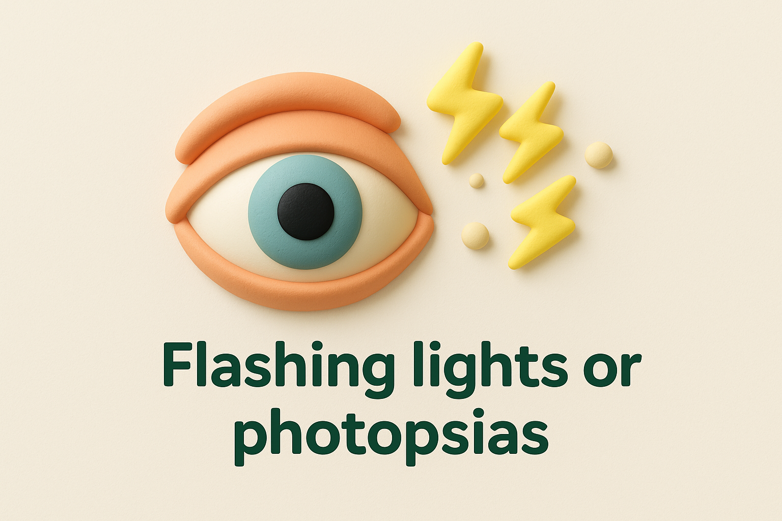
Flashes accompanying new floaters create a high-risk combination for retinal tears. Your vitreous gel pulls away from the retina, and this traction can create holes or tears in the delicate tissue. These symptoms warrant urgent ophthalmology assessment within 24 hours to prevent progression to detachment.
Shadow or curtain across vision
A grey veil or shadow spreading across your visual field indicates that your retina is detaching from its normal position. This curtain typically starts at the edges of your vision and gradually extends toward the centre. The shadow moves as fluid accumulates beneath the detached retina, separating it from the layer that provides nutrients.
When to go to emergency
Visit an emergency department or eye casualty immediately if you experience any combination of warning signs listed above. Head to emergency care outside normal clinic hours, particularly if you notice a rapidly expanding shadow or severe vision loss. Bring a list of your current medications and any relevant medical history. Time matters critically in these situations, as surgical success rates decline when treatment delays beyond 48 hours.
How eye specialists diagnose black spots
Your eye specialist follows a systematic approach when you report a little black spot in vision. The diagnostic process begins with detailed questions about your symptoms and progresses through specialised examinations that reveal what’s happening inside your eye. This thorough evaluation ensures accurate identification of harmless floaters versus serious retinal problems. Your specialist combines multiple assessment techniques to build a complete picture of your eye health.
Taking your medical history
Your appointment starts with questions about when you first noticed the black spots and how they’ve changed. Your specialist asks about the number and pattern of floaters, whether you’ve seen flashing lights, and if any shadows block your peripheral vision. They need to know about existing health conditions like diabetes or high blood pressure, previous eye injuries or surgeries, and your family history of retinal disease. This conversation provides crucial clues that guide the physical examination and help predict which conditions might be present.
Dilated fundus examination
Your specialist places drops in your eyes to widen your pupils, allowing a clear view of your retina and vitreous. This dilation takes 15 to 30 minutes to reach full effect and enables examination of the entire retinal surface. Using a bright light and magnifying lenses, your doctor inspects your vitreous for floating debris, checks for retinal tears or holes, and assesses whether your retina remains properly attached. The examination reveals the exact location and severity of any problems, determining whether you need immediate treatment or routine monitoring.
Dilated examination remains the gold standard for detecting retinal tears and detachment before they progress to vision loss.
Optical coherence tomography and ultrasound
Some situations require advanced imaging to supplement the clinical examination. Optical coherence tomography (OCT) creates detailed cross-sectional images of your retina, revealing subtle changes in its layers. Your specialist uses this technology when they suspect macular involvement or need to track progression over time. B-scan ultrasound becomes necessary when blood or dense floaters prevent a clear view of your retina. Sound waves map the internal eye structures, showing retinal detachment or vitreous abnormalities that can’t be seen directly. These imaging tools provide objective documentation of your condition and help guide treatment decisions.
Treatment options and what to expect
Your treatment path depends entirely on what’s causing that little black spot in vision and how much it affects your daily life. Benign floaters often require no intervention beyond regular monitoring, while serious retinal problems demand immediate surgical repair. Your eye specialist tailors the approach to your specific condition, balancing the risks of treatment against the benefits of clearer vision. Understanding each option helps you make informed decisions about your eye care.
Observation and monitoring for benign floaters
Most people with simple age-related floaters need only regular check-ups rather than active treatment. Your brain gradually adapts to these spots through a process called neural habituation, where you become less consciously aware of them over time. This adaptation typically takes several weeks to months, and most patients find the floaters eventually stop bothering them. Your specialist schedules follow-up examinations at three to six month intervals to ensure the floaters remain stable and no new complications develop. You can return to normal activities immediately, with no restrictions on work, exercise, or daily tasks.
Laser vitreolysis for troublesome floaters
Laser vitreolysis offers a non-invasive option when floaters significantly disrupt your quality of life. Your specialist uses a focused laser beam to break apart the larger collagen clumps casting shadows on your retina. The procedure takes place in the clinic using topical anaesthetic drops, and you can return home the same day. Success rates vary depending on floater location and size, with some patients experiencing complete relief while others notice partial improvement. Your specialist assesses whether your floaters sit in a safe position away from the retina before recommending this treatment.
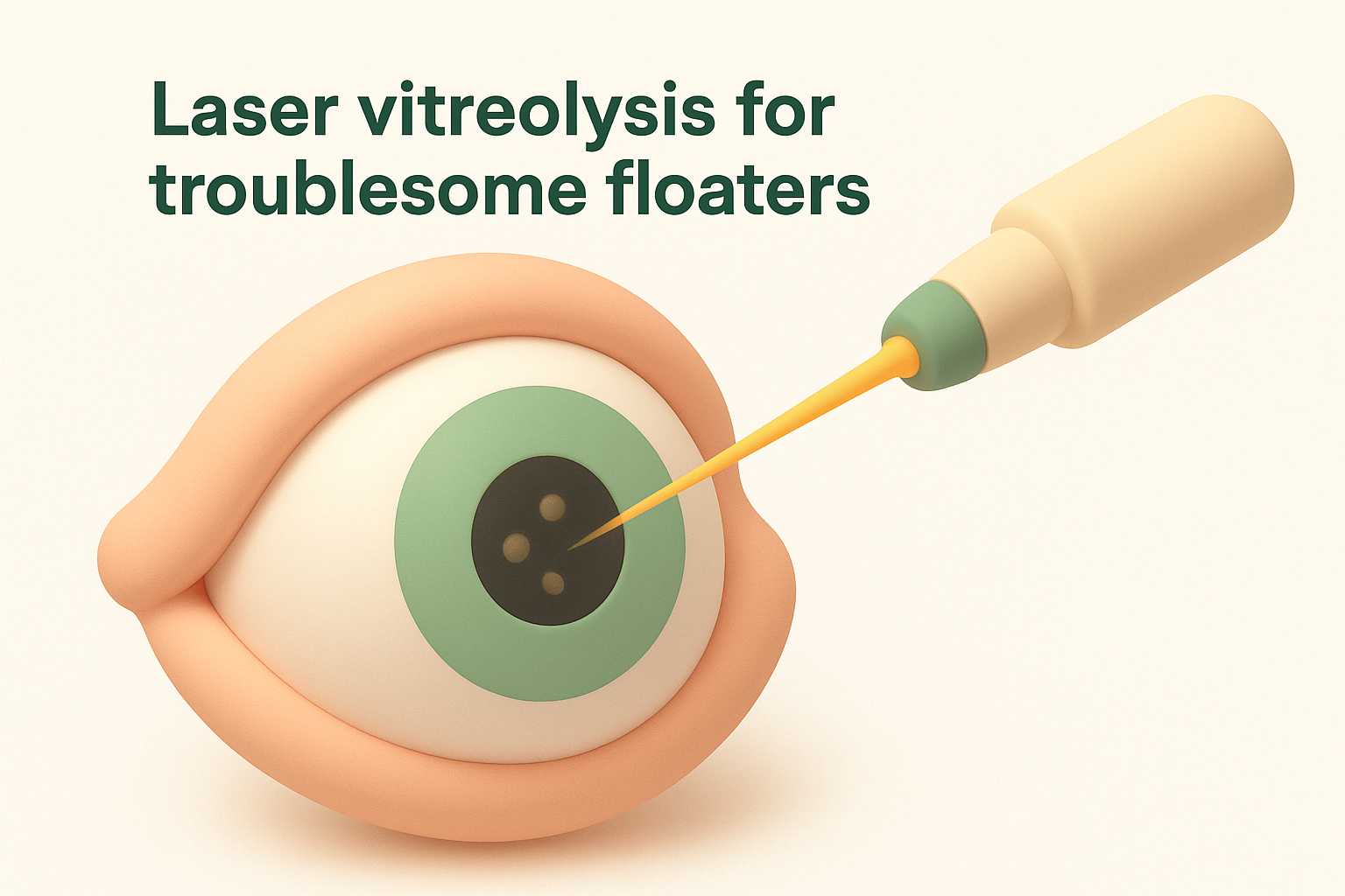
Laser vitreolysis works best for larger, well-defined floaters located at least 2 millimetres from the retina.
Vitrectomy surgery for severe cases
Vitrectomy becomes necessary when dense floaters severely impair your vision or when complications arise. The surgeon makes tiny incisions in your eye wall, removes the cloudy vitreous gel, and replaces it with a clear saline solution. This procedure requires local or general anaesthetic and takes place in a day surgery unit. Recovery involves several weeks of restricted activity, and you’ll need regular post-operative reviews to monitor healing. Your vision gradually improves as your eye adjusts to the new fluid, though some patients develop cataracts months to years later as a known complication of vitrectomy.
Emergency procedures for retinal tears and detachment
Retinal tears need laser photocoagulation or cryotherapy to seal the break and prevent fluid from collecting beneath your retina. These procedures take minutes to perform in the clinic and create a protective scar around the tear. Detached retinas require surgical reattachment using techniques like scleral buckle or vitrectomy with gas tamponade. You’ll need to maintain specific head positioning for several days after surgery to keep the gas bubble pressing against the repaired area. Success rates exceed 90 per cent when treatment occurs promptly, though some patients need additional procedures if the retina detaches again.
Looking after your eyes and preventing problems
You can’t prevent all causes of that little black spot in vision, but you can reduce your risk of serious complications through regular care and healthy habits. Proactive eye health involves more than just responding to symptoms; it requires consistent monitoring and attention to factors that affect your retina and vitreous. Taking steps now protects your vision for decades to come.
Schedule regular eye examinations
Your eyes need routine professional assessment even when they feel perfectly healthy. Book comprehensive eye exams every two years if you’re under 50 and have no risk factors, but increase this to annual visits once you reach middle age. People with diabetes, high myopia, or previous eye surgery require more frequent monitoring, often every six to twelve months. These examinations allow your specialist to detect early warning signs of vitreous changes or retinal problems before they cause symptoms. Your optometrist or ophthalmologist can spot subtle changes during dilated exams that you’d never notice in daily life.
Manage underlying health conditions
Systemic diseases like diabetes and high blood pressure directly affect your retinal blood vessels and increase your risk of serious floaters. Keep your blood sugar levels within target ranges through medication, diet, and regular exercise. Control hypertension with prescribed medications and lifestyle modifications, as uncontrolled blood pressure damages the delicate vessels supplying your retina. Regular blood tests and monitoring help you track how well you’re managing these conditions.
Maintaining optimal blood sugar and blood pressure reduces your risk of vitreous haemorrhage and diabetic retinopathy by up to 50 per cent.
Adopt protective lifestyle habits
Simple daily choices strengthen your eye health over time. Wear quality sunglasses that block UV radiation whenever you’re outdoors, as cumulative sun exposure accelerates vitreous degeneration. Stop smoking immediately if you currently use tobacco, since smoking restricts blood flow to your retina and increases inflammation. Eat a balanced diet rich in leafy greens, colourful vegetables, and omega-3 fatty acids from fish. Stay well hydrated throughout the day to maintain proper vitreous consistency. Protect your eyes during sports and work activities by wearing appropriate safety glasses, as trauma can trigger sudden vitreous changes or retinal tears.

Next steps for your eye health
Noticing a little black spot in vision requires action, not just worry. Start by recording when the spot appeared, how often you notice it, and whether the number has changed since you first saw it. Track any accompanying symptoms like flashing lights or shadows spreading across your field of view. This detailed information helps your eye specialist make an accurate diagnosis during your examination.
Book a comprehensive eye assessment if you haven’t had one recently, particularly if you’re over 50, have diabetes, or wear strong glasses for short-sightedness. Prompt evaluation ensures minor problems stay manageable rather than progressing to sight-threatening emergencies. Your specialist examines your retina under dilation to identify the exact cause and determine whether you need treatment or simple monitoring.
Don’t wait for symptoms to worsen before seeking professional help. Bright Sight Eye Specialists offers expert assessment and treatment for floaters and retinal conditions, with transparent pricing and same-day appointments available for urgent concerns. Taking action now protects your vision for the years ahead.