


Your eyes depend on a thin, transparent layer called the cornea to focus light and shield against dust, germs, and debris. When this protective barrier becomes inflamed, a condition known as keratitis, the consequences can range from mild discomfort to serious vision impairment if left untreated.
Keratitis affects thousands of Australians each year, with contact lens wearers facing a particularly elevated risk. Whether caused by bacteria, viruses, fungi, or parasites, this condition demands prompt recognition and appropriate care. Catching the warning signs early can make the difference between a straightforward recovery and lasting corneal damage.
At Bright Sight Eye Specialists, Dr Rahul Dubey and the team regularly assess patients throughout Sydney’s Eastern Suburbs who present with corneal concerns requiring expert evaluation. This guide covers everything you need to know about keratitis, from recognising symptoms and understanding what causes the condition to exploring treatment pathways and realistic recovery timelines.
Why keratitis needs urgent attention
Your cornea contains no blood vessels, which means it relies on tears and oxygen to stay healthy and heal. When keratitis develops, this unique structure makes the cornea particularly vulnerable to rapid deterioration. Delaying treatment by even a few days can allow infectious organisms to penetrate deeper layers of corneal tissue, transforming what might have been a straightforward infection into a medical emergency requiring intensive intervention.
Rapid progression of untreated keratitis
The cornea can deteriorate with alarming speed when infection takes hold. Bacterial keratitis, particularly strains like Pseudomonas aeruginosa commonly found in contaminated contact lens cases, can destroy corneal tissue within 24 to 48 hours if you don’t receive proper treatment. Fungal and amoebic varieties progress more slowly but prove equally devastating, often burrowing deeper into the cornea while creating symptoms that patients initially dismiss as minor irritation.
You might notice your symptoms worsen dramatically overnight. What begins as mild redness and a scratchy sensation can escalate to severe pain, light sensitivity, and blurred vision by the following morning. Infectious keratitis doesn’t follow a predictable timeline, and waiting to see if symptoms improve on their own gives harmful microorganisms critical hours to multiply and spread.
The cornea’s lack of blood supply means it cannot fight infection as effectively as other tissues, making early intervention essential to prevent irreversible damage.
Permanent vision complications
Corneal scarring represents the most common long-term consequence of untreated or inadequately treated keratitis. These scars form as your body attempts to repair damaged tissue, but unlike healthy corneal cells that remain transparent, scar tissue appears opaque and permanently obstructs your line of sight. The location and density of scarring determine how severely your vision becomes affected, with central scars causing more significant impairment than peripheral ones.
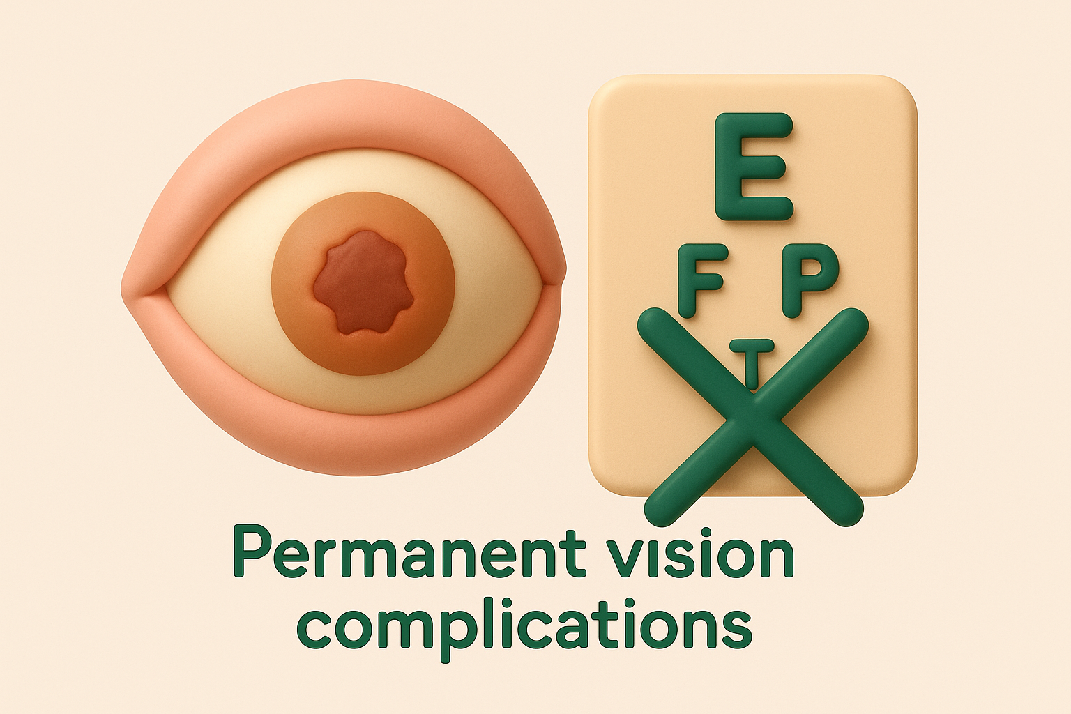
Corneal ulceration develops when infection eats through layers of corneal tissue, creating an open wound on the eye’s surface. Deep ulcers can perforate the cornea entirely, allowing the clear gel inside your eye to leak out and potentially leading to total vision loss in the affected eye. Even when perforation doesn’t occur, severe ulcers often require surgical intervention such as corneal transplantation to restore vision.
Beyond direct corneal damage, untreated keratitis can trigger secondary complications throughout your eye. The infection may spread to adjacent structures, causing endophthalmitis (infection inside the eye), glaucoma from increased pressure, or cataract formation. Each additional complication multiplies the complexity of treatment and reduces the likelihood of achieving your previous level of visual clarity. Patients who delay seeking specialist care also face extended recovery periods, often requiring months of follow-up appointments and medication rather than the weeks needed when treatment begins promptly.
Impact on daily function and quality of life
Your ability to drive, work, and perform routine activities depends heavily on clear, comfortable vision. Keratitis symptoms such as photophobia (light sensitivity) and excessive tearing can make it impossible to focus on screens, read documents, or navigate outdoors during daylight hours. Contact lens wearers must cease lens use entirely during treatment and recovery, which creates immediate practical challenges for those who don’t have an up-to-date prescription for glasses.
Pain and discomfort from keratitis often prove severe enough to disrupt sleep, concentration, and overall wellbeing. The constant awareness of foreign body sensation and the need for frequent medication administration throughout the day demand significant time and attention. Early treatment minimises these disruptions and helps you return to normal activities faster.
Types of keratitis
Doctors classify keratitis into two broad categories based on whether microorganisms cause the inflammation or other factors trigger the condition. Understanding which type affects your eye shapes treatment decisions and determines how quickly you can expect to recover. Infectious varieties typically demand more aggressive intervention than non-infectious forms, though both require professional assessment to prevent complications.
Infectious keratitis
Bacteria, viruses, fungi, and parasites can all invade your cornea and cause infectious keratitis. Bacterial keratitis represents the most common form, often developing when you wear contact lenses overnight or fail to clean them properly. Staphylococcus aureus and Pseudomonas aeruginosa rank among the most frequent culprits in Australia, with the latter particularly dangerous due to its rapid tissue destruction.

Viral keratitis commonly stems from the herpes simplex virus, the same pathogen responsible for cold sores. This variant creates distinctive branching patterns on your cornea visible during examination and tends to recur periodically throughout your lifetime once initial infection occurs. Varicella-zoster virus, which causes chickenpox and shingles, can also affect your cornea when reactivation occurs near your eyes.
Fungal infections develop less frequently but prove notoriously difficult to treat, often requiring months of intensive medication. Fusarium and Aspergillus species thrive in warm, humid environments and typically enter your eye through plant material injuries or contaminated contact lens solutions. Acanthamoeba keratitis, caused by a microscopic parasite found in tap water, hot tubs, and swimming pools, affects primarily contact lens users who rinse lenses with water rather than proper disinfecting solutions.
Acanthamoeba keratitis accounts for roughly 5% of contact lens-related infections in Australia but causes disproportionate vision loss due to delayed diagnosis and treatment resistance.
Non-infectious keratitis
Your cornea can become inflamed without any microbial invasion through several mechanisms. Physical trauma from scratches, chemical burns, or foreign objects creates immediate inflammation that mimics infectious presentations. You might develop keratitis from prolonged UV exposure without protective eyewear, a condition sometimes called photokeratitis or "arc eye" among welders.
Dry eye disease causes a form of keratitis when your tear film fails to adequately lubricate and protect your corneal surface. The resulting inflammation creates similar discomfort to infectious varieties but requires fundamentally different treatment approaches focused on tear supplementation rather than antimicrobial medication. Autoimmune conditions such as rheumatoid arthritis or Sjögren’s syndrome can trigger non-infectious corneal inflammation as part of broader systemic disease patterns.
Allergic reactions and exposure to irritants like chlorine or smoke produce temporary corneal inflammation that resolves once you remove the triggering substance. These variants rarely cause lasting damage when you address them promptly.
Symptoms and early warning signs
Your eyes provide clear signals when keratitis develops, though you might initially mistake these warnings for less serious conditions like allergies or dry eyes. Recognising the specific pattern of symptoms helps you determine whether your discomfort requires urgent specialist evaluation or simple home care. The intensity and combination of symptoms offer crucial clues about the severity and underlying cause of corneal inflammation.
Common initial symptoms
Most people first notice a persistent feeling that something sits in their eye, similar to the sensation of a trapped eyelash or grain of sand. This foreign body feeling refuses to resolve with blinking or eye rubbing and often worsens throughout the day. Excessive tearing accompanies this irritation as your eye attempts to flush away what it perceives as a threat, though the tears provide little relief from discomfort.
Redness develops as blood vessels in the white part of your eye dilate in response to inflammation. You’ll typically see concentrated redness around the cornea rather than generalised pink eye appearance. Light sensitivity (photophobia) emerges early in keratitis, making it uncomfortable to look at bright screens, go outdoors during daylight, or sit in well-lit rooms. Your vision may appear slightly blurred or hazy, particularly if discharge accumulates on your corneal surface or if swelling affects the central visual zone.
Early keratitis symptoms often mirror common eye irritations, but the combination of persistent foreign body sensation, light sensitivity, and progressive pain signals the need for professional assessment.
Progressive symptoms requiring immediate attention
Pain represents a critical escalation that distinguishes keratitis from minor eye irritation. The discomfort intensifies from mild scratchiness to severe, sharp pain that penetrates deep into your eye socket. This pain typically worsens with eye movement and can radiate to your forehead and surrounding facial areas. Contact lens wearers who experience sudden pain upon insertion should remove lenses immediately and seek evaluation.
Discharge production increases as infection advances, with bacterial keratitis often creating thick, yellow or green pus that crusts your eyelids shut overnight. Fungal infections tend to produce white or grey infiltrates visible on your cornea’s surface. Your vision deterioration accelerates beyond simple blurriness to include cloudy patches or dark spots in your visual field, indicating deeper corneal involvement.
Severe photophobia that makes it impossible to open your eyes in normal lighting conditions signals advanced inflammation. You might develop involuntary eyelid spasms (blepharospasm) as your body attempts to protect the damaged cornea from further light exposure. Any sudden vision loss, particularly when accompanied by intense pain, requires immediate emergency care to prevent permanent damage.
Causes and risk factors in Australia
Understanding what triggers keratitis helps you take preventive steps and recognise situations that increase your vulnerability. Multiple factors contribute to corneal inflammation, with some causes particularly prevalent in Australia’s climate and lifestyle patterns. Contact lens wear dominates as the leading risk factor, but environmental exposures, eye injuries, and underlying health conditions also create opportunities for infection and inflammation to develop.
Contact lens-related causes
Contact lens wear accounts for 80 to 90 percent of microbial keratitis cases in Australia, with improper lens hygiene creating ideal conditions for bacterial and parasitic growth. You significantly increase your risk when you sleep in lenses not designed for overnight wear, as this practice reduces oxygen supply to your cornea and allows microorganisms to multiply. Tap water exposure represents another major hazard, with Acanthamoeba parasites lurking in household water supplies and swimming pools throughout Australia.
Extended wear schedules beyond manufacturer recommendations create microscopic tears in your corneal surface where pathogens can enter. Using old or contaminated lens solution, sharing lenses with others, or wearing lenses while swimming or showering all multiply your infection risk substantially. Your lens case itself becomes a breeding ground for bacteria if you don’t clean and replace it regularly, with studies showing that most contaminated cases belong to users who rinse them with water rather than fresh disinfecting solution.
Contact lens users who swim or shower while wearing lenses face up to 16 times greater risk of developing Acanthamoeba keratitis compared to those who remove lenses before water exposure.
Environmental and lifestyle factors
Australia’s intense UV radiation levels damage your corneal surface when you spend extended periods outdoors without proper eye protection, creating vulnerability to subsequent infection. Agricultural and construction workers face elevated risk from plant material injuries and flying debris that can carry fungal spores directly into corneal tissue. Beach activities expose your eyes to sand particles and saltwater that may harbour microorganisms.
Your occupation matters significantly if you work as a welder, gardener, or farmer. Metal fragments, chemical splashes, and organic matter contact create entry points for pathogens. Australia’s warm, humid climate in northern regions provides optimal conditions for fungal growth, increasing fungal keratitis incidence compared to cooler southern areas.
Underlying health conditions
Dry eye disease compromises your cornea’s natural defence mechanisms, making you more susceptible to keratitis when tear production proves inadequate. Diabetes weakens your immune response and slows corneal healing, creating longer windows for infection to establish. Immunosuppressive medications for autoimmune conditions or following organ transplants reduce your body’s ability to fight off corneal invaders. Cold sore sufferers carry permanent risk of herpes simplex keratitis, as the virus can reactivate and spread to corneal tissue during periods of stress or illness.
How to get keratitis diagnosed
Diagnosing keratitis requires specialised equipment and expertise that only an ophthalmologist or optometrist can provide. Your general practitioner cannot perform the detailed corneal examination necessary to identify the specific cause and severity of inflammation. Prompt professional assessment becomes essential when you experience symptoms, as the diagnostic process not only confirms keratitis but determines which treatment approach will work most effectively for your particular case.
Initial examination and patient history
Your eye specialist begins by asking about symptom onset and progression, including when discomfort started, whether pain worsened suddenly, and what activities preceded your symptoms. Revealing your contact lens habits proves particularly important, as specialists need to know your wearing schedule, cleaning routine, and whether you’ve exposed lenses to water recently. Details about recent eye injuries, cold sore history, or outdoor activities help narrow potential causes.
The specialist examines your visual acuity using a standard eye chart to establish how much keratitis has affected your vision. External observation allows them to assess redness patterns, discharge type, and eyelid swelling. You’ll receive anaesthetic drops to minimise discomfort during detailed examination, as the specialist needs to keep your eye open for extended viewing without causing excessive pain.
Slit-lamp examination and diagnostic tests
A slit-lamp microscope provides magnified, illuminated views of your cornea that reveal inflammation, ulceration, or foreign material invisible to the naked eye. Your specialist directs a narrow beam of light across your corneal surface while viewing through the microscope, allowing them to assess tissue layers individually and identify characteristic patterns that indicate specific infections. Herpes simplex creates branching ulcers, while fungal infections produce dense white infiltrates with irregular edges.
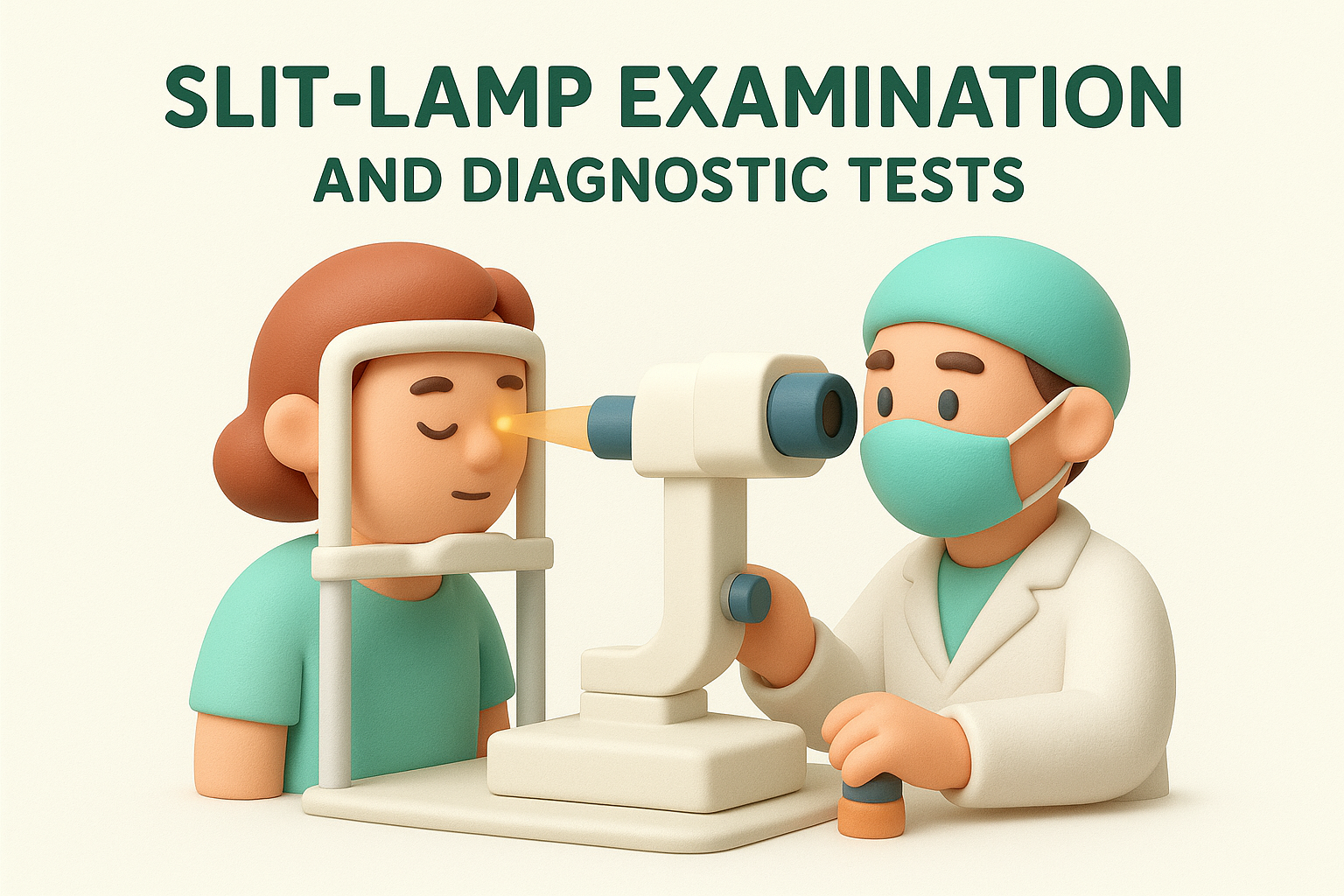
Fluorescein staining makes corneal damage visible under blue light. Your specialist applies orange dye drops that collect in areas where your corneal surface has broken down, highlighting ulcers, scratches, or erosions. The staining pattern helps determine infection depth and extent, guiding treatment intensity.
Corneal cultures obtained during your diagnostic appointment identify the specific organism causing infection, ensuring you receive targeted antimicrobial treatment rather than broad-spectrum medications.
Specimens collected by gently scraping your corneal surface go to laboratories for culture and sensitivity testing. Results typically arrive within 48 to 72 hours, though your specialist starts treatment immediately based on clinical appearance rather than waiting for confirmation. You may undergo additional tests such as corneal confocal microscopy if Acanthamoeba infection seems likely, as this parasitic keratitis requires specific imaging to confirm diagnosis and monitor treatment response.
Treatment options and what to expect
Treatment for keratitis varies dramatically depending on which microorganism or condition causes your corneal inflammation. Your specialist tailors medication choices to match your specific diagnosis, with bacterial infections requiring different approaches than viral, fungal, or parasitic varieties. Infectious keratitis typically demands intensive medication schedules during the first few days, while non-infectious forms respond to anti-inflammatory treatments and addressing underlying triggers.
Antimicrobial medications and application schedules
Bacterial keratitis responds to antibiotic eye drops that you’ll apply every 30 to 60 minutes around the clock during the initial 48 hours. Your specialist prescribes broad-spectrum antibiotics initially, then adjusts medication once culture results identify the specific bacteria and its sensitivities. Fortified antibiotics, prepared by hospital pharmacies at concentrations stronger than commercial preparations, treat severe infections that threaten your vision.
Viral keratitis caused by herpes simplex requires antiviral drops or oral medications such as acyclovir taken for 7 to 14 days. Fungal infections need antifungal drops like natamycin applied hourly initially, with treatment extending for weeks or months depending on infection depth. Acanthamoeba keratitis proves most challenging, requiring dual antimicrobial drops administered hourly for the first week, then gradually reduced over a treatment course that often spans six months or longer.
Treatment intensity decreases as your cornea heals, but stopping medication prematurely allows infection to resurge and potentially cause worse damage than the original presentation.
Supportive care and pain management
Your specialist prescribes cycloplegic drops that dilate your pupil and paralyse focusing muscles, reducing pain from muscle spasms and preventing complications like iris adhesion to your lens. These drops cause temporary vision blurring and light sensitivity but significantly improve comfort during the acute phase. Oral pain relievers such as paracetamol or ibuprofen help manage discomfort between eye drop applications.
You must cease contact lens wear completely throughout treatment and recovery. Your specialist may recommend lubricating drops to supplement natural tears and protect your healing corneal surface. Wearing sunglasses outdoors and dimming indoor lighting reduces photophobia discomfort while inflammation resolves.
Monitoring progress and adjusting treatment
Follow-up appointments occur daily or every few days initially, allowing your specialist to assess whether infection is responding to prescribed medications. Your doctor examines your cornea under the slit lamp at each visit, looking for reduced inflammation, healing ulcers, and clearing infiltrates. Culture results guide treatment adjustments if your initial improvement seems inadequate or symptoms worsen despite medication compliance.
Severe cases that don’t respond to medical treatment may require surgical intervention such as corneal debridement to remove infected tissue or, in extreme situations, therapeutic corneal transplantation. Your specialist discusses these possibilities if your keratitis proves resistant to standard therapies or threatens corneal perforation.
Recovery time and long-term outlook
Recovery timelines for keratitis vary widely based on which microorganism caused your infection, how quickly you received treatment, and how deeply inflammation penetrated your corneal tissue. Mild bacterial keratitis caught early typically resolves within one to two weeks, though you’ll need to continue using medication for the full prescribed duration even after symptoms improve. Severe or delayed cases may require months of treatment and monitoring, with healing complicated by scarring or persistent inflammation that extends your recovery period.
Expected healing timelines by keratitis type
Bacterial keratitis responds fastest to treatment when you start appropriate antibiotics within the first 24 to 48 hours of symptoms appearing. Your corneal surface usually begins healing within three to five days, with complete resolution occurring in seven to fourteen days for superficial infections. Deep bacterial ulcers take longer, potentially requiring four to six weeks before your cornea fully heals and your specialist clears you to resume normal activities including contact lens wear.

Viral keratitis caused by herpes simplex follows a longer timeline, with initial healing taking two to three weeks even with antiviral treatment. Fungal infections prove notably stubborn, demanding intensive treatment for six to twelve weeks or longer depending on organism type and infection depth. Acanthamoeba keratitis requires the most extended treatment course, often lasting six months to a year, with healing occurring gradually as antimicrobial drops slowly eliminate parasitic cysts embedded in your corneal layers.
Recovery from infectious keratitis doesn’t mean your treatment ends, as many patients require maintenance medication or ongoing monitoring to prevent recurrence and manage residual corneal changes.
Factors influencing your healing speed
Your overall health status significantly affects how quickly your cornea recovers from infection. Diabetes, immunosuppression, and autoimmune conditions slow healing processes and increase complication risks. Smoking impairs corneal repair mechanisms and oxygen delivery to healing tissues, potentially doubling your recovery time compared to non-smokers.
Treatment compliance determines outcomes more than any other controllable factor. Missing doses or stopping medication early allows infection to resurge and cause additional damage that extends your recovery substantially. You heal faster when you attend all scheduled follow-up appointments, enabling your specialist to adjust treatment as needed and catch complications before they worsen.
Long-term vision and recurrence considerations
Most people recover good vision after keratitis if treatment begins promptly and proceeds without complications. Minor corneal scarring may leave subtle visual disturbances like glare or slight blurring that you adapt to over time. Central scars affecting your visual axis create more significant problems, potentially requiring corneal transplantation to restore clear vision if scarring proves dense enough to obstruct light transmission.
Herpes simplex keratitis recurs in approximately 25 to 50 percent of patients within two years, as the virus remains dormant in nerve tissue and reactivates periodically. Your specialist may prescribe long-term suppressive antiviral medication to reduce recurrence frequency. Contact lens wearers can usually resume lens wear three to six months after complete healing, though you’ll need to demonstrate improved hygiene practices and attend regular monitoring appointments to verify your cornea remains healthy.
Prevention and contact lens hygiene
Preventing keratitis requires consistent attention to hygiene practices and environmental awareness rather than relying on occasional caution. Contact lens wearers bear the highest prevention responsibility, as proper lens care eliminates most infection risks that lead to corneal inflammation. You can substantially reduce your vulnerability by implementing straightforward protective measures that address the most common pathways through which microorganisms reach your corneal surface.
Daily contact lens care practices
Your hands harbour bacteria, oils, and debris that transfer directly to your lenses during handling. Wash your hands thoroughly with soap and water before touching your eyes or lenses, then dry them with a lint-free towel to avoid introducing fibres onto lens surfaces. Never rinse lenses with tap water, saliva, or any substitute for proper disinfecting solution, as these alternatives contain Acanthamoeba parasites and other pathogens that cause severe infections.
Discard used solution from your lens case immediately after removing lenses each evening rather than topping up old solution with fresh product. This practice prevents biofilm formation where bacteria and parasites multiply despite the presence of disinfectant. Rub and rinse your lenses for at least five seconds per side even when using "no-rub" solutions, as mechanical cleaning removes protein deposits and microorganisms more effectively than chemical disinfection alone.
Studies show that contact lens wearers who consistently rub their lenses during cleaning reduce their infection risk by up to 90% compared to those who skip this step.
Store lenses in fresh disinfecting solution for the minimum time specified by your solution manufacturer, typically four to six hours, ensuring complete disinfection occurs before next use. Your lens case needs cleaning after each use by rinsing with fresh solution (never water), then leaving it open and inverted to air dry completely.
Water exposure and environmental precautions
Remove your contact lenses before swimming, showering, or entering hot tubs regardless of water source. Chlorinated pools contain Acanthamoeba despite chemical treatment, while untreated bodies of water harbour numerous pathogens that adhere to lens surfaces and become trapped against your cornea. You should also avoid wearing lenses during water sports, steam rooms, or activities where water splashing might occur.
Protective eyewear becomes essential during activities involving dust, chemicals, or flying debris. Gardeners, tradies, and outdoor workers face elevated fungal keratitis risk from plant material and soil particles, making safety glasses or goggles necessary protective equipment rather than optional accessories.
When to replace lenses and equipment
Follow your prescribed wearing schedule strictly, replacing daily disposable lenses each day and monthly lenses exactly every 30 days regardless of how frequently you’ve worn them. Extended use beyond recommended timelines allows protein deposits and bacterial colonisation to accumulate, creating infection risks that no amount of cleaning eliminates. Your lens case requires replacement every three months at minimum, as plastic surfaces develop microscopic scratches where microorganisms hide from disinfecting solutions.
Replace your disinfecting solution bottle once opened for longer than the manufacturer’s specified period, typically 90 days, as preservatives degrade over time and allow contamination. Never decant solution into travel-sized containers, as this introduces contamination and dilutes preservative concentration.

What to do now
Your cornea cannot wait when keratitis symptoms appear. Early recognition and professional assessment within the first 24 to 48 hours determine whether you experience straightforward recovery or face complications like scarring and permanent vision loss. Remove contact lenses immediately if you develop pain, redness, or light sensitivity, and avoid the temptation to self-treat with over-the-counter drops that might mask worsening infection.
Contact lens wearers should review their hygiene practices today rather than waiting for problems to develop. Replace old lens cases, discard expired solutions, and commit to never exposing lenses to water. Your vision deserves the few extra minutes that proper care requires each day.
If you’re experiencing corneal symptoms or want specialist assessment of your eye health, book a consultation at Bright Sight Eye Specialists. Dr Rahul Dubey and the team provide comprehensive corneal evaluations and treatment throughout Sydney’s Eastern Suburbs, combining advanced diagnostic technology with personalised care that addresses your specific concerns.