


Your eye contains a clear, gel substance that fills most of the space between the lens and retina. This substance is called the vitreous, though many people refer to it as the jelly inside the eye. The vitreous gives your eye its round shape and helps light pass through to the retina. As you age, this gel can change in ways that affect your vision, sometimes causing floaters or other symptoms.
This article explains what the vitreous is and why understanding it matters for your eye health. You’ll learn how to recognise when changes to this gel might signal a problem, what causes common symptoms like floaters, and how conditions like posterior vitreous detachment develop. We’ll cover when you should see an eye specialist, what tests might be needed, and what treatment options exist. Whether you’re experiencing new floaters or simply want to understand more about your eyes, this guide gives you the facts you need.
Why the eye has jelly inside
The vitreous exists in your eye for several important reasons that directly affect your vision and eye health. This gel substance makes up about 80% of your eye’s volume and plays a crucial role in keeping everything working properly. Understanding why your eye needs this jelly helps explain what happens when age-related changes occur and why certain symptoms develop over time.

The vitreous maintains your eye’s shape and function
Your eye needs the vitreous to maintain its round, globe-like shape throughout your life. Without this gel filling the space, your eye would collapse inward like a deflated balloon. The vitreous applies gentle, consistent pressure against the retina, keeping it pressed against the back wall of your eye where it belongs. This pressure ensures your retina stays in the right position to receive and process light signals that your brain converts into vision. The gel also acts as a shock absorber, protecting your delicate internal eye structures from sudden movements or impacts.
The vitreous helps keep your retina in place while allowing light to pass through clearly to create sharp vision.
Composition and properties that support vision
The jelly inside eye consists of 98% water mixed with collagen fibres and other proteins that give it a gel-like consistency. This composition makes the vitreous completely transparent, allowing light to travel through it without distortion or blockage. Hyaluronic acid molecules within the gel help bind water and maintain the right thickness, while the network of collagen fibres provides structure. The gel’s optical clarity matches the other clear structures in your eye, like the cornea and lens, creating an uninterrupted path for light. This transparency explains why changes to the vitreous, such as condensation or liquefaction, become so noticeable. When the gel’s structure breaks down, those previously invisible collagen fibres can cast shadows on your retina.
How to recognise problems with eye jelly
Most changes to the jelly inside eye happen gradually as you age, but certain symptoms signal that you need professional evaluation. You might notice visual disturbances that appear suddenly or changes that worsen over days or weeks. Recognising these warning signs early helps your eye specialist determine whether you need treatment or simply monitoring. While many vitreous changes cause no lasting harm, some symptoms indicate serious problems that require immediate attention to prevent vision loss.
Common warning signs to watch for
You should pay attention when new floaters appear in your vision, especially if they develop suddenly rather than gradually. These floaters might look like dots, strings, cobwebs, or shadows that drift across your field of view. Flashes of light represent another key symptom, typically appearing as brief sparks or lightning-like streaks in your peripheral vision, particularly when you move your eyes quickly or look to the side. These flashes occur when the vitreous gel tugs on your retina during age-related changes.
New floaters or flashes warrant an eye examination, even if they seem minor, because they can indicate vitreous changes affecting your retina.
Many people also notice a slight increase in blurred vision or difficulty seeing in certain lighting conditions. Cloudy or hazy vision might develop if blood leaks into the vitreous cavity or if debris accumulates in the gel. You might experience a sensation that something blocks part of your vision, like a curtain or veil covering one area. Changes to your colour perception or contrast sensitivity can also signal problems with the vitreous. Some people describe seeing shapes or patterns that move when they shift their gaze, then slowly drift downward due to gravity pulling on clumped collagen fibres.
When symptoms need urgent attention
Certain warning signs demand immediate medical evaluation within 24 hours because they suggest retinal tears or detachment. Contact an eye specialist straight away if you see a sudden shower of new floaters, experience persistent flashing lights that don’t fade, or notice a dark shadow spreading across your vision. Loss of peripheral vision in one eye particularly concerns eye specialists because it often indicates the retina has detached from the back wall of your eye.
You should also seek urgent care if floaters become dense enough to obstruct your central vision or if you notice a sudden decrease in visual sharpness. These symptoms might signal vitreous haemorrhage or retinal damage that needs prompt treatment to preserve your sight. Any rapid change in your vision deserves professional assessment, even if the symptoms improve temporarily.
Eye floaters and posterior vitreous detachment
Floaters and posterior vitreous detachment represent the most common changes that happen to the jelly inside eye as you age. These two conditions often occur together, though you can experience floaters without developing posterior vitreous detachment straight away. Understanding how these conditions develop and relate to each other helps you recognise normal ageing changes versus symptoms that need medical attention. Most people over 50 experience some degree of these changes, though the severity and timing vary considerably between individuals.
What causes eye floaters to appear
Eye floaters develop when the collagen fibres within your vitreous gel clump together instead of staying evenly distributed throughout the jelly. These clumps cast shadows on your retina, which you perceive as dark spots, strings, or cobweb-like shapes drifting through your vision. The vitreous gel starts to liquefy and shrink with age, typically beginning in your 40s or 50s, causing previously invisible collagen strands to stick together and become visible.
Several factors accelerate floater formation beyond normal ageing. People with severe nearsightedness experience floaters earlier and more frequently because their elongated eyeballs stretch the vitreous gel, speeding up its breakdown. Eye injuries, inflammation, or surgery can also trigger floater development by disrupting the gel’s structure. Diabetes, which affects blood vessels throughout your body including those in your eyes, increases your risk of developing floaters due to bleeding into the vitreous cavity. While most floaters remain harmless, a sudden increase in their number or density might signal more serious problems.
Understanding posterior vitreous detachment
Posterior vitreous detachment occurs when the gel completely separates from your retina’s surface, moving forward into the main cavity of your eye. This separation happens because the vitreous shrinks and liquefies over time, eventually pulling away from the back wall where it attached throughout your younger years. The process typically begins around age 60, though it can happen earlier in people with certain risk factors like nearsightedness or previous eye surgery.
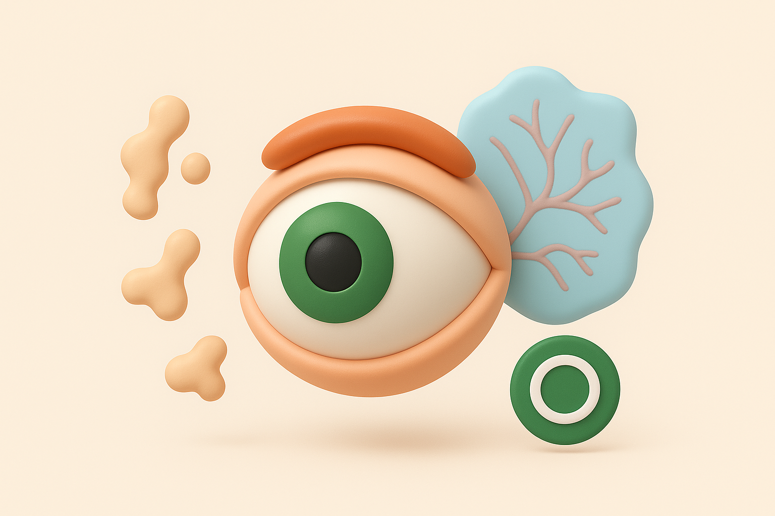
Most people experience posterior vitreous detachment in at least one eye by age 70, making it a natural part of eye ageing rather than a disease.
The detachment itself causes distinct symptoms that differ from simple floaters. You might see flashing lights, particularly when moving your eyes or in dim lighting, because the separating gel tugs on your retina and stimulates light-sensitive cells. Many people notice one large floater that looks like a ring or cobweb, which represents the point where the gel separated from the optic nerve. These symptoms typically improve over weeks or months as the vitreous settles into its new position away from the retina.
How floaters and PVD connect
Floaters almost always accompany posterior vitreous detachment because the separation process itself creates new clumps and debris in the vitreous gel. When the jelly pulls away from your retina, it can tear small blood vessels or lift microscopic pieces of retinal tissue, adding to the floating particles you see. The flashing lights that characterise PVD gradually fade as the gel stops pulling on your retina, but the floaters often persist indefinitely.
Your eye specialist monitors both conditions together because the real concern involves retinal tears or detachment during the separation process. About 10 to 15 percent of people with posterior vitreous detachment develop a retinal tear that could lead to vision loss without treatment. Following an examination that confirms uncomplicated PVD, you can expect the floaters to become less noticeable over time as your brain learns to ignore them, though they rarely disappear completely.
Tests and diagnosis by an eye specialist
Your eye specialist uses several examination techniques to assess the jelly inside eye and determine whether changes to your vitreous pose any risk to your vision. These tests allow your doctor to see the structure and condition of your vitreous gel and check whether any changes have affected your retina. Most examinations take place during a single appointment, though complex cases might require follow-up visits or additional imaging. Understanding what happens during these tests helps you prepare for your appointment and know what to expect.
Initial examination procedures
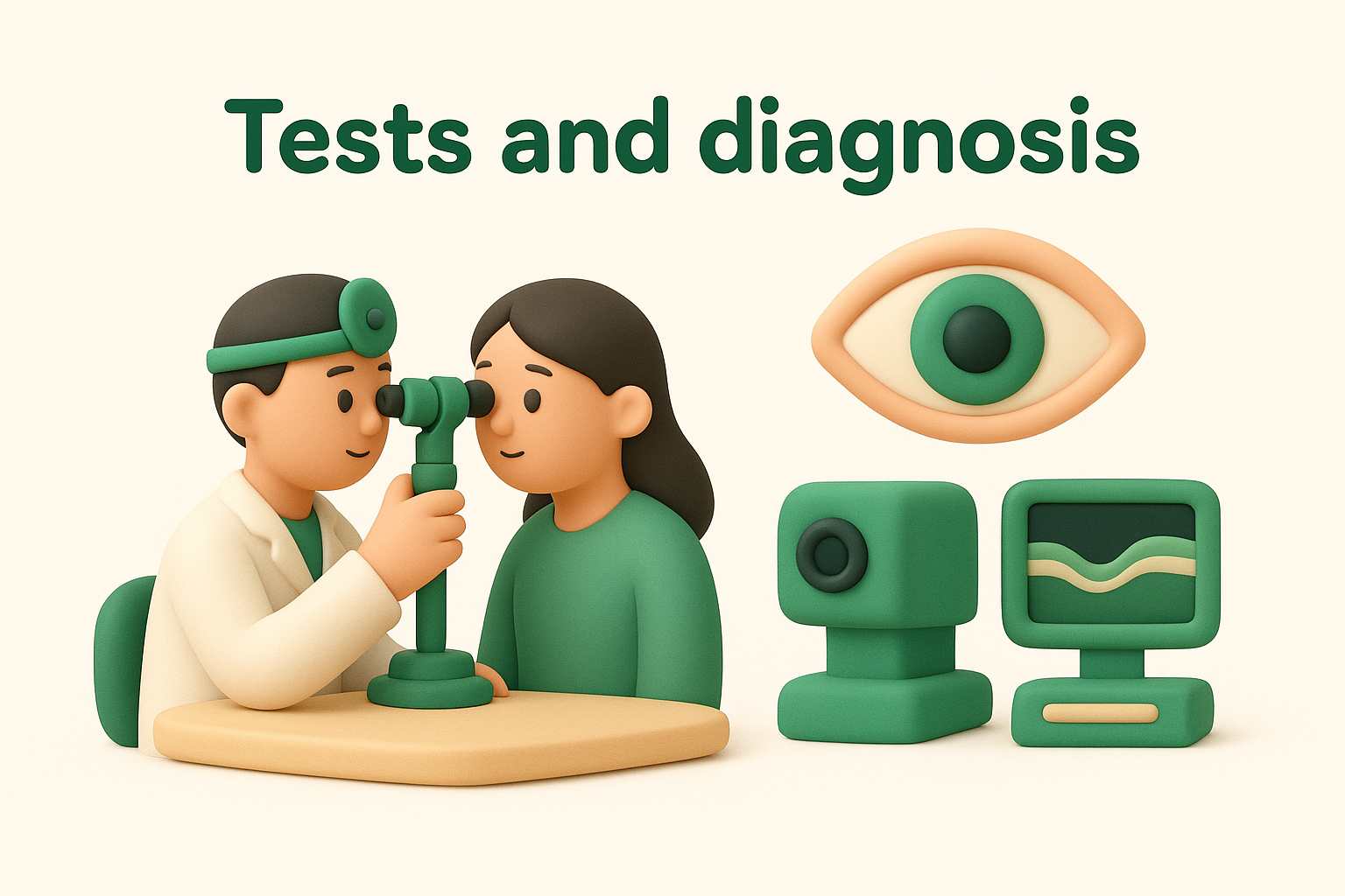
Your eye specialist begins by dilating your pupils with special eye drops that temporarily enlarge the openings at the centre of your eyes. This dilation takes about 20 to 30 minutes and allows your doctor to see through to the back of your eye where the vitreous gel sits. The drops blur your vision and make your eyes sensitive to light for several hours, so you should arrange transport home rather than driving yourself.
Once your pupils dilate fully, your specialist performs a comprehensive dilated eye examination using a slit lamp and special lenses. This equipment provides a magnified, three-dimensional view of your vitreous cavity, retina, and other internal structures. Your doctor looks for signs of vitreous detachment, retinal tears, bleeding, or inflammation that might explain your symptoms. The examination also reveals whether floaters come from harmless clumps of collagen or from more serious sources like blood or retinal tissue.
Your specialist checks not just the vitreous changes but also examines your entire retina to ensure no tears or detachment have occurred during the separation process.
Advanced imaging when needed
Some situations require your eye specialist to order optical coherence tomography (OCT) scans that create detailed cross-sectional images of your retina and vitreous. These scans help detect subtle problems that might not appear during standard examination, particularly when symptoms suggest possible retinal damage. Your doctor might also use ultrasound imaging if bleeding or clouding prevents clear visualisation of your vitreous and retina through the pupil. This painless test uses sound waves to create images of structures inside your eye, revealing whether vitreous haemorrhage or detachment has occurred.
Treatment options and self care
Most changes to the jelly inside eye require no medical treatment beyond regular monitoring by your eye specialist. Your doctor focuses on watching for complications rather than treating the vitreous changes themselves, since the gel’s natural ageing process cannot be reversed. However, certain situations call for active intervention, particularly when floaters severely impair your vision or when the vitreous detachment causes retinal problems. Understanding your options helps you make informed decisions about managing symptoms and knowing when treatment might benefit you.
When treatment becomes necessary
Your eye specialist might recommend laser treatment or surgery if floaters significantly obstruct your central vision and interfere with daily activities like reading or driving. A procedure called vitrectomy removes the vitreous gel and replaces it with a clear saline solution, eliminating floaters completely. This surgery carries risks including cataract formation, retinal detachment, and infection, so doctors typically reserve it for severe cases where symptoms substantially impact your quality of life. YAG laser vitreolysis offers a less invasive option that uses targeted laser pulses to break up large floaters into smaller, less noticeable particles, though not all floaters respond well to this treatment.
Retinal tears discovered during your examination require immediate treatment to prevent progression to retinal detachment. Your doctor uses laser photocoagulation or cryotherapy to seal the tear and create a bond between your retina and the underlying tissue. These procedures typically take place in the clinic with local anaesthetic, allowing you to return home the same day. Following treatment, you need regular follow-up appointments to ensure the seal holds and no new tears develop.
Self-care approaches for managing symptoms
You can help your brain adapt to floaters by avoiding sudden eye movements that make floaters more noticeable and giving yourself time to adjust to their presence. Most people find that floaters become less bothersome after several months as their visual system learns to filter them out. Moving your eyes up and down sometimes shifts floaters out of your central vision temporarily, providing relief when you need to focus on detailed tasks.
Your brain gradually learns to ignore persistent floaters, making them fade into the background of your awareness even though they remain physically present.
Protecting your eyes from further changes involves maintaining good overall health through regular exercise, controlling blood sugar if you have diabetes, and scheduling routine eye examinations. You should avoid activities that might injure your eyes and wear protective eyewear during sports or work that poses impact risks. Report any sudden increases in floaters, new flashes of light, or vision changes to your eye specialist promptly, as these symptoms might indicate complications requiring urgent attention.

Final thoughts
Changes to the jelly inside eye represent a normal part of ageing that affects most people eventually. Understanding what the vitreous does and recognising when symptoms need attention helps you protect your vision and respond appropriately to concerning changes. Most floaters and posterior vitreous detachment remain harmless, requiring only observation rather than treatment, though sudden increases in symptoms always warrant professional evaluation.
Your eye health benefits from regular examinations that catch problems early, particularly after age 50 when vitreous changes become more common. While you cannot prevent age-related changes to your vitreous gel, prompt attention to warning signs prevents serious complications like retinal detachment. If you notice new floaters, flashes of light, or any vision changes, you should schedule an examination rather than waiting for symptoms to worsen.
Book an appointment with our eye specialists in Randwick to discuss any concerns about floaters, vitreous changes, or other aspects of your eye health. Our team provides comprehensive examinations and treatment options tailored to your specific needs.