


A haemorrhage inside the eye means blood has leaked into one of the fluid-filled spaces within your eyeball. This can happen in the vitreous gel that fills most of your eye, in the layers of your retina, or in the front chamber behind your cornea. The bleeding might appear as a sudden burst of floaters, a red patch on the white of your eye, or vision that dims without warning. Some people notice symptoms immediately. Others discover the problem only during a routine eye examination.
This article explains what causes bleeding inside the eye, the different types of haemorrhage you might experience, and the symptoms that signal you need urgent care. You’ll learn how eye specialists diagnose the condition, what treatment options exist, and practical steps to protect your vision. Whether you’re experiencing symptoms now or want to understand your risk factors, this guide gives you the information you need to make informed decisions about your eye health.
Why a haemorrhage inside the eye matters
Your eye relies on transparent tissues and fluids to focus light precisely onto your retina. When blood enters these spaces, it blocks the light path and disrupts the delicate chemistry your photoreceptor cells need to function. Even a small amount of blood can scatter light rays, blur your central vision, or create blind spots that make reading and driving dangerous. The problem extends beyond temporary vision loss. Blood contains iron and other substances that become toxic to your retina if they remain too long.
Immediate vision threats
A haemorrhage inside eye tissue can rob you of sight within hours or days. Blood in your vitreous gel creates a red or dark curtain across your field of vision, and you might lose the ability to see faces or recognise objects. Bleeding in the front chamber of your eye (hyphema) can raise your intraocular pressure to dangerous levels, causing severe pain and risking permanent damage to your optic nerve. Some haemorrhages signal retinal tears or detachment, conditions that require surgical repair within 24 to 48 hours to preserve your vision.
The longer blood remains in contact with your retinal tissue, the higher your risk of permanent scarring and vision loss.
Long-term complications
Blood that persists in your eye triggers inflammatory responses that damage surrounding tissues. Your body sends white blood cells to clean up the haemorrhage, but these cells release chemicals that can harm your retina’s delicate layers. Repeated bleeding episodes, common in diabetic retinopathy and other vascular diseases, create scar tissue that contracts and pulls your retina away from its supporting layer. This tractional detachment can progress slowly, destroying your central vision cell by cell. Iron deposits from old haemorrhages also stain your cornea and lens, reducing clarity even after the blood itself clears.
How to respond if you suspect bleeding in the eye
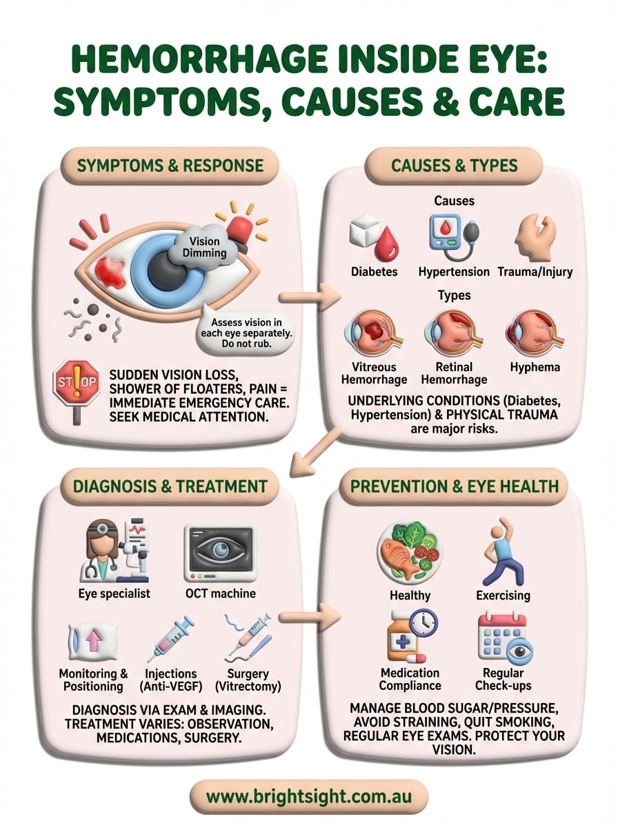
Your first action when you notice blood in your eye or sudden vision changes should be to assess the severity of your symptoms. Stop what you’re doing and find a safe, well-lit space where you can evaluate your vision in each eye separately. Cover one eye at a time and check whether you can see shapes, read text, or detect movement. Note any pain, light sensitivity, or visible blood in the white part of your eye. These observations will help medical professionals determine how urgently you need treatment.
Recognise the emergency signs
You need immediate emergency care if you experience sudden, severe vision loss in one eye, especially if it feels like a curtain has dropped across your field of vision. Go straight to your nearest hospital emergency department if you see a sudden shower of new floaters combined with flashing lights, or if you have a haemorrhage inside eye tissue accompanied by intense pain and nausea. Head injuries that result in visible blood in your eye also require emergency assessment, as they can indicate more serious internal damage. Don’t wait to see if symptoms improve on their own.
Any sudden loss of vision or visible blood inside your eye after trauma requires immediate medical attention, not a "wait and see" approach.
Take protective measures
While arranging medical care, avoid rubbing or pressing on your affected eye. Don’t lie completely flat if you can help it; instead, keep your head elevated at about 30 degrees to help blood settle away from your central vision. Stop taking aspirin or anti-inflammatory medications unless your doctor has prescribed them for a serious condition, as these can increase bleeding. If you wear contact lenses, remove them carefully. Protect your eye from bright light by wearing sunglasses, but don’t apply any patches, medications, or home remedies.
Book an urgent specialist appointment
Less dramatic symptoms still demand prompt professional assessment within 24 to 48 hours. Contact your eye specialist if you notice gradual dimming of vision, new floaters that don’t improve, or a small red patch that appeared without injury. Your optometrist can provide an initial assessment and referral if needed, but retinal haemorrhages often require evaluation by an ophthalmologist who specialises in retinal conditions. Don’t assume minor symptoms will resolve without treatment, as early intervention often prevents permanent damage.
What causes a haemorrhage inside the eye
Blood vessels inside your eye can rupture or leak for dozens of reasons, ranging from sudden physical trauma to progressive diseases that weaken your vessel walls over months or years. The specific cause determines not only your immediate symptoms but also your risk of repeated bleeding episodes. Understanding what triggered your haemorrhage inside eye tissue helps your specialist choose the right treatment and prevent future complications. Some causes require urgent surgical intervention, while others respond well to medication or lifestyle changes.
Vascular and metabolic diseases
Diabetes ranks as the leading cause of bleeding inside the eye in adults under 65. High blood sugar levels damage the tiny vessels in your retina, making them fragile and prone to leaking. These weakened vessels can burst spontaneously or develop abnormal new blood vessels that bleed easily. Hypertension creates similar problems by increasing pressure within your retinal arteries, causing them to narrow, harden, and eventually rupture. Your risk rises sharply if you have both conditions together.
Retinal vein occlusion occurs when a blood clot blocks one of the veins draining your retina, causing pressure to build behind the blockage until vessels burst. Age-related macular degeneration can also trigger bleeding when abnormal blood vessels grow under your macula and leak into your retinal layers. Other vascular causes include sickle cell disease, which creates misshapen blood cells that clog your retinal vessels, and inflammatory conditions like vasculitis that weaken vessel walls throughout your body.
Physical trauma and injury
Direct blows to your eye or head can tear blood vessels instantly, causing visible bleeding in your anterior chamber or hidden haemorrhage in your vitreous. You might experience this after car accidents, sports injuries, falls, or physical assaults. Even minor trauma like rubbing your eye too hard can rupture fragile vessels if you have underlying vascular disease. Surgical complications during cataract removal or retinal procedures also occasionally cause bleeding, though modern techniques have reduced this risk significantly.
Certain blood-thinning medications increase your bleeding risk even from minor trauma that wouldn’t normally cause a haemorrhage.
Medications and systemic factors
Anticoagulant drugs like warfarin and direct oral anticoagulants prevent dangerous clots but make spontaneous eye bleeding more likely. Aspirin and anti-inflammatory medications have similar effects, particularly in older adults with fragile retinal vessels. Your specialist needs to know every medication you take, including herbal supplements that affect clotting. Blood disorders such as leukaemia, anaemia, and clotting deficiencies can all trigger spontaneous haemorrhages. Sudden increases in pressure from severe coughing, vomiting, or heavy lifting occasionally rupture healthy vessels in people with no underlying disease.
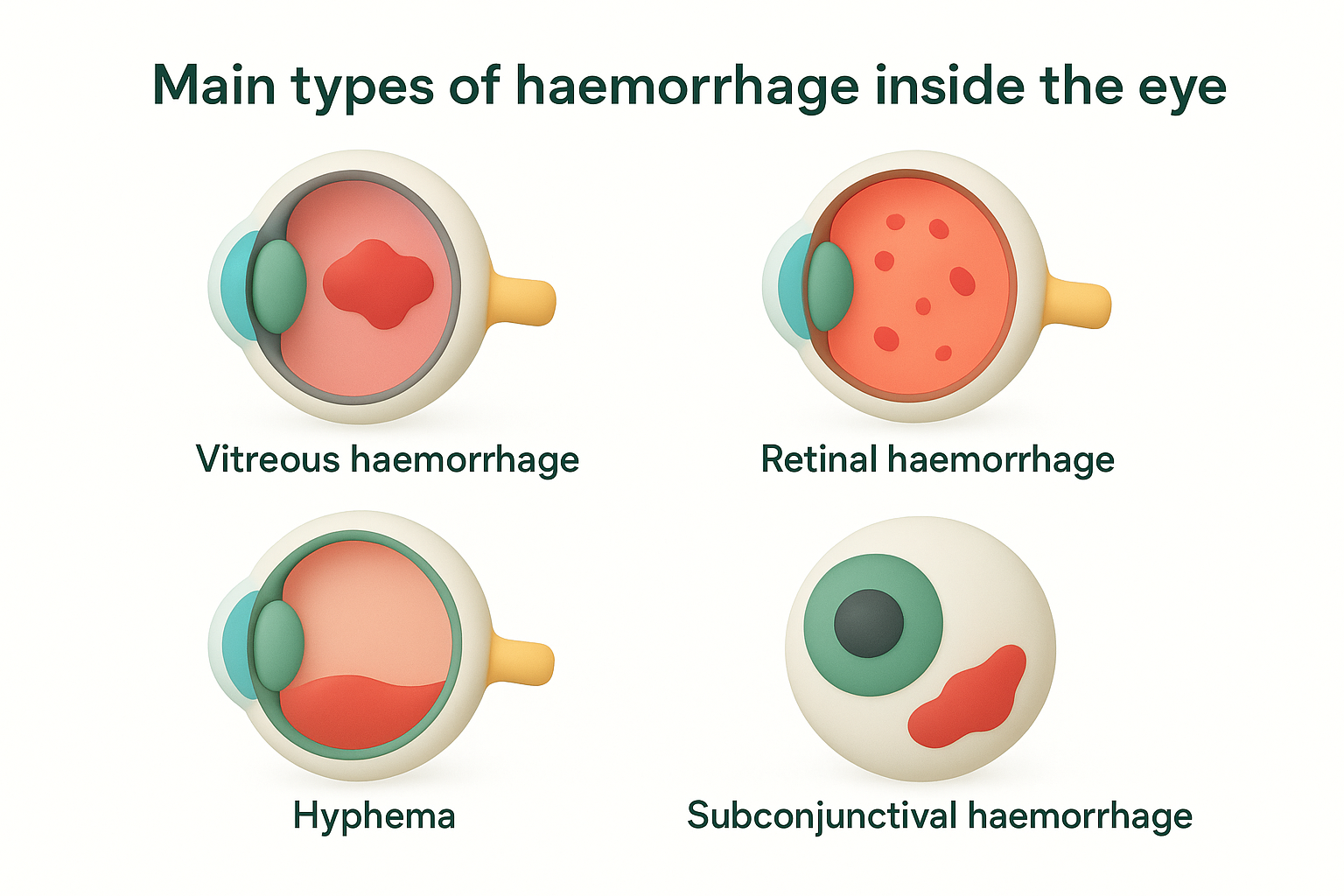
Main types of haemorrhage inside the eye
Eye specialists classify bleeding based on which anatomical space contains the blood, as each location produces distinct symptoms and requires different treatments. The structure of your eye creates several separate compartments, and blood in one area behaves quite differently from blood in another. Understanding these types helps you recognise your symptoms and communicate clearly with your eye care team. Your specialist will use specific terms to describe where the bleeding has occurred, ranging from surface vessels that look dramatic but heal easily to deep haemorrhages that threaten permanent vision loss.
Vitreous haemorrhage
Blood that leaks into the vitreous gel filling the main cavity of your eye creates what specialists call vitreous haemorrhage. This clear, jelly-like substance normally allows light to pass through to your retina without any obstruction. When blood enters this space, it disperses throughout the gel, creating floaters, cobwebs, or a red haze across your vision. The bleeding typically comes from torn retinal vessels, abnormal new vessels growing from your retina, or vessels damaged by diabetic retinopathy.
Your symptoms depend on how much blood has leaked and where it settles. Small vitreous haemorrhages might produce only a few new floaters that drift across your vision, while massive bleeding can block light completely, leaving you able to see only bright lights or hand movements. The blood usually settles toward the bottom of your eye over hours or days, which is why keeping your head elevated helps clear your central vision faster. This type of haemorrhage inside eye tissue rarely resolves without medical intervention if the underlying cause remains untreated.
Retinal haemorrhage
Blood can accumulate in several different layers within your retina itself, each producing characteristic patterns that help specialists identify the underlying cause. Flame-shaped haemorrhages occur in your nerve fibre layer and often indicate high blood pressure, appearing as red streaks that follow your nerve fibres. Dot and blot haemorrhages sit deeper in your retinal tissue and commonly result from diabetes or vein occlusions. These look like small red spots scattered across your retina during examination.
Sub-retinal bleeding occurs between your photoreceptor layer and the supporting tissue beneath, often from age-related macular degeneration or trauma. This type of haemorrhage directly damages your light-sensing cells and requires urgent treatment to prevent permanent scarring. Pre-retinal haemorrhages pool between your retina and vitreous gel, creating boat-shaped collections that block light but cause less direct damage to your retinal cells than bleeding within the retinal layers themselves.
Retinal haemorrhages often signal serious underlying diseases that affect your entire body, not just your eyes.
Hyphema
Bleeding into the anterior chamber, the fluid-filled space between your cornea and iris, produces a visible layer of blood that settles at the bottom of your eye due to gravity. You can often see this type of haemorrhage in a mirror as a red or dark brown level behind your cornea. Hyphema usually results from blunt trauma that tears vessels in your iris or surrounding structures, though spontaneous bleeding can occur in patients taking blood thinners or those with abnormal vessels growing from severe retinal disease.
The blood cells can block your drainage channels and cause dangerous pressure spikes that require immediate treatment. Unlike other types, hyphema creates obvious visible signs that bring patients to medical attention quickly. The condition requires careful monitoring because bleeding can recur within the first week, particularly between days three and five after the initial injury.
Subconjunctival haemorrhage
Blood that collects under the clear membrane covering the white of your eye creates bright red patches that look alarming but rarely affect your vision. These haemorrhages occur when tiny vessels rupture from sneezing, coughing, straining, or minor trauma, and the blood spreads between your conjunctiva and the underlying sclera. The bleeding appears suddenly and looks worse than it actually is, often causing more concern to others who see your red eye than to you experiencing it.
Most subconjunctival haemorrhages resolve within two weeks without any treatment as your body reabsorbs the blood. They don’t obstruct light reaching your retina and cause no pain or vision changes. However, frequent recurrences warrant investigation for underlying blood disorders or uncontrolled hypertension.
Signs and symptoms to watch for
Your ability to recognise the warning signs of bleeding inside your eye determines how quickly you get treatment and how well your vision recovers. Some symptoms appear suddenly and unmistakably, while others develop gradually over days or weeks. The type and location of the haemorrhage dictates which symptoms you’ll experience, and some people notice no symptoms at all until a routine eye examination reveals the problem. Learning to identify these signs helps you respond appropriately and avoid the mistake of dismissing serious symptoms as minor irritations.
Visual disturbances that signal bleeding
Sudden appearance of floaters ranks among the most common first symptoms of haemorrhage inside eye tissue. These might look like small dark spots, squiggly lines, cobwebs, or clouds that drift across your vision when you move your eyes. A few floaters appearing gradually over years are normal, but a sudden shower of dozens or hundreds of new floaters, particularly if accompanied by flashing lights, demands immediate medical attention. The flashes typically appear in your peripheral vision and resemble lightning streaks or camera flashes, indicating that your retina is under tension or tearing.
Your vision might become hazy, blurred, or dim, as if you’re looking through dirty glass or smoke. Some people describe a red or brown tint to their vision, while others report seeing shadows or dark curtains that obscure parts of their visual field. These symptoms often worsen when you first wake up because blood has settled across your central vision while you slept lying flat. Small haemorrhages might affect only your peripheral vision, but larger ones can reduce your sight to hand movements or light perception only.
If you notice a sudden curtain or shadow moving across your vision, treat it as an emergency requiring immediate hospital attention.
Physical sensations and discomfort
Pain severity varies dramatically depending on where the bleeding occurs. Vitreous and retinal haemorrhages typically cause no pain at all, which is why many people delay seeking treatment. Your eye might feel completely normal even as blood accumulates inside. However, bleeding in your anterior chamber (hyphema) or bleeding that raises your intraocular pressure can cause throbbing pain that worsens with movement or bright light. You might also experience a dull ache around your eye or in your temple.
Visible redness provides another clue, though its appearance differs by type. Subconjunctival haemorrhages create bright red patches on the white of your eye that you can see in a mirror, while bleeding deeper inside your eye produces no external redness at all. Your eye might feel scratchy, irritated, or sensitive to light, particularly if inflammation accompanies the haemorrhage. Some people notice decreased colour perception or loss of contrast sensitivity, making it harder to distinguish similar shades. Persistent symptoms, worsening vision, or any combination of these signs warrant urgent professional assessment.
How eye specialists diagnose eye bleeding
Your eye specialist begins diagnosis by gathering detailed information about when your symptoms started and what you were doing when they appeared. They’ll ask about recent injuries, medical conditions like diabetes or hypertension, and every medication you take, including over-the-counter products and supplements. This conversation helps them form an initial hypothesis about the type and location of your haemorrhage before they examine your eyes. Accurate diagnosis requires sophisticated equipment and specialised training, as blood hidden deep within your eye structures won’t show on a simple external examination.
Initial examination and history
During your first consultation, your specialist tests your visual acuity by asking you to read letters on a standard eye chart with each eye separately. This establishes baseline measurements that help track your recovery later. They measure your intraocular pressure using a device that gently touches your cornea or blows a puff of air, checking whether blood cells have blocked your drainage channels and caused dangerous pressure spikes. Pupil reactions are assessed with a bright light to ensure your optic nerve and retinal pathways remain functional despite the bleeding.
Your specialist then examines your eye’s internal structures using a slit lamp microscope, which provides magnified, illuminated views of your anterior chamber, lens, and vitreous cavity. They can see blood floating in your anterior chamber or suspended in your vitreous gel during this examination. After dilating your pupils with eye drops, they use special lenses to view your retina in fine detail, looking for tears, detachments, bleeding sources, and signs of underlying vascular disease. This thorough examination typically reveals not only the haemorrhage inside eye tissue but also its probable cause.
Imaging tests that reveal hidden bleeding
Optical coherence tomography (OCT) creates detailed cross-sectional images of your retinal layers, showing exactly where blood has accumulated and whether it has damaged your photoreceptor cells. The test takes only minutes and uses light waves rather than radiation, producing images so precise that your specialist can measure fluid and blood down to micrometres. OCT helps distinguish between different types of retinal haemorrhages and guides treatment decisions by revealing hidden tractional forces or macular oedema.
Advanced imaging technology allows your specialist to see blood and structural damage that would remain invisible during standard examination.
Ultrasonography becomes essential when blood clouds your vitreous so heavily that your specialist cannot see through to your retina. This painless test uses sound waves to create images of your eye’s internal structures, detecting retinal detachments, foreign bodies, and large clots that require surgical removal. Fluorescein angiography involves injecting a yellow dye into your arm vein and photographing it as it flows through your retinal vessels, revealing leaking areas, blocked vessels, and abnormal new vessel growth that caused your bleeding. Your specialist selects tests based on your specific symptoms and initial examination findings.
Treatment options for eye haemorrhage
Treatment for bleeding inside your eye depends on where the blood has accumulated, how much vision you’ve lost, and what caused the haemorrhage in the first place. Your eye specialist selects from a range of approaches, from careful observation to urgent surgical intervention. Some haemorrhages resolve naturally as your body reabsorbs the blood, while others require active treatment to prevent permanent damage. The underlying cause often determines your treatment path more than the bleeding itself, which is why accurate diagnosis proves so essential for recovery.
Conservative management and monitoring
Small vitreous haemorrhages that don’t threaten your central vision often clear on their own within weeks or months without any intervention. Your specialist monitors your progress with regular examinations, checking that the blood is reabsorbing steadily and that no complications have developed. During this period, you keep your head elevated while sleeping, avoid heavy lifting or straining, and stop taking any blood-thinning medications that aren’t medically essential. This approach works best when the bleeding remains mild and your underlying condition is well controlled.
Positioning your head correctly helps blood settle away from your central visual axis, allowing clearer vision while your body removes the blood cells. Your specialist might prescribe eye drops to reduce inflammation or control intraocular pressure if needed. Regular follow-up appointments every one to two weeks ensure that your haemorrhage is improving rather than worsening. However, if your vision doesn’t improve after several weeks, or if new bleeding occurs, your treatment plan will shift toward more active interventions.
Medical treatments and injections
Intravitreal injections deliver medication directly into your vitreous cavity to stop bleeding and prevent recurrence. Anti-VEGF drugs such as ranibizumab, aflibercept, and bevacizumab target abnormal blood vessel growth that causes haemorrhage inside eye tissue in diabetic retinopathy, age-related macular degeneration, and vein occlusions. These injections reduce swelling, dry up leaking vessels, and prevent new fragile vessels from forming. You typically receive injections monthly at first, then less frequently as your condition stabilises.
Medical therapy often prevents progression and recurrent bleeding more effectively than surgical removal of blood alone.
Your specialist might combine injections with laser photocoagulation to seal leaking vessels and destroy areas of your retina that are triggering abnormal vessel growth. The laser creates tiny burns that reduce oxygen demand in your peripheral retina, eliminating the chemical signals that promote bleeding. Corticosteroid injections provide an alternative for cases where anti-VEGF therapy proves ineffective, though they carry a higher risk of raising intraocular pressure and accelerating cataract formation.
Surgical interventions
Vitrectomy surgery becomes necessary when blood doesn’t clear after several months, when you have severe vision loss that prevents daily activities, or when retinal detachment accompanies the haemorrhage. Your surgeon makes tiny incisions in your eye wall and uses specialised instruments to remove the blood-filled vitreous gel, replacing it with a clear saline solution or gas bubble. During the procedure, they also repair any retinal tears, remove scar tissue pulling on your retina, and apply laser treatment to prevent future bleeding. Most patients undergo this microsurgical procedure under local anaesthesia as a day case.
Recovery from vitrectomy requires strict positioning if your surgeon uses a gas bubble, often meaning you keep your head face-down for several days to ensure the bubble holds your retina in place. Your vision improves gradually over weeks as inflammation settles and your eye heals. Anterior chamber washout treats hyphema when blood raises your intraocular pressure dangerously high or begins staining your cornea. Your surgeon flushes the blood from your anterior chamber through a small incision, immediately relieving pressure and clearing your visual axis.
Living with and preventing eye haemorrhage
Once you’ve experienced bleeding inside your eye, your focus shifts to preventing recurrence and managing the underlying conditions that caused it. Recovery requires more than just treating the immediate haemorrhage. You need to address the root causes, protect your eyes from future damage, and maintain vigilant monitoring. Your daily habits and medical compliance directly influence whether you’ll face repeated episodes or preserve your vision long term. Prevention strategies vary based on what triggered your initial bleeding, but several universal principles apply to everyone at risk.
Control underlying health conditions
Managing your blood sugar levels becomes essential if diabetes caused your haemorrhage inside eye tissue. You need to check your glucose regularly, take medications as prescribed, and keep your HbA1c below your target level. Poor diabetic control accelerates retinal vessel damage and guarantees future bleeding episodes. Similarly, if hypertension triggered your haemorrhage, you must monitor your blood pressure at home and work with your doctor to achieve consistent readings below 130/80 mmHg through medication, diet, and stress management.
Your medication regimen requires regular review with both your eye specialist and general practitioner. Blood thinners like warfarin or anticoagulants serve important purposes but increase bleeding risk, so you need to maintain the lowest effective dose. Never stop these medications without medical supervision, as doing so might trigger strokes or heart attacks. Instead, discuss with your doctors whether alternative treatments might balance your cardiovascular needs against your eye bleeding risk.
Preventing the first haemorrhage through good health management is far easier than recovering from repeated bleeding episodes.
Lifestyle modifications that protect your eyes
You should avoid heavy lifting, straining, and activities that create sudden pressure spikes in your blood vessels. This includes intense weightlifting, high-impact sports, and even prolonged periods of bending with your head below your heart. Protect your eyes from physical trauma by wearing appropriate safety glasses during sports, using protective eyewear for DIY projects, and exercising caution in situations where eye injury could occur. Your retinal specialist can advise which activities remain safe based on your specific condition.
Stopping smoking dramatically reduces your haemorrhage recurrence risk because tobacco damages blood vessels throughout your body, including those in your eyes. Maintaining a healthy weight, eating a diet rich in leafy greens and omega-3 fatty acids, and staying physically active through moderate exercise all support vascular health and reduce inflammation that weakens your retinal vessels.
Regular monitoring and early detection
Schedule follow-up examinations according to your specialist’s recommendations, typically every three to six months for patients with diabetic retinopathy or vein occlusions. These appointments catch early warning signs before major bleeding occurs. Report new floaters, flashes, or vision changes immediately rather than waiting for your next scheduled visit. Your proactive monitoring and quick response to symptoms often mean the difference between simple treatment and extensive surgical intervention.
Next steps for your eye health
Understanding the signs and risks of haemorrhage inside eye tissue empowers you to protect your vision and respond appropriately when symptoms appear. You now recognise that sudden floaters, flashing lights, or vision loss demand immediate medical attention, while less urgent symptoms still require prompt specialist assessment within 24 to 48 hours. Your knowledge of the different haemorrhage types, from vitreous bleeding to retinal haemorrhages, helps you communicate effectively with your healthcare team and understand your treatment options.
Prevention remains your most powerful tool. Controlling diabetes, hypertension, and other vascular conditions reduces your bleeding risk substantially, while regular eye examinations catch warning signs before major haemorrhages occur. If you experience any concerning symptoms or have risk factors like diabetes that increase your haemorrhage risk, schedule a comprehensive assessment with a specialist who can evaluate your retinal health thoroughly.
Book a consultation at Bright Sight Eye Specialists to discuss your eye health concerns with experienced retinal specialists who use advanced diagnostic technology to detect and treat haemorrhages before they threaten your vision.