


Diabetes can quietly damage blood vessels throughout the body, and the eye is one of the first places where that damage shows up. A hemorrhage behind eye diabetes, bleeding into the retina or vitreous cavity, is a serious complication that can cause sudden vision changes or, in some cases, permanent sight loss if left untreated.
The good news is that early detection and timely treatment can make a real difference. Understanding why these haemorrhages happen, what symptoms to watch for, and when to seek urgent care puts you in a stronger position to protect your vision long-term. That’s especially true if you already have a diagnosis of diabetic retinopathy or have been living with diabetes for several years.
At Bright Sight Eye Specialists in Randwick, Dr Rahul Dubey manages diabetic eye disease daily, from routine monitoring and intravitreal injections through to advanced retinal surgery for vitreous haemorrhage and other complications. This article explains what causes bleeding behind the eye in diabetes, how it’s diagnosed, and the treatment options available to preserve and restore your sight.
What bleeding behind the eye means
When doctors talk about a haemorrhage behind the eye, they are referring to bleeding that occurs in the posterior segment, the back portion of the eye that includes the retina and the vitreous cavity. This is distinct from a surface-level bleed on the white of the eye, which usually looks alarming but rarely affects vision. A haemorrhage deeper in the eye is a different matter entirely, and in people with diabetes, it is one of the most significant threats to long-term sight.
Retinal haemorrhage versus vitreous haemorrhage
The back of the eye contains two main areas where bleeding can occur, and understanding the difference matters for both diagnosis and treatment. A retinal haemorrhage refers to bleeding within or beneath the layers of the retina itself, the light-sensitive tissue that processes everything you see. These bleeds can be small and localised, or they can spread across a wider area of the retina and directly damage the cells responsible for central and detailed vision.
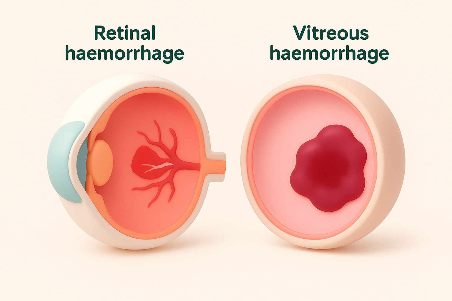
Bleeding can also occur inside the vitreous, the clear gel that fills most of the interior of the eye. Because the vitreous sits in the direct line of sight between the lens and the retina, even a small amount of blood can scatter light and cause significant vision disruption. In cases of haemorrhage behind eye diabetes, both types can occur together, particularly when fragile new blood vessels rupture and bleed into multiple layers at once.
If blood enters the vitreous cavity, it can block light from reaching the retina entirely, causing vision to drop suddenly and without warning.
Why location matters for your prognosis
Where the bleeding occurs and how much of the retina is affected directly shapes the outlook for your vision. A small retinal haemorrhage away from the central macula may resolve on its own with careful monitoring. Bleeding that involves the macula or produces a large vitreous haemorrhage, however, requires prompt intervention to prevent permanent damage to the cells that give you sharp, detailed sight.
Why diabetes can cause eye bleeding
High blood sugar damages small blood vessels throughout the body over time, and the tiny vessels that supply your retina are particularly vulnerable. When these vessels are repeatedly exposed to elevated glucose levels, their walls weaken, swell, and leak, allowing fluid and blood to spill into the surrounding retinal tissue.
The role of diabetic retinopathy
Diabetic retinopathy is the underlying condition that sets the stage for a haemorrhage behind eye diabetes. In its early stage, known as non-proliferative diabetic retinopathy, weakened vessel walls develop small bulges called microaneurysms. These microaneurysms can rupture and bleed into the retina, sometimes without causing any immediate symptoms you would notice.
The longer you live with diabetes, and the less consistently your blood sugar is controlled, the greater the likelihood that this damage will progress. Poor management of blood pressure and cholesterol alongside blood sugar significantly accelerates the rate of retinal vessel damage.
When new blood vessels become the problem
As retinopathy advances, the retina responds to its reduced blood supply by triggering the growth of new blood vessels, a process called neovascularisation. These new vessels are structurally abnormal and far more fragile than healthy ones. They grow across the retinal surface and into the vitreous cavity, where they can rupture with little warning and cause a sudden, large bleed.
This stage, proliferative diabetic retinopathy, carries the highest risk of severe and rapid vision loss from haemorrhage.
Symptoms and when to seek urgent care
The symptoms of a haemorrhage behind eye diabetes vary depending on where the bleeding occurs and how much blood is involved. Some bleeds develop gradually and produce subtle changes you might dismiss, while others cause a dramatic and sudden drop in vision that is impossible to ignore.
What you might notice in your vision
Small retinal haemorrhages can be completely symptom-free, which is why regular diabetic eye screening matters so much even when your vision feels normal. Larger bleeds into the vitreous typically cause more obvious warning signs that appear quickly.
Common symptoms include:
- Sudden floaters or dark spots moving across your field of view
- A red or reddish tint to your vision
- Blurry or hazy vision that comes on without explanation
- Shadows or dark patches that block part of your sight
- A sudden and significant drop in overall visual clarity
When to treat it as an emergency
Any sudden change in your vision requires prompt medical attention, particularly if you have a known diagnosis of diabetic retinopathy or have been managing diabetes for a number of years. Do not wait to see if the symptoms resolve on their own.
A sudden loss of vision in one eye is a medical emergency and you should contact an eye specialist or present to an emergency department the same day.
If you notice multiple new floaters, a shadow spreading across your vision, or a rapid reduction in sight, contact your eye specialist immediately.
How doctors diagnose it
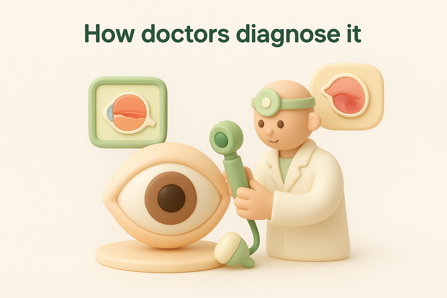
When you see an eye specialist with suspected diabetic eye bleeding, the diagnostic process combines clinical examination with imaging to build a clear picture of what is happening inside your eye. The aim is to confirm the location and extent of any haemorrhage behind eye diabetes, assess which stage of retinopathy you have reached, and identify what treatment is needed.
What the examination involves
Your specialist will start with a dilated fundus examination, using drops to widen your pupil so they can examine the retina, vitreous, and optic nerve directly. This gives an immediate view of any visible bleeding, microaneurysms, or abnormal new vessel growth. For cases where blood in the vitreous blocks the direct view, an ultrasound scan of the eye (B-scan) allows the doctor to assess the retina behind the blood without relying on optical clarity.
Optical coherence tomography (OCT) provides detailed cross-sectional images of the retinal layers, helping your doctor detect swelling or bleeding that is not visible through standard examination alone.
Fluorescein angiography may also be used, where a dye injected into your bloodstream highlights how blood moves through the retinal vessels. This test pinpoints areas of leakage, blocked circulation, and abnormal new vessel growth with precision, giving your specialist the information needed to plan the most appropriate treatment for your specific situation.
Treatment options and what to expect
The right treatment for a hemorrhage behind eye diabetes depends on how much blood is present, whether the retina is intact, and how far your retinopathy has progressed. Your specialist will match the approach to your specific situation, balancing the urgency of the bleed with the safest path to preserving your sight.
Laser and injection therapy
Laser photocoagulation treats the abnormal blood vessels that caused the bleed, sealing leaking vessels and reducing the stimulus for further neovascularisation. Intravitreal injections of anti-VEGF medication are often used alongside or instead of laser, directly targeting the chemical signal that drives new vessel growth. Both treatments are performed in a clinical setting and do not require general anaesthesia.
Anti-VEGF injections have significantly improved outcomes for proliferative diabetic retinopathy, reducing the risk of recurrent haemorrhage when given consistently over time.
Surgery for large vitreous bleeds
When blood fills the vitreous and blocks the view of the retina, vitrectomy surgery becomes necessary. Your surgeon removes the blood-filled vitreous gel and replaces it with a clear solution, restoring the light path to the retina. If a retinal detachment has occurred alongside the bleed, your surgeon addresses this at the same time, which is critical for recovering useful vision.
Recovery after vitrectomy typically takes several weeks, and your final visual outcome depends on how much retinal damage occurred before treatment, which is why acting quickly on any sudden vision change matters so much.

What to do now
A hemorrhage behind eye diabetes is not something to monitor from the sidelines. If your vision has changed suddenly, if you have been living with diabetes for years without a recent eye check, or if you already have a diagnosis of diabetic retinopathy, now is the right time to act. Early treatment consistently produces better outcomes than waiting until the damage is severe.
Your first step is to book a comprehensive diabetic eye examination with a specialist who understands both the medical and surgical sides of retinal disease. At Bright Sight Eye Specialists in Randwick, Dr Rahul Dubey assesses and manages diabetic eye disease across all stages, from early monitoring through to vitrectomy surgery for serious vitreous haemorrhage. The practice also offers intravitreal injections and laser treatment in a patient-centred setting with transparent pricing.
Do not wait for symptoms to worsen. Book an appointment with Dr Rahul Dubey and take the next step toward protecting your sight.