


A full thickness macular hole is exactly what it sounds like. It’s a complete break in your macula, the tiny central area of your retina responsible for sharp, detailed vision. When this happens, the delicate layers of tissue that capture light and send visual signals to your brain develop a gap. Think of it like a hole in the fabric of your central vision.
This isn’t just a minor inconvenience. A full thickness macular hole affects your ability to read, recognise faces, drive, and perform tasks that require fine detail. The condition typically develops gradually, often starting with mild distortion before progressing to a noticeable blind spot in the centre of your vision.
The good news is that modern surgical techniques can repair most macular holes successfully, especially when caught early. This article explains how these holes form and progress through different stages, what symptoms to watch for, and how ophthalmologists use OCT imaging to diagnose and plan treatment. You’ll also learn about vitrectomy surgery, recovery expectations, and the factors that influence your prognosis. Understanding the urgency of treatment can make all the difference to your visual outcome.
Why a full thickness macular hole is serious
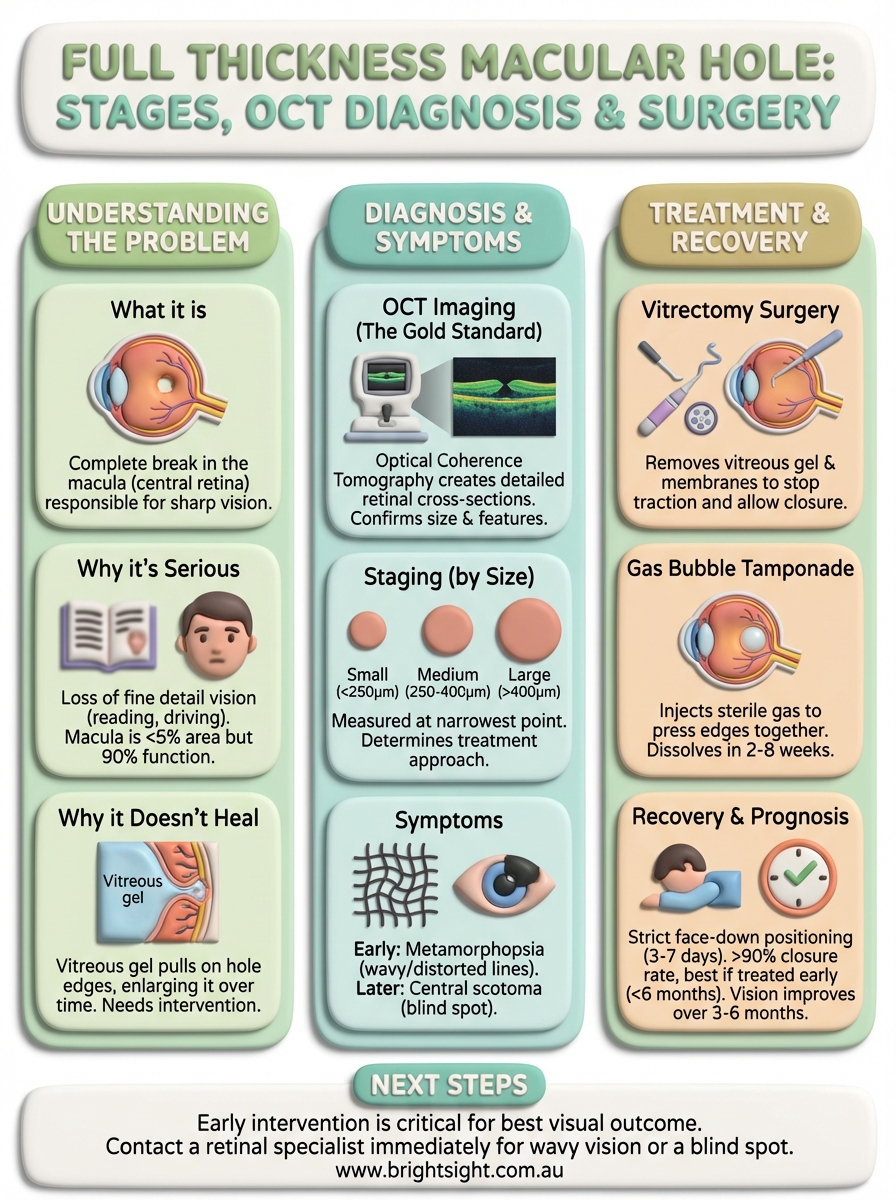
Your macula occupies less than 5% of your retina, yet it handles 90% of your detailed vision. When a full thickness macular hole develops, you lose the ability to see fine details in your central field of view. This creates immediate problems with reading, driving, recognising faces, and any task requiring precision. Unlike peripheral vision loss, which often goes unnoticed, central vision loss dramatically impacts your independence and quality of life.

Why macular holes don’t heal themselves
A full thickness macular hole rarely closes without intervention. The vitreous gel in your eye continues to pull on the edges of the hole, often making it larger over time. Left untreated, the hole typically enlarges and your central vision worsens progressively. The longer you wait, the more photoreceptor cells you lose around the damaged area.
Early treatment gives you the best chance of recovering useful central vision and preventing permanent damage.
The time-sensitive nature of treatment
Surgery becomes less effective as the hole ages. Holes present for less than six months respond far better to vitrectomy surgery than chronic holes that have existed for years. Your retinal specialist will assess how long the hole has been present, as this directly affects your surgical prognosis and expected visual recovery.
How to diagnose and stage a full thickness macular hole
Your ophthalmologist can’t diagnose a full thickness macular hole by looking at your retina alone. The diagnosis requires optical coherence tomography (OCT), a sophisticated imaging technique that captures cross-sectional views of your retina at microscopic resolution. This scan creates detailed images showing all the retinal layers, making it easy to identify breaks, measure hole size, and assess surrounding tissue damage. The entire scan takes less than five minutes and requires no injections or uncomfortable procedures.
OCT imaging reveals the complete picture
OCT technology works like ultrasound for your eye, using light waves instead of sound to build precise images. Your specialist examines these scans to confirm whether you have a true full thickness macular hole or a similar condition like a lamellar hole or pseudohole. The scan shows the exact dimensions of the hole, including its diameter at the narrowest point and whether the internal limiting membrane has separated from the retina. This information directly influences treatment planning and helps predict your surgical outcome.

OCT imaging is the gold standard for diagnosing macular holes and monitoring them over time.
The staging system explained
Ophthalmologists classify macular holes by size and anatomical features visible on OCT. Small holes measure 250 micrometres or less at their narrowest diameter. Medium holes range from 250 to 400 micrometres, whilst large holes exceed 400 micrometres. Your specialist also notes whether you have vitreomacular traction, where the vitreous gel still pulls on the retinal surface around the hole.
Stage classification helps determine your treatment approach and likely prognosis. Smaller, newer holes generally respond better to surgery than larger, chronic ones. Your ophthalmologist may also identify features like cystoid oedema in the surrounding retina or changes at the base of the hole. These details affect surgical technique decisions, such as whether to use a special dye to peel the internal limiting membrane or whether an inverted flap technique might improve your chances of hole closure.
Diagnostic accuracy matters because treatment timing significantly impacts outcomes. Your specialist uses OCT measurements to track any progression if you’re monitoring a smaller hole or to plan the most effective surgical approach when intervention becomes necessary.
Symptoms and early warning signs
The earliest symptom of a full thickness macular hole is metamorphopsia, which means straight lines appear wavy or distorted. You might notice this when reading, where text lines curve or bend unexpectedly. Many patients first spot the problem when they cover their good eye and realise something looks wrong in their affected eye. The distortion often appears subtle initially, making it easy to dismiss as tiredness or normal changes.
What you’ll notice first
Wavy vision typically precedes any blind spot by weeks or months. You see this distortion most clearly when looking at objects with straight edges like doorframes, window frames, or printed text. Your brain can’t compensate for the missing tissue at your macula, so you perceive these geometric distortions directly. Some people describe it as looking through slightly rippled water.

When distortion becomes a blind spot
As the hole enlarges, you develop a central scotoma, a grey or dark patch in your direct line of sight. This blind spot makes reading nearly impossible without turning your head to use peripheral vision. You lose the ability to see fine details like facial features or small print. Tasks requiring precision become frustrating or dangerous.
If you notice persistent wavy vision or a developing blind spot, contact your ophthalmologist immediately, as early treatment significantly improves outcomes.
Treatment options and macular hole surgery
Surgical repair remains the only effective treatment for a full thickness macular hole. Whilst some early-stage holes occasionally close spontaneously, any confirmed full thickness defect requires vitrectomy surgery to restore the retinal layers and improve your vision. Your retinal specialist performs this microsurgical procedure under local anaesthetic, usually as day surgery at a hospital or surgical centre. The operation takes about one hour, and you go home the same day with specific positioning instructions.
Vitrectomy: the gold standard procedure
Vitrectomy removes the vitreous gel from your eye to eliminate traction on the macula. Your surgeon makes three tiny incisions in the white of your eye, each less than 1 millimetre wide. Through these openings, microscopic instruments remove the vitreous and carefully peel away the internal limiting membrane around the hole. This membrane removal stops any remaining traction and creates conditions for the hole to close.

Modern techniques often use brilliant blue dye to stain the internal limiting membrane, making it visible during surgery. Some surgeons employ an inverted flap technique for larger holes, where they fold the membrane back over the defect to act as a scaffold for healing. Your specialist chooses the technique based on your hole size and OCT characteristics.
Gas bubble tamponade and its role
After removing the vitreous and membranes, your surgeon injects sterile gas into your eye. This gas bubble presses against the macula, holding the edges of the hole together whilst your retina heals. The bubble gradually dissolves over two to eight weeks, depending on which gas type your surgeon uses. Sulfur hexafluoride typically lasts two weeks, whilst longer-acting gases remain for up to two months.
The gas bubble creates the pressure needed for your macular hole to close, making it essential to maintain proper head positioning after surgery.
You cannot fly or travel to high altitudes whilst the gas remains in your eye, as pressure changes cause the bubble to expand dangerously. Your surgeon explains exactly how long positioning restrictions apply and when you can resume normal activities.
Recovery, prognosis and protecting your vision
Recovery from macular hole surgery requires strict face-down positioning for three to seven days after your operation. This positioning keeps the gas bubble pressed against your macula, holding the hole closed whilst healing occurs. Your surgeon provides specific instructions about how many hours per day you need to maintain this position, and you’ll receive equipment like a face-down chair or mirror system to make compliance easier. Most patients find the positioning challenging but manageable, and the temporary discomfort proves worthwhile when their vision improves.
What to expect during recovery
Your vision remains severely blurred whilst the gas bubble fills your eye. As the gas gradually dissolves, you see it as a moving line that drops lower each day. Full visual recovery takes three to six months after the gas completely disappears. During this time, you cannot drive, fly, or undergo general anaesthesia involving nitrous oxide, as these activities risk dangerous pressure changes in your eye.
Success rates for full thickness macular hole surgery
Modern vitrectomy techniques achieve closure rates exceeding 90% for most macular holes. Smaller holes caught early show success rates approaching 95%, whilst larger chronic holes have slightly lower closure rates. Even when the hole closes successfully, your final vision depends on hole size, duration, and baseline retinal health. Many patients regain reading vision, though complete restoration to pre-hole acuity remains uncommon.
Surgery performed within six months of hole formation typically delivers the best visual outcomes.
Next steps
If you experience wavy vision, distorted straight lines, or a developing blind spot, contact a retinal specialist immediately. Full thickness macular hole treatment success depends heavily on early intervention, and waiting often reduces your chances of recovering useful central vision. The longer you delay, the larger the hole typically becomes and the more photoreceptor damage you accumulate around your macula.
Regular eye examinations help detect macular problems before symptoms become severe. Your ophthalmologist uses OCT imaging to identify retinal changes before they affect your daily activities. If you’re over 60, have high myopia, or notice any vision changes in the Eastern Suburbs of Sydney, schedule a consultation at Bright Sight Eye Specialists for comprehensive retinal assessment. Dr Rahul Dubey provides advanced diagnostic imaging and surgical expertise to preserve your vision.