


Those tiny shadows drifting across your field of view, specks, threads, or cobweb-like shapes that seem to move when you try to focus on them. If you’ve noticed floaters in your vision, you’re far from alone. Most people experience them at some point, and while they’re often harmless, they can sometimes signal something that needs prompt attention.
At Bright Sight Eye Specialists in Randwick, Dr Rahul Dubey and Dr Capucine Odouard regularly help patients understand what’s happening inside their eyes. Knowing the difference between a normal age-related change and a warning sign can make all the difference to preserving your sight.
This article explains what causes floaters, the symptoms to watch for, and when you should seek urgent care. Whether you’ve just noticed your first floater or have lived with them for years, you’ll find clear, practical guidance to help you decide on your next steps.
What floaters are and what they look like
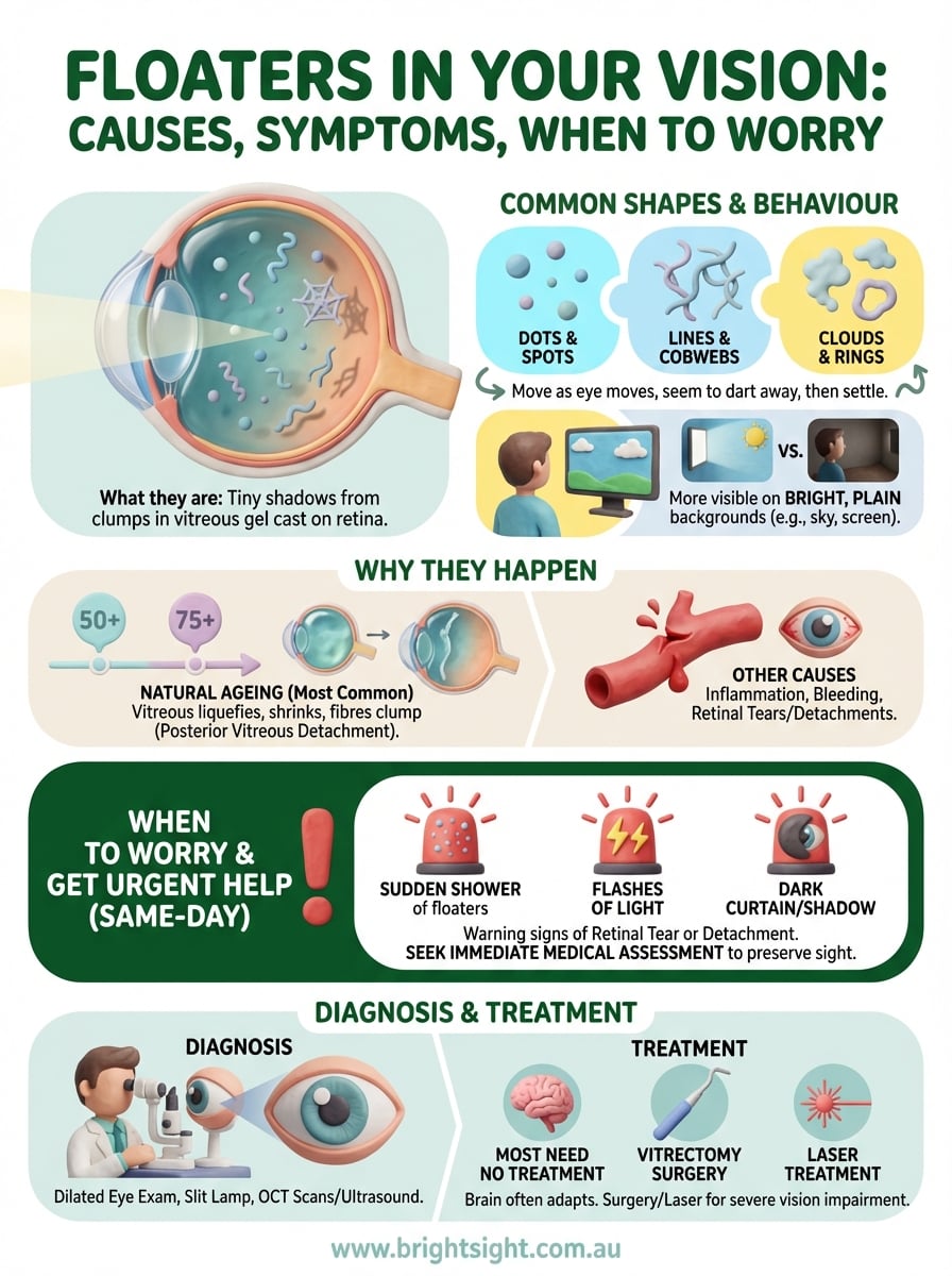
Floaters are tiny shadows cast onto your retina by small clumps of gel or cells inside the vitreous, the clear, jelly-like substance that fills the space between your lens and retina. You see them because light passing through your eye projects these internal structures onto the light-sensitive tissue at the back of your eye. They appear to float across your vision because they’re suspended in the vitreous gel and move as your eye moves.
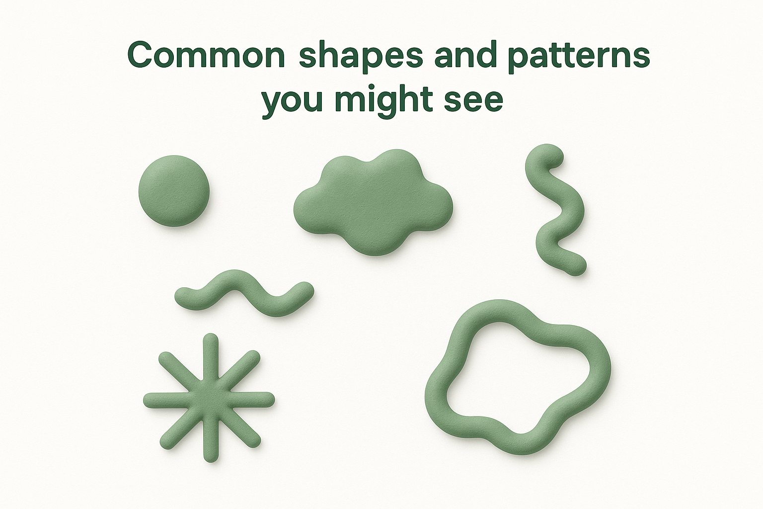
Common shapes and patterns you might see
You might notice floaters in your vision appearing as dots, circles, lines, or clouds. Some people describe them as looking like flies buzzing around, while others see cobwebs, squiggly lines, or thread-like strands. The shapes depend on the size and position of the clumps inside your eye. Larger floaters tend to cast more defined shadows, while smaller ones create fainter, more subtle spots.
Your floaters may look grey, black, or even slightly translucent, and they’re usually most noticeable against bright, plain backgrounds like a clear sky, a white wall, or a computer screen. Reading or looking at something evenly lit often makes them more obvious. In dim lighting, you might not notice them at all because the contrast between the floater and the background reduces.
Floaters become more visible when you look at uniform, bright surfaces because the contrast makes their shadows stand out against the background.
How floaters move and behave
Floaters drift slowly through your field of view, and they seem to dart away when you try to look at them directly. This happens because they move with the vitreous gel inside your eye. When you shift your gaze quickly, the vitreous swirls slightly, carrying the floaters with it. They settle gradually once your eye stops moving, though they never stay completely still.
You can’t control where floaters go or make them disappear by blinking or rubbing your eyes. They’re inside your eye, not on the surface, so external actions won’t shift them. Over time, your brain often learns to ignore floaters that remain stable, making them less bothersome even though they’re still there. Some floaters fade or move out of your central vision naturally, while others remain visible for years.
Why floaters happen in the eye
The vitreous inside your eye changes as you age, and this natural process accounts for most floaters people notice. Your vitreous starts out firm and gel-like, keeping its shape and staying transparent. Over time, it begins to liquefy and shrink, pulling away from the retina in a process called posterior vitreous detachment. As the vitreous gel breaks down, tiny fibres clump together, casting shadows that appear as floaters in your vision.
The natural ageing process and vitreous changes
Most people develop floaters between the ages of 50 and 75, though they can appear earlier, particularly if you’re short-sighted or have had eye surgery. The vitreous contains collagen fibres that hold its structure together. When these fibres degenerate, they cluster into strands or lumps that drift through the liquid part of the vitreous. This process happens gradually, and the floaters you see today might have formed weeks or months ago before drifting into your line of sight.
Posterior vitreous detachment is a normal part of ageing and affects nearly everyone eventually, though the timing varies between individuals.
Other causes beyond normal ageing
Inflammation inside your eye can introduce white blood cells and debris into the vitreous, creating floaters that appear suddenly. Bleeding from damaged blood vessels, often linked to diabetes or injury, releases red blood cells that show up as numerous small dots or hazy clouds. Retinal tears or detachments can also release pigment cells into the vitreous, producing new floaters alongside flashes of light. These non-age-related causes require prompt medical assessment to prevent permanent vision loss.
When to worry and get urgent help
Most floaters in your vision develop slowly and pose no threat to your sight, but certain warning signs demand immediate medical attention. If you notice a sudden increase in floaters, especially accompanied by flashes of light or a shadow spreading across your vision, you need to see an eye specialist the same day. These symptoms suggest your retina may have torn or started to detach, and prompt treatment can prevent permanent vision loss.
Emergency symptoms that need same-day attention
A sudden shower of new floaters appearing within hours or days requires urgent assessment, particularly if you also see bright flashes of light in your peripheral vision. These flashes often look like lightning streaks or camera flashes and indicate that your vitreous is tugging on your retina. A dark curtain or shadow moving across any part of your visual field signals that your retina may already be detaching, making immediate intervention critical.
Retinal tears and detachments progress rapidly, and treatment within hours or days significantly improves the chances of preserving your sight.
Why immediate assessment matters
Retinal tears can progress to full detachment within days if left untreated, and once the central part of your retina detaches, your vision may not recover fully even with surgery. Dr Dubey and Dr Odouard at Bright Sight Eye Specialists assess urgent cases promptly, using advanced imaging to examine your retina thoroughly. Acting quickly gives your specialist the best opportunity to seal tears with laser treatment or prevent detachment from spreading, protecting your long-term vision.
How eye specialists diagnose the cause
Your eye specialist starts by asking detailed questions about when you first noticed floaters in your vision, how many appeared, and whether you’ve experienced flashes of light or vision loss. This information helps them determine whether your floaters stem from normal ageing or require urgent intervention. They’ll also review your medical history, checking for conditions like diabetes, high blood pressure, or previous eye surgery that might increase your risk of complications.
Examination techniques used during assessment
Dr Dubey and Dr Odouard perform a dilated eye examination to see inside your eye clearly. They place drops in your eyes to widen your pupils, allowing them to examine your vitreous and retina thoroughly. Using a slit lamp microscope and specialised lenses, they can see the vitreous gel, any clumps causing floaters, and the entire surface of your retina.
The dilated examination takes about 20 minutes, and your vision remains blurry for several hours afterwards, so you’ll need someone to drive you home. Your specialist checks for retinal tears, detachments, bleeding, or inflammation that might explain new or concerning floaters. They examine the peripheral retina carefully because tears often occur in the outer areas that you can’t see directly.
Dilating your pupils allows your specialist to examine areas of your retina that remain hidden during a standard eye test.
Imaging tests for detailed assessment
Advanced imaging provides additional information when your specialist needs a closer look. Optical coherence tomography (OCT) scans create detailed cross-sectional images of your retina, revealing any subtle changes that might cause symptoms. Ultrasound imaging helps assess your vitreous and retina if bleeding or dense floaters block the view during examination.
Treatment options and what helps day to day
Most floaters require no medical treatment because they stem from normal ageing and pose no threat to your sight. Your brain often adapts over time, learning to ignore stable floaters so they become less noticeable in daily life. However, when floaters result from bleeding, inflammation, or retinal problems, your specialist treats the underlying condition rather than the floaters themselves. Surgery becomes an option only in rare cases where floaters severely impair your vision and affect your quality of life.
When medical treatment becomes necessary
Vitrectomy surgery removes the vitreous gel containing floaters and replaces it with a clear saline solution, eliminating the shadows from your vision. Dr Dubey performs this procedure for patients whose floaters in your vision significantly interfere with daily activities despite conservative management. The surgery carries risks including cataract formation, retinal detachment, and infection, so your specialist reserves it for severe cases. Laser treatment can sometimes break up large floaters, though this approach works for specific types and positions only.
Vitrectomy effectively removes floaters but remains reserved for severe cases due to the surgical risks involved.
Practical strategies for day-to-day comfort
Moving your eyes up and down shifts floaters temporarily out of your central vision, providing brief relief when they obscure something important. Adjusting lighting in your workspace reduces the contrast between floaters and backgrounds, making them less prominent during computer work or reading. Wearing sunglasses outdoors helps because bright sky makes floaters more visible, and reducing glare improves comfort. Your floaters may drift to the periphery naturally over months or years, becoming less bothersome without intervention.
Next steps for peace of mind
Understanding floaters in your vision helps you distinguish between normal age-related changes and symptoms requiring urgent care. Most floaters develop gradually and pose no risk to your sight, remaining stable for months or years without intervention. However, sudden increases accompanied by flashes or shadows demand same-day assessment to rule out retinal tears or detachment. Knowing these warning signs puts you in control of protecting your vision.
If you’re concerned about new floaters or experiencing any worrying symptoms, Dr Dubey and Dr Odouard at Bright Sight Eye Specialists provide thorough assessments at their Randwick practice. They use advanced imaging techniques to examine your retina completely, giving you clear answers about what’s happening inside your eyes. Their transparent approach means you’ll understand your diagnosis and treatment options fully before making any decisions. Book a consultation to discuss your symptoms and receive expert guidance tailored to your individual situation and concerns.