


You notice small shapes drifting across your vision when you look at a bright surface or the sky. These floaters and spots in vision appear as dark specks, threads, or cobweb-like shadows that seem to float with your eye movements. They’re actually tiny clumps of cells or protein inside the clear gel that fills your eyeball. Most people experience them at some point, particularly as they age. While usually harmless, they can sometimes indicate a more serious condition affecting your retina.
This article explains why these spots appear in your vision, how to distinguish normal floaters from warning signs that require immediate attention, and what medical treatments exist for persistent cases. You’ll discover the specific risk factors that increase your likelihood of developing floaters, what to expect during a comprehensive retinal examination, and practical steps to protect your vision over time. Understanding when floaters are harmless and when they signal danger helps you make confident decisions about seeking professional eye care.
Why floating spots appear in your field of vision
Your eye contains a clear, jelly-like substance called the vitreous that fills the space between your lens and retina. This gel consists mainly of water and collagen fibres arranged in a precise structure that keeps your eye’s shape and allows light to pass through cleanly. When these fibres clump together or the gel begins to liquefy, they cast shadows on your retina, creating the floaters and spots in vision that drift across your sight. The process happens gradually in most cases, though sudden changes can occur.
The role of vitreous gel changes
As you age, the vitreous gel naturally shrinks and becomes more liquid, pulling away from the back wall of your eye in a process called posterior vitreous detachment. This separation typically begins after age 50, though it can happen earlier in people who are near-sighted or have experienced eye trauma. The collagen fibres that once spread evenly throughout the gel now cluster together, forming the distinct shapes you observe floating in your vision. These clumps move when you shift your gaze because they’re suspended in the liquefied vitreous.
The vitreous gel occupies about two-thirds of your eye’s volume and maintains its clear structure through a delicate balance of proteins and water molecules.
Your brain eventually learns to ignore stable floaters that remain consistent over time, making them less noticeable during daily activities. However, the initial appearance of new floaters often causes concern because they seem prominent and distracting. Most people notice them most clearly against bright, uniform backgrounds like a white wall, blue sky, or computer screen.
Common triggers beyond ageing
Several conditions accelerate vitreous changes beyond normal ageing processes. Eye inflammation (uveitis) causes white blood cells and inflammatory debris to float in the vitreous, creating spots that appear suddenly. Bleeding inside the eye from diabetes, torn blood vessels, or injury releases red blood cells into the vitreous gel, producing numerous dark floaters that can significantly obscure your vision. Near-sightedness stretches your eyeball, creating mechanical stress on the vitreous that promotes earlier gel liquefaction and detachment. Eye surgery, particularly cataract removal, also disrupts the vitreous structure and frequently leads to new floaters appearing in the months following the procedure.
How to distinguish normal floaters from emergencies
Most floaters and spots in vision pose no threat to your sight and represent normal vitreous changes. You can safely monitor these common floaters that appear gradually, move predictably with your eye movements, and don’t interfere significantly with your vision. However, certain patterns signal retinal damage that requires immediate medical attention to prevent permanent vision loss. Knowing the difference between these two situations helps you respond appropriately when new floaters appear.
Warning signs requiring immediate attention
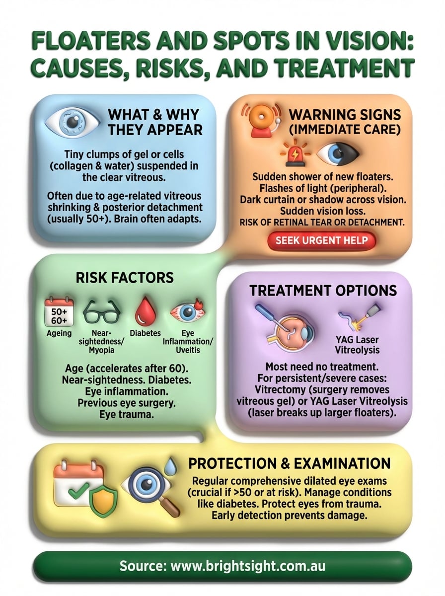
Sudden onset of numerous new floaters appearing together within hours or days indicates potential retinal tears or detachment. You should seek emergency eye care if you notice flashes of light accompanying the floaters, particularly in your peripheral vision, as this suggests your retina is being tugged or torn. A dark curtain or shadow spreading across your vision from any direction represents advanced retinal detachment and demands immediate surgical intervention. Any sudden vision loss in one eye, even if accompanied by just a few new floaters, warrants urgent professional examination.
Retinal detachment progresses rapidly without treatment, and delays of even 24 to 48 hours can result in irreversible vision damage.
Characteristics of harmless floaters
Normal floaters develop slowly over months or years and remain relatively stable in number and appearance. They appear as translucent or grey shapes rather than solid black objects blocking your vision. Benign floaters move smoothly when you shift your gaze and settle back into predictable positions when your eyes stop moving. You’ll notice them most against bright backgrounds but they don’t obscure your central vision or prevent you from reading or recognising faces.
Risk factors that increase the likelihood of spots
Your chances of developing floaters and spots in vision increase significantly based on specific biological and environmental factors. Understanding these risk factors helps you recognise whether you belong to a higher-risk group that requires more vigilant monitoring. Some factors you can’t control, like your age or genetic predisposition, while others relate to existing health conditions or lifestyle choices that affect your eye structure.
Age-related vitreous changes
People over 50 years old face substantially higher risks of developing floaters because the vitreous gel naturally liquefies and separates from the retina during this stage of life. Your risk accelerates after age 60, when posterior vitreous detachment occurs in approximately 50% of individuals. By age 80, nearly all people experience some degree of vitreous separation and the accompanying floaters. The process happens earlier in women than men, though researchers haven’t definitively explained why this gender difference exists.
Nearly 70% of people develop noticeable floaters by age 70, making it one of the most common age-related eye changes.
Medical conditions affecting the vitreous
Near-sightedness (myopia) ranks among the strongest predictors of early floater development because elongated eyeballs create mechanical stress on the vitreous gel. Diabetes damages blood vessels throughout your body, including those in your eyes, leading to bleeding and inflammatory debris in the vitreous. Eye inflammation conditions like uveitis release white blood cells and protein into the vitreous fluid.
Previous eye surgery, particularly cataract removal, disrupts the normal vitreous architecture and frequently produces new floaters within months of the procedure. Eye injuries that cause trauma to the vitreous cavity also increase your risk substantially, regardless of your age at the time of injury.
Medical treatments for persistent eye floaters
Most floaters and spots in vision don’t require treatment because your brain gradually adapts to ignore them. However, when floaters significantly impair your daily activities or result from serious conditions like vitreous haemorrhage, your ophthalmologist may recommend intervention. Two primary treatments exist for persistent floaters that resist natural resolution, each with distinct advantages and limitations based on your specific situation.
Vitrectomy surgery for severe cases
Vitrectomy removes the vitreous gel containing the floaters and replaces it with a clear saline solution that permanently eliminates the shadows in your vision. Surgeons perform this procedure under local anaesthesia, making tiny incisions in your eye to access and extract the problematic vitreous. The operation effectively resolves severe floaters but carries risks including retinal detachment, cataract formation, and infection. Your surgeon will only recommend vitrectomy when floaters substantially reduce your quality of life and no safer alternatives exist.

Vitrectomy achieves success rates exceeding 90% for floater removal, though most ophthalmologists reserve it for cases where vision impairment justifies the surgical risks.
Recovery typically requires several weeks during which you must avoid strenuous activities and protect your eye from injury.
Laser vitreolysis as a less invasive option
YAG laser vitreolysis targets individual floaters with precise laser energy that breaks them into smaller, less noticeable pieces. This office-based procedure requires no incisions and causes minimal discomfort, making it attractive for people seeking non-surgical solutions. However, laser treatment works best on larger, well-defined floaters positioned away from your retina and lens. Smaller, diffuse floaters often remain too difficult to target safely with current laser technology. Your ophthalmologist evaluates your floater characteristics to determine whether laser treatment offers realistic benefits in your case.
What to expect during a professional retinal exam
Your ophthalmologist performs a comprehensive retinal examination to identify the source of floaters and spots in vision and assess whether they indicate retinal damage. The procedure typically takes 30 to 45 minutes and involves several diagnostic steps that allow your specialist to view the interior structures of your eye in detail. You’ll receive dilating drops that widen your pupils, making it possible to examine your retina, vitreous, and optic nerve thoroughly. Understanding what happens during this examination helps reduce anxiety and ensures you arrive prepared.
Initial diagnostic procedures
Your examination begins with pupil dilation using medicated eye drops that take approximately 20 to 30 minutes to work fully. These drops temporarily blur your near vision and increase light sensitivity, so you should bring sunglasses and arrange transport home if possible. Once your pupils dilate, your ophthalmologist uses a specialised microscope called a slit lamp combined with condensing lenses to examine your retina’s surface, blood vessels, and surrounding structures.
The specialist looks specifically for retinal tears, detachments, or areas of thinning that might produce the floaters you’re experiencing. They’ll ask you to look in different directions whilst holding your eye steady with gentle pressure, allowing them to view your peripheral retina where tears most commonly occur. Your doctor may perform optical coherence tomography (OCT) to capture detailed cross-sectional images of your retina and vitreous interface.
Dilated eye examinations detect 90% of sight-threatening retinal conditions before symptoms worsen, making them essential for anyone experiencing new or changing floaters.
This painless scan provides precise measurements of any structural changes affecting your vision.
Protecting your long-term vision
You protect your vision by scheduling regular comprehensive eye examinations that detect retinal problems before they cause permanent damage. Annual dilated eye exams become essential once you reach age 50 or develop risk factors like near-sightedness or diabetes. Early detection of conditions causing floaters and spots in vision allows your ophthalmologist to intervene before serious complications develop. You should contact your eye specialist immediately whenever you notice sudden changes in your floaters, flashes of light, or vision loss in any part of your visual field.
Managing underlying health conditions like diabetes and high blood pressure reduces your risk of vitreous haemorrhage and other sight-threatening complications. Protecting your eyes from trauma during sports or work activities prevents injury-related floaters and retinal damage. Your retinal health depends on prompt professional attention when warning signs appear. Bright Sight Eye Specialists provides comprehensive retinal examinations and advanced treatment options in Sydney’s Eastern Suburbs, helping you maintain clear vision through expert, patient-centred care.