


You’ve noticed a small shadow drifting across your vision, maybe a speck, a thread, or a cobweb-like shape, and it’s still there days or weeks later. When a floater in eye won’t go away, it’s natural to wonder whether something more serious is happening. Most people experience floaters at some point, particularly after the age of 50, but persistent floaters that don’t fade can feel distracting and, understandably, a bit worrying.
The good news is that the majority of floaters are harmless, caused by age-related changes inside the eye. However, in some cases they can signal conditions like retinal tears, retinal detachment, or vitreous haemorrhage, all of which need prompt attention. Knowing the difference matters, and that’s where a thorough eye examination becomes essential.
At Bright Sight Eye Specialists in Randwick, Dr Rahul Dubey and Dr Capucine Odouard regularly assess and treat patients dealing with persistent floaters, including those requiring retinal and macular surgery. This article explains why floaters develop, when they’re a red flag, and what treatment options are available, from observation through to surgical intervention, so you can make an informed decision about your eye health.
Why a floater can stick around
Most people expect floaters to disappear within a few days, so when they don’t, the natural question is why. The vitreous, a clear gel filling the inside of your eye, is the source of virtually every floater you see. Understanding what happens to this gel over time explains why so many people end up dealing with a floater in eye won’t go away situations that last for months or even years.
The vitreous gel and what changes inside your eye
Your eye is filled from the inside with a gel called the vitreous humour, which is about 99% water held together by fine collagen fibres. When you’re young, this gel is uniform and transparent, so light passes through it cleanly. As you age, the collagen fibres begin to clump together, creating small opaque strands and particles that cast shadows on your retina. Those shadows are what you perceive as floaters.

The process accelerates from around age 50, though it can happen earlier in people who are shortsighted (myopic), have had eye surgery, or experienced eye inflammation. Over time, the vitreous gel can also shrink and pull away from the retina at the back of the eye, an event called posterior vitreous detachment (PVD). PVD is the single most common cause of new floaters in adults, and while it sounds alarming, it is usually a normal part of ageing.
Once a PVD is complete, floaters often become less noticeable over months, but the collagen clumps causing them rarely disappear entirely.
Why floaters don’t simply dissolve
When the collagen fibres clump together, your body has no reliable mechanism to break them down or reabsorb them. Unlike a bruise that fades because the immune system clears damaged tissue, the vitreous interior sits in a largely avascular environment, meaning it has very limited blood supply. Without circulation reaching the clumps, the material just sits there, suspended in what remains of the gel.
Your brain does try to help. Over time, it learns to ignore the shadow cast by a stable floater, which is why many people find they stop noticing a floater they’ve had for a long time. This is called neural adaptation. However, if the floater is large, sits close to the centre of your vision, or if your visual demands are high, such as reading fine print or working on screens for long hours, adaptation may never be complete.
Conditions that cause floaters to linger or worsen
Not all persistent floaters come from normal ageing. Several medical conditions can introduce floaters or make existing ones more significant, and these tend to stick around because the underlying problem is still active. Understanding these causes helps you assess whether your floaters fall into the routine category or need more urgent investigation.
Common causes beyond normal ageing include:
- Retinal tear or detachment – a break in the retina can release cells or blood into the vitreous
- Vitreous haemorrhage – bleeding into the vitreous from fragile or damaged blood vessels
- Uveitis – inflammation inside the eye caused by infection, autoimmune disease, or injury
- Diabetic retinopathy – abnormal new blood vessels that bleed into the vitreous
- High myopia – a very long eyeball puts mechanical stress on the vitreous, accelerating gel breakdown
Each of these conditions requires a different management approach, which is why a professional eye examination is essential when floaters persist or change suddenly. Floaters from inflammation or bleeding are often denser and more disruptive than those from a straightforward PVD, and they will not settle until the cause is addressed.
What floaters look like and what they are not
Before you can assess whether your floater in eye won’t go away situation is worth worrying about, it helps to understand what you’re actually seeing. Floaters are shadows cast on your retina by particles inside the vitreous gel, not marks on your eye’s surface or lens. They move because the gel is fluid, so they drift when your eye moves and slide a little further when you stop, which is one of their most recognisable characteristics.
The shapes and patterns you typically see
Floaters appear in a wide range of forms, and knowing the common ones can help you describe them accurately to your eye specialist. The most frequent shapes include:
- Dots or specks – small, round shadows that drift across your central vision
- Threads or strands – thin wiggly lines that move with your eye
- Cobwebs or nets – a broader, mesh-like shadow, often associated with posterior vitreous detachment
- Rings – a circular floater known as a Weiss ring, which forms when the vitreous detaches from around the optic nerve
- Clumps or clusters – multiple small shapes grouped together, sometimes from bleeding or inflammation
Most floaters are most obvious against a bright, uniform background, such as a white wall, a clear sky, or a computer screen. In lower light, you are far less likely to notice them, which is why many people find floaters more bothersome during the day or when reading.
What floaters are not
It is worth being clear about what floaters are not, because people sometimes confuse them with other visual symptoms. Floaters are distinct from flashing lights, which are brief sparks or streaks of light caused by the vitreous tugging on the retina. Flashes suggest mechanical traction and always warrant a prompt check.
If you see a sudden shower of new floaters combined with flashing lights, this combination is a warning sign, not a normal variation.
Floaters are also not the same as phosphenes (the coloured patterns you see when you press on a closed eye) or visual disturbances caused by migraine, which typically form spreading, shimmering arcs that resolve within an hour. Understanding these distinctions helps you give your specialist an accurate history, which directly influences how quickly you get the right assessment.
When a persistent floater is urgent
Most floaters that develop gradually over weeks are unlikely to signal danger, but certain changes in your floaters or vision require urgent attention, ideally on the same day. Knowing which signs to act on immediately can be the difference between preserving your sight and losing it permanently. When a floater in eye won’t go away and is accompanied by other symptoms, do not wait for a routine appointment or monitor it over a few more days.
Warning signs that need same-day attention
Some symptoms alongside floaters indicate a potential retinal tear, retinal detachment, or significant vitreous haemorrhage, all of which are time-sensitive emergencies. If you notice any of the following, contact an eye specialist or emergency department without delay:
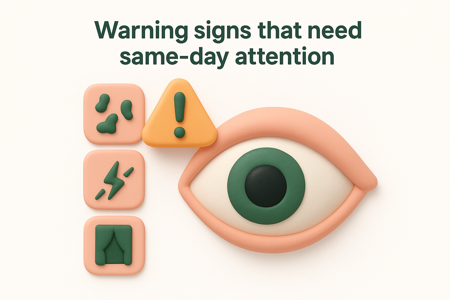
- A sudden increase in the number of floaters, particularly a shower of new dark spots appearing within minutes or hours
- Flashing lights (photopsia) in your peripheral vision, which suggest the vitreous is pulling on or tearing your retina
- A dark curtain, shadow, or veil spreading across part of your visual field
- Sudden blurring or loss of central vision that was not there before
- Any floaters that appear dense, red, or blood-tinged, which can indicate vitreous haemorrhage
A retinal detachment is a medical emergency. Without treatment within hours to days, permanent vision loss is the likely outcome.
Why timing matters so much
When a retinal tear occurs before the retina lifts away, laser treatment or cryotherapy can seal the break quickly and prevent progression to a full detachment. This is a straightforward procedure that takes only minutes and preserves vision effectively. Once the retina detaches, however, surgery becomes significantly more complex, recovery is considerably longer, and the chance of achieving a full visual outcome drops.
Your risk of a retinal detachment is higher if you are highly myopic, have had previous eye surgery such as cataract removal, or have a family history of retinal problems. People with diabetes face additional risk from abnormal new blood vessel growth that can bleed suddenly into the vitreous, clouding vision rapidly. Acting quickly when your floaters change character gives your retina the best possible chance of staying intact and your vision the best chance of remaining clear.
What to expect at an eye exam in Australia
If a floater in eye won’t go away situation has brought you to an eye specialist, the examination itself is straightforward and painless. Knowing what is involved ahead of time helps you feel prepared and ensures you get the most out of your appointment. Most assessments for persistent floaters follow a structured process that covers both the front and back of your eye in detail.
What happens during the examination
Your specialist will begin by asking you a detailed history of your symptoms, including when the floaters started, whether they changed suddenly, and whether you have noticed any flashing lights or changes to your peripheral vision. This information guides how urgently they examine particular areas. Your visual acuity will also be measured using a standard chart to establish a baseline.
The most important part of the examination for floaters is a dilated fundus examination, where eye drops are used to widen your pupil so the specialist can see clearly to the back of your eye. Once dilated, a slit-lamp or indirect ophthalmoscope allows a detailed view of the vitreous, retina, optic nerve, and macula. The drops take roughly 20 to 30 minutes to work, and your vision will remain blurry and light-sensitive for a few hours afterwards, so bring sunglasses and arrange a lift home if possible.
If the specialist suspects a retinal tear or detachment, they may also use an ultrasound scan of the eye to see through any blood or dense floaters that block the direct view.
Additional imaging such as optical coherence tomography (OCT) may be used to examine the macula and retinal layers in fine detail, particularly if there is concern about macular conditions contributing to your symptoms.
Understanding your results and next steps
After the examination, your specialist will explain exactly what they found and whether your floaters are linked to a normal posterior vitreous detachment, an underlying retinal condition, or another cause. You will receive a clear explanation of whether monitoring, treatment, or urgent intervention is required. Most patients in Australia can access this examination through a GP or optometrist referral, and Medicare covers a significant portion of specialist consultation fees, making it accessible without large out-of-pocket costs.
How to cope day to day without treatment
When a floater in eye won’t go away but your specialist has confirmed it is not linked to a retinal tear or detachment, you face a different challenge: learning to live with something that sits in your line of sight. The good news is that most people find their floaters become far less noticeable over time, even without any intervention, as the brain gradually learns to filter them out. There are practical steps you can take right now to speed up that process and reduce the impact floaters have on your daily life.
Adjust how you use your eyes
One of the simplest things you can do is move your eyes deliberately before you try to focus. A quick downward glance followed by returning your gaze forward shifts the floater away from your central vision temporarily, using gravity to drop the particle below your line of sight. This works especially well when you are reading or using a screen, because your field of view stays fixed for long periods.
Repeatedly chasing a floater with your eyes actually keeps it in view longer, so try to look past it rather than directly at it.
Taking regular breaks from close work also reduces the frustration. The 20-20-20 rule, looking at something roughly six metres away for 20 seconds every 20 minutes, gives your visual system a reset and makes individual floaters far less likely to dominate your attention throughout the day.
Change your environment to reduce distraction
Lighting plays a significant role in how prominent your floaters appear. Bright, uniform light sources such as white walls, clear skies, or unshaded windows make floaters stand out sharply, while dimmer or more varied environments tend to minimise their visibility. Using warmer-toned lighting indoors or switching to a dark mode display on your devices can make a noticeable difference if you spend long hours at a screen.
Wearing sunglasses outdoors on bright days reduces the intense glare that makes floaters most obvious. Polarised lenses are particularly helpful because they cut reflective light from roads and water, reducing the contrast that makes floaters pop against your field of vision. These adjustments do not remove your floaters, but they significantly reduce the number of moments during your day when floaters pull your attention away from what actually matters.
Treatment options for bothersome floaters
When coping strategies are not enough and a floater in eye won’t go away despite months of trying to adapt, two treatment options are available through a specialist: surgical removal and laser treatment. Neither is offered routinely for mild floaters, but for patients whose quality of life is significantly affected, both are legitimate and proven approaches worth discussing with your eye surgeon.
Vitrectomy
A vitrectomy is a surgical procedure where the vitreous gel, along with the floaters suspended in it, is removed from inside your eye and replaced with a clear saline solution. Your eye gradually produces its own fluid to maintain normal pressure over time. Vitrectomy is highly effective at eliminating floaters because the source of the problem is physically removed, rather than treated indirectly. It is performed under local anaesthetic as a day procedure, and most patients in Australia return home the same day.

Vitrectomy carries small but real surgical risks, including cataract development, retinal detachment, and infection, so your surgeon will only recommend it when floaters are causing significant, measurable disruption to your daily life.
The procedure is most appropriate when floaters are large, dense, or central, and when they have already been present for several months without any sign of natural adaptation. Your surgeon will assess your overall eye health, lens status, and retinal condition before recommending vitrectomy.
YAG laser vitreolysis
YAG laser vitreolysis is a non-surgical option where a focused laser beam is directed at individual floaters inside the vitreous, breaking them into smaller fragments that are either reabsorbed or become too small to notice. It is performed in a clinic setting without any incisions, making it a less invasive alternative to vitrectomy. Sessions are typically short, taking around 15 to 20 minutes, and most patients need more than one session for meaningful results.
This approach works best for well-defined, accessible floaters such as a Weiss ring, which forms after a posterior vitreous detachment. Diffuse or numerous small floaters are harder to target effectively with a laser. Your specialist will be direct with you about whether your floaters are good candidates for this technique, and what realistic outcomes look like based on the size, location, and density of what is present.
How to reduce your risk of future problems
You cannot stop the natural ageing of your vitreous gel, but you can take deliberate steps to lower the chance that a floater in eye won’t go away situation develops into something more serious. Certain lifestyle choices and health habits directly affect the health of your retina and vitreous, and getting on top of them now reduces your risk of the more dangerous causes of floaters down the track.
Protect your eyes from UV light and injury
Ultraviolet radiation accelerates oxidative damage inside the eye, contributing to vitreous breakdown and increasing the risk of cataract development, which itself can affect how floaters are perceived. Wearing UV-blocking sunglasses outdoors, particularly during high-exposure periods between 10am and 2pm in Australia, is one of the most straightforward preventive measures available. Look for lenses rated to Australian Standard AS/NZS 1067, which confirms they provide adequate protection for local conditions.
If your work or sport puts you at risk of eye injury, protective eyewear is non-negotiable. Blunt trauma and penetrating injuries can trigger sudden vitreous haemorrhage or retinal tears, both of which produce severe, sudden floaters and carry a high risk of permanent vision loss without prompt treatment.
Manage your systemic health conditions
Diabetes is one of the most significant preventable causes of vitreous haemorrhage, the type of bleeding that fills your eye with dense, vision-obscuring floaters almost overnight. Keeping your blood glucose within your target range, attending regular medical reviews, and having your eyes checked at least annually if you have diabetes all reduce the risk of diabetic retinopathy reaching the stage where bleeding occurs.
High blood pressure also damages retinal blood vessels over time, so controlling it through diet, exercise, and medication where prescribed protects your retina as much as your heart.
Stay up to date with eye examinations
Regular comprehensive eye checks allow your specialist to detect early retinal changes before they cause symptoms, giving you the chance to intervene before floaters or more serious problems develop. If you are over 50, highly myopic, or have a family history of retinal disease, an annual dilated eye examination is a reasonable baseline. Your optometrist can refer you on if anything warrants specialist attention, keeping your care connected and consistent over time.

Next steps for peace of mind
A floater in eye won’t go away situation rarely turns out to be sinister, but the only way to know for certain is to have your eyes examined by a qualified specialist. Leaving persistent floaters unchecked carries real risk, particularly if there is an underlying retinal condition quietly worsening in the background. Acting sooner rather than later gives you accurate information, removes the guesswork, and puts you in control of your eye health.
Both Dr Rahul Dubey and Dr Capucine Odouard at Bright Sight Eye Specialists in Randwick assess and treat persistent floaters, from straightforward posterior vitreous detachment through to complex retinal and vitreous conditions. You will receive a thorough dilated examination, a clear explanation of your findings, and honest advice about whether monitoring or active treatment is the right path for you. To book your assessment, contact Bright Sight Eye Specialists today.