


You’re reading a book or looking at a clear blue sky when you notice small dark shapes drifting across your vision. Then you catch brief flashes of light in the corner of your eye. These are floaters and flashes, and they happen when the gel-like substance inside your eye changes or pulls on the sensitive retina. Most of the time they’re harmless signs of ageing. But sometimes they can signal something more serious that needs urgent medical attention.
This article explains what causes flashing floaters, why they matter, and how to tell the difference between normal age-related changes and symptoms that require immediate care. You’ll learn which warning signs demand same-day assessment, what happens during diagnosis, and how conditions like retinal tears or detachment are treated at specialist centres. Understanding these symptoms helps you protect your vision and know exactly when to act quickly.
Why flashing floaters in vision matter
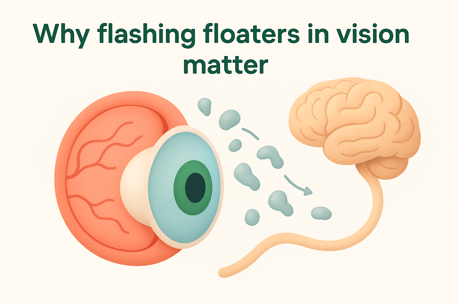
Your retina is a thin layer of light-sensitive tissue at the back of your eye that sends visual signals to your brain. When you experience flashing floaters in vision, these symptoms tell you something is happening with the vitreous gel that fills your eye or how it’s interacting with your retina. The vitreous normally sits firmly against the retina, but as you age it begins to liquefy and shrink. This process pulls the vitreous away from the retina, causing mechanical stimulation that you perceive as flashes of light. You see floaters because tiny clumps of collagen fibres cast shadows on your retina as they drift through the liquefied vitreous.
The connection to your retina
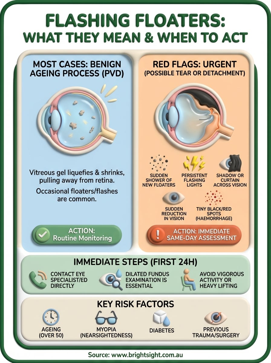
Most cases of flashing floaters represent benign age-related changes called posterior vitreous detachment (PVD). This affects roughly two-thirds of people over 70 and rarely causes lasting problems. However, the same pulling forces that create harmless flashes can also tear your retina. Research shows that approximately 14% of posterior vitreous detachments produce a retinal tear during the separation process. When the vitreous tugs forcefully enough, it can rip the delicate retinal tissue, creating an opening that allows fluid to seep underneath.
The challenge is that you cannot reliably distinguish between harmless PVD and a sight-threatening retinal tear based on symptoms alone.
What happens when symptoms progress
Untreated retinal tears progress to retinal detachment in 33 to 46% of cases. Once fluid passes through the tear and lifts the retina away from its underlying blood supply, the photoreceptor cells begin to die. You’ll notice this as a shadow or curtain spreading across your field of vision. If the detachment reaches your macula (the central part responsible for sharp vision), you may lose your ability to read, recognise faces, or drive permanently. Time matters critically here because surgical success rates drop significantly once the macula detaches.
How to respond when you notice flashing floaters
When you first notice flashing floaters in vision, your immediate response determines whether you protect your sight or risk permanent damage. The key is distinguishing between symptoms that need same-day attention and those that can wait for a routine appointment. You should contact an ophthalmologist immediately if you see a sudden shower of new floaters, multiple flashes, or any shadow spreading across your vision. These symptoms suggest possible retinal tear or detachment, conditions that require urgent examination with dilated pupils and specialised equipment.
Assess the urgency
You need same-day assessment if you experience any of the following warning signs: a dramatic increase in the number of floaters appearing within hours, persistent flashing lights (especially if they continue when your eyes are closed), a curtain or shadow moving across your peripheral or central vision, or sudden reduction in visual clarity. Research shows that approximately 70% of patients with vitreous haemorrhage accompanying posterior vitreous detachment have at least one retinal tear present. Blood in your vitreous appears as tiny black or red spots that don’t fade, indicating a torn blood vessel that needs immediate attention.

If symptoms appear suddenly or intensify rapidly, treat this as an emergency rather than waiting to see if they improve.
Longstanding floaters that haven’t changed over weeks or months typically represent benign vitreous changes and can be assessed through routine examination. However, any new symptoms or changes to existing floaters warrant professional evaluation.
What to do in the first 24 hours
Contact your eye specialist or emergency department directly rather than waiting for a referral from your general practitioner. Time-critical conditions like retinal detachment require examination within hours, not days. An ophthalmologist will perform a comprehensive dilated fundus examination to visualise your entire retina, including the peripheral areas where tears most commonly occur. This examination takes roughly 10 to 15 minutes once the dilating drops take effect.
Avoid vigorous physical activity, heavy lifting, or actions that jar your head until you’ve been examined. These activities can worsen an existing tear or accelerate progression to detachment. Keep track of any changes in your symptoms, including the specific location of shadows or whether flashes increase in frequency.
Causes and risk factors for flashing floaters
Understanding what triggers flashing floaters in vision helps you recognise whether your symptoms represent normal ageing or indicate elevated risk. The vitreous gel inside your eye undergoes continuous changes throughout your life, but certain factors accelerate these changes or make complications more likely. Your individual risk profile depends on a combination of age, eye structure, medical conditions, and previous eye trauma or surgery.
Natural age-related changes
The most common cause of flashing floaters is posterior vitreous detachment (PVD), a natural process where the vitreous gel liquefies and separates from your retina. This affects nearly 66% of people over 70 years old and occurs because the collagen fibres within the vitreous break down over time. When these fibres clump together, they create the shadows you perceive as floaters. The mechanical pulling during separation stimulates your retina, producing the sensation of flashes or lightning streaks in your peripheral vision. Most people notice these symptoms becoming more pronounced when looking at bright, uniform backgrounds like white walls or clear skies.
Vitreous syneresis describes the gradual liquefaction of the vitreous that begins as early as your twenties but accelerates significantly after age 50. This process creates pockets of liquid within the gel, causing light to bend at these junctions and generating additional floater sensations.
Medical and structural risk factors
Your eye’s physical structure and overall health significantly influence your likelihood of experiencing flashing floaters. Myopia (nearsightedness) ranks among the most important risk factors because elongated eyes create greater vitreoretinal forces and thinner retinal tissue. Research consistently shows that people who need glasses to see distant objects face substantially higher rates of both benign floaters and serious complications like retinal tears.

Diabetes increases your risk through proliferative diabetic retinopathy, where abnormal blood vessels grow and can bleed into your vitreous. This causes vitreous haemorrhage, which you experience as sudden showers of dark spots or a red haze across your vision. Previous cataract surgery accelerates vitreous detachment by several years because removing your natural lens disturbs the normal eye structure.
Trauma to your eye creates compression and decompression forces strong enough to tear your retina even years after the initial injury.
Inflammatory conditions like uveitis cause debris to accumulate in your vitreous, while a family history of retinal problems suggests you carry genetic susceptibility. If you’ve had retinal detachment in one eye, your other eye faces significantly elevated risk.
When flashing floaters signal an emergency
Recognising the difference between harmless flashing floaters in vision and genuine emergencies can save your sight. Certain combinations of symptoms indicate that your retina has torn or begun detaching, conditions that demand immediate professional assessment. You have a narrow window to prevent permanent vision loss once these warning signs appear. The challenge is that early symptoms of serious retinal problems often feel similar to benign posterior vitreous detachment, which is why same-day examination becomes essential when specific red flags emerge.
Critical warning signs that require immediate action
You need to contact an eye specialist urgently if you notice a sudden shower of new floaters appearing within hours rather than gradually over weeks. This dramatic increase suggests that cells or blood have released into your vitreous, often indicating a fresh retinal tear. Persistent flashing lights that continue even when you close your eyes signal ongoing mechanical stimulation of your retina, which happens when vitreous strands pull on torn tissue.
A dark shadow or curtain spreading across your peripheral or central vision represents the most serious warning sign. This shadow shows that fluid has passed through a retinal tear and lifted your retina away from its blood supply. Research demonstrates that superior retinal tears (in the upper part of your eye) cause macular detachment more frequently because gravity pulls fluid downward. Once your macula detaches, you lose central vision permanently in many cases, even after successful surgery.
You cannot accurately judge the severity of your symptoms at home, which is why professional examination within 24 hours becomes non-negotiable when warning signs appear.
Vitreous haemorrhage produces a distinctive pattern of symptoms. You’ll see countless tiny black or red spots that don’t fade, sometimes accompanied by a red haze obscuring your vision. This bleeding occurs when a retinal tear disrupts blood vessels, and approximately 70% of these cases involve retinal tears that need urgent treatment. Sudden reduction in visual acuity or the sensation that you’re looking through a veil also demands immediate assessment, as does any combination of multiple warning signs appearing together within a short timeframe.
How quickly retinal detachment progresses
Retinal detachment can progress from a small tear to complete detachment within days or even hours. The speed depends on the tear’s location and size, the amount of vitreous liquefaction present, and your activity level. Superior tears progress faster than inferior ones because fluid naturally flows downward with gravity. You might notice the shadow expanding across your vision hour by hour, with permanent damage occurring in the areas that remain detached beyond approximately one week. This rapid progression explains why ophthalmologists treat suspected retinal tears as true emergencies requiring same-day or next-day intervention.
Diagnosis and treatment at Bright Sight
When you arrive at Bright Sight Eye Specialists with flashing floaters in vision, Dr. Rahul Dubey and his team prioritise thorough assessment using advanced diagnostic technology. Your examination begins with a detailed discussion of your symptoms, including when they started, their frequency, and any associated visual changes. The team then performs a comprehensive dilated fundus examination to visualise your entire retina, particularly the peripheral areas where tears most commonly develop. This careful approach, combined with Dr. Dubey’s training at London’s Moorfields Eye Hospital, ensures accurate diagnosis and appropriate treatment recommendations tailored to your specific condition.
The examination process
Your diagnostic appointment at Bright Sight involves several precise steps designed to identify any retinal problems. After applying dilating drops, Dr. Dubey examines your retina using specialised lenses and ophthalmoscopy to detect tears, detachment, or vitreous haemorrhage. The clinic uses advanced imaging technology to capture detailed photographs of your retina, creating a permanent record that helps monitor changes over time. This thorough examination typically takes 30 to 45 minutes and allows the team to distinguish between benign posterior vitreous detachment and conditions requiring intervention. You receive clear explanations throughout the process, with Dr. Dubey showing you images of your retina and discussing findings in straightforward language you can understand.
Treatment approaches for retinal conditions
Treatment depends entirely on what your examination reveals. Uncomplicated posterior vitreous detachment requires monitoring rather than intervention, with follow-up appointments scheduled at six weeks to check for delayed tears. If Dr. Dubey identifies retinal tears, he applies laser or cryotherapy to create protective scars around the break, preventing fluid from seeping underneath your retina. These procedures typically occur as outpatient treatments with local anaesthetic.
Early treatment of retinal tears prevents progression to detachment, preserving your vision and avoiding more complex surgery.
Established retinal detachment requires vitreoretinal surgery, which may involve vitrectomy, scleral buckling, or pneumatic retinopexy depending on your specific situation. Bright Sight offers transparent pricing for all procedures, with many treatments available as "no gap" options for privately insured patients. The practice serves patients across Sydney’s Eastern Suburbs and maintains connections with rural communities, ensuring you access specialist care when you need it most.
Protecting your sight
Recognising flashing floaters in vision as potential warning signs empowers you to act quickly when necessary. Most episodes represent harmless age-related changes that require monitoring rather than treatment, but you cannot safely make this distinction yourself at home. The symptoms of benign posterior vitreous detachment and serious retinal tears appear remarkably similar, which is why professional examination becomes essential whenever new floaters or flashes appear. Your vision depends on prompt diagnosis and treatment when tears or detachment occur, with surgical success rates highest when problems are caught early.
Schedule regular eye examinations if you have risk factors like myopia, diabetes, or previous eye surgery, as these conditions increase your likelihood of developing complications. Contact an ophthalmologist immediately if you notice sudden changes in your symptoms, including multiple new floaters, persistent flashes, shadows across your vision, or reduced clarity. Book your comprehensive eye assessment at Bright Sight to ensure any retinal problems receive expert attention before they threaten your sight permanently.