


You notice something odd while looking at a clear sky or white wall. Small specks drift across your vision, or brief flashes of light appear in the corner of your eye. These are floaters and flashes, visual phenomena that happen when the gel-like substance inside your eye (the vitreous) changes or pulls on your retina. Most people experience them at some point, particularly after age 50, and they’re usually harmless. But sometimes they signal something more serious that needs immediate attention.
This article explains what causes flashes and floaters, how to tell when they’re a warning sign of retinal problems, and what treatment options exist. You’ll learn the difference between normal age-related changes and symptoms that require urgent care. We’ll cover the examination process, what your specialist looks for during a dilated eye exam, and practical guidance on when you can safely monitor floaters at home versus when you need same-day assessment. By the end, you’ll know exactly how to respond if you experience these symptoms.
Why flashes in eyes and floaters matter
Your vision depends on your retina staying attached to the back of your eye, and flashes in eyes and floaters can signal when that attachment is at risk. Retinal detachment is one of the few true eye emergencies, and the symptoms often begin with sudden increases in floaters or new flashes of light. When you catch these warning signs early, treatment can preserve your vision. Delay even a few days, and you may face permanent vision loss that no surgery can reverse.

The window for preventing vision loss
Time-sensitive treatment makes the difference between keeping your sight and losing it forever. A retinal tear takes only hours or days to progress into full detachment, particularly if the tear occurs in the upper part of your retina where gravity pulls fluid downward behind the retina. Your macula (the central part of your retina responsible for sharp, detailed vision) can detach within this timeframe. Once your macula detaches, even successful reattachment surgery typically leaves you with reduced central vision. You’ll struggle with reading, recognising faces, and driving.
Early detection and treatment of retinal tears can prevent up to 98% of retinal detachments.
Why age makes this relevant to you
Posterior vitreous detachment affects nearly two-thirds of people over 70, making floaters and flashes increasingly common as you age. Your vitreous gel naturally shrinks and pulls away from your retina, and this process creates the floaters you see. While most vitreous detachments cause no problems, about 14% create retinal tears during the separation. Your risk increases if you’re nearsighted, have had cataract surgery, or experienced eye inflammation. These factors stretch or weaken your retina, making tears more likely when the vitreous pulls away.
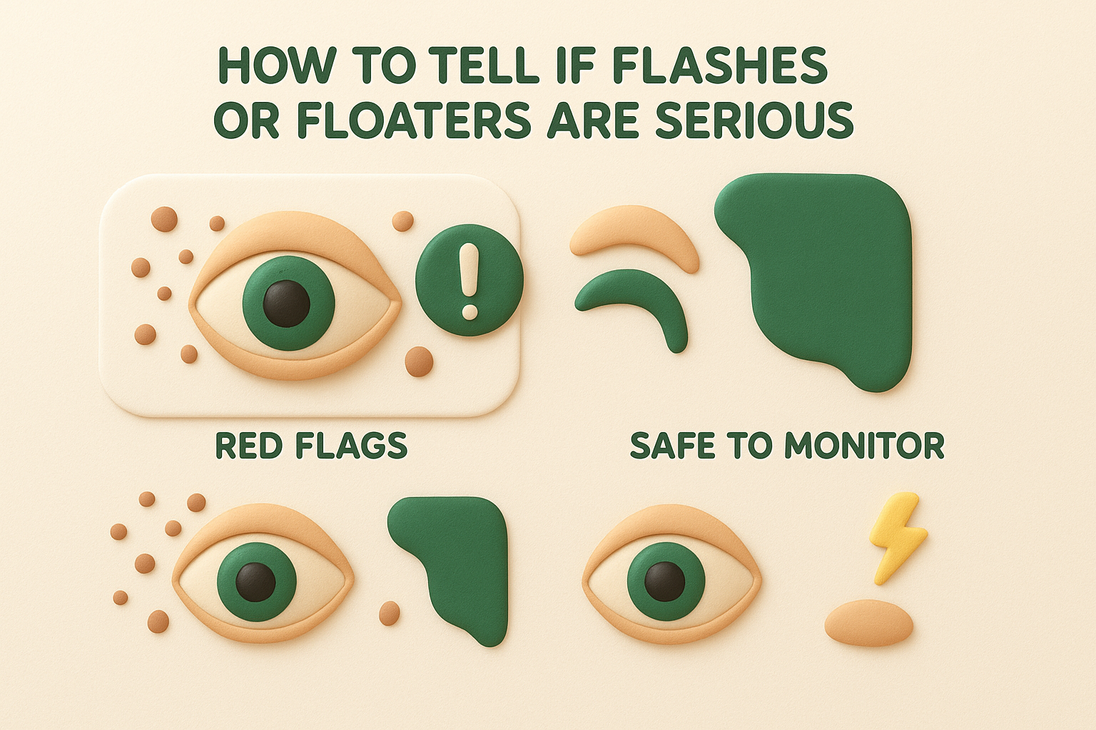
How to tell if flashes or floaters are serious
You need to distinguish between normal floaters that require no action and urgent warning signs that demand same-day assessment. A few occasional floaters you’ve had for years pose no threat. But sudden changes, particularly a shower of new floaters appearing within hours, signal potential retinal problems. Your brain’s ability to adapt makes this tricky as well, since longstanding floaters fade from your awareness over time whilst new ones appear obvious and alarming.
Red flags requiring same-day examination
Sudden onset of many new floaters (often described as hundreds of tiny dots or a cloud) means you need an eye examination within hours. This symptom often indicates vitreous haemorrhage, where blood from a torn retinal vessel creates the appearance of multiple dark spots. Persistent flashes of light, especially if they continue for more than a few minutes or recur throughout the day, suggest ongoing traction on your retina. You should seek immediate care if you notice a dark shadow or curtain moving across your vision from any direction. This curtain represents retinal detachment already in progress. Vision loss in any part of your visual field makes assessment truly urgent.
If you see a curtain or shadow in your vision, you’re already experiencing retinal detachment and need emergency care within hours.
When monitoring at home remains safe
Longstanding floaters that haven’t changed in months or years rarely indicate serious problems. You can safely observe occasional flashes that occur only when you move your eyes quickly in dim lighting, particularly if you’ve had them for weeks without change. Gradual increases in floater number over months, rather than sudden appearance within hours or days, typically reflect normal vitreous changes. One or two new floaters without accompanying flashes or vision loss generally warrant a routine examination within a few weeks rather than emergency assessment.
Pattern changes signal trouble
Sudden increases in either the number or size of flashes in eyes and floaters indicate worsening conditions. Symptoms that initially seemed mild but progressively worsen over hours or days require prompt evaluation. New symptoms after an initial vitreous detachment (even weeks later) suggest delayed complications like retinal tears. Your symptoms becoming more frequent, more persistent, or accompanied by vision changes all warrant immediate specialist review rather than continued home monitoring.
What causes flashes in eyes and floaters
Age-related changes in your vitreous gel cause most floaters and flashes, but several serious conditions can produce identical symptoms. Your vitreous fills the space between your lens and retina, and it remains clear and firm in childhood. As you age, this gel undergoes chemical changes that make it more liquid, allowing collagen fibres to clump together and cast shadows on your retina. These shadows create the floaters you see. Understanding the specific cause helps you and your specialist determine whether you need treatment or can safely monitor the condition at home.
Age-related vitreous degeneration
Vitreous syneresis describes the natural breakdown of gel structure that occurs in everyone over time. Your vitreous loses its uniform consistency and develops pockets of liquid mixed with strands of collagen. These condensed collagen fibres appear as dots, threads, or cobweb shapes drifting through your vision. The process typically begins in your fifties and progresses gradually. You notice floaters more easily when looking at bright, uniform backgrounds like white walls or clear skies because the contrast makes the shadows more visible.
Posterior vitreous detachment
Posterior vitreous detachment causes the majority of sudden-onset flashes in eyes and floaters in people over 50. Your vitreous gel shrinks away from your retina, creating a complete separation that happens relatively quickly over days or weeks. During this separation, the mechanical tugging on your retina triggers photoreceptor cells that interpret any stimulation as light, creating the flashes you see. The separated vitreous itself forms a large floater (often called a Weiss ring) that you notice as a prominent circle or arc. Around 66% of people over 70 experience this separation, and it occurs naturally without any underlying disease.

Posterior vitreous detachment occurs in two-thirds of people over 70 and causes most cases of new floaters.
Retinal tears and their progression
Vitreous traction sometimes pulls hard enough during detachment to tear your retina, which happens in about 14% of posterior vitreous detachments. Your retina tears more easily if you’re nearsighted because your elongated eyeball stretches the retina thinner. Retinal breaks release pigment cells and sometimes blood into your vitreous, creating a sudden shower of new floaters. Fluid then seeps through the tear underneath your retina, causing retinal detachment that progresses over hours or days. Superotemporal tears (in the upper outer part of your eye) prove most dangerous because gravity pulls fluid downward, rapidly detaching your macula.
Less common but serious causes
Vitreous haemorrhage from diabetes, trauma, or torn blood vessels creates hundreds of tiny dark spots that appear suddenly. Diabetic retinopathy weakens retinal blood vessels, making them prone to bleeding into your vitreous. Inflammation (uveitis) produces floaters through inflammatory cells floating in your vitreous rather than from vitreous changes. Recent eye surgery or trauma can trigger both haemorrhage and inflammation. These conditions require different treatments than simple posterior vitreous detachment, making accurate diagnosis essential for appropriate care.
Getting a diagnosis and eye examination
Your eye specialist performs a dilated fundus examination to diagnose the cause of flashes in eyes and floaters accurately. This examination remains the gold standard for detecting retinal tears, detachment, and vitreous changes because it provides a clear view of your entire retina. You’ll typically receive your diagnosis during this single appointment, though complex cases may require additional imaging. The process takes about 30 to 45 minutes from arrival to completion, including the time needed for your pupils to dilate fully.
The dilated eye examination process
Your specialist instils dilating drops into both eyes at the start of your appointment, which take 10 to 15 minutes to work fully. These drops temporarily paralyse the muscles that constrict your pupils, allowing your specialist to see past your iris to examine your retina. Direct and indirect ophthalmoscopy follows, where your specialist uses specialised lenses and lights to inspect your retina in sections. The bright light feels uncomfortable but causes no harm. Your specialist pays particular attention to your peripheral retina (where most tears occur) by asking you to look in different directions whilst examining each area carefully.

Dilating your pupils carries a small risk of triggering acute angle-closure glaucoma, but this risk is far outweighed by the danger of missing a retinal tear.
What your specialist examines
Vitreous assessment shows whether you have posterior vitreous detachment, haemorrhage, or inflammatory cells floating in the gel. Your specialist looks for the characteristic Weiss ring (a circular floater) that confirms complete vitreous separation. Retinal inspection identifies any tears, holes, or areas of detachment, noting their size, location, and whether your macula remains attached. Pigment dispersal (called tobacco dust) and fresh or old haemorrhage provide clues about when damage occurred. Your specialist also checks your retinal periphery systematically, examining all four quadrants to ensure no tears hide in areas you haven’t noticed symptoms from yet.
Treatment options and living with floaters
Most flashes in eyes and floaters require no medical treatment at all, whilst serious retinal problems demand urgent surgical intervention to preserve your sight. Your specialist determines the appropriate approach based on examination findings rather than symptom severity alone. Benign floaters from posterior vitreous detachment gradually become less noticeable over weeks or months as your brain adapts to filter them out. You’ll likely forget about them most of the time, though they remain permanently in your eye. Treatment decisions depend entirely on whether your retina has tears or detachment rather than how bothersome the floaters feel.
When treatment becomes necessary
Retinal tears need prompt laser or cryotherapy treatment to prevent progression to detachment. Your specialist applies laser burns or freezing treatment around the tear, creating a scar that seals the edges and stops fluid seeping underneath your retina. These procedures take 10 to 20 minutes in the consulting room, cause minimal discomfort, and prevent detachment in most cases. Retinal detachment requires surgery within days, using vitrectomy, scleral buckling, or gas bubble injection to reattach your retina. Surgery success rates exceed 90% for first operations, though you may need repeat procedures. Your visual recovery depends critically on whether your macula detached before surgery, which is why same-day assessment matters so much for new symptoms.
Laser treatment of retinal tears prevents detachment in over 95% of cases when performed promptly.
Managing harmless floaters at home
Observation alone proves sufficient for uncomplicated posterior vitreous detachment without retinal tears. Your floaters will fade from your awareness over three to six months as neurological adaptation occurs, where your brain learns to ignore the constant shadows. Moving your eyes quickly when floaters obstruct your central vision shifts them temporarily out of your direct line of sight. Adequate lighting reduces floater visibility since they show up most against bright backgrounds. Severe floaters that persistently interfere with daily activities may warrant vitrectomy surgery, though this carries risks of cataract formation and retinal detachment. Most specialists recommend exhausting all adaptation strategies before considering surgical removal of benign floaters.

Bringing it all together
You now understand that flashes in eyes and floaters usually stem from age-related vitreous changes, but they sometimes signal retinal tears or detachment requiring urgent treatment. New symptoms demand same-day assessment, whilst longstanding floaters typically need only routine monitoring. Your vision depends on recognising warning signs like sudden showers of floaters, persistent flashes, or dark shadows moving across your visual field.
Immediate action preserves sight when retinal problems occur, because delays of even days can result in permanent vision loss. Most posterior vitreous detachments resolve without complications, allowing you to adapt to floaters over time. However, you shouldn’t attempt self-diagnosis when symptoms first appear. Contact Bright Sight Eye Specialists for a thorough dilated examination if you experience any sudden changes in your vision, ensuring early detection and treatment of potentially sight-threatening conditions.