


The eye vitreous body is the clear, gel-like substance that fills the space between your lens and your retina. It makes up roughly 80% of the eye’s total volume, yet most people never think about it, until something goes wrong. Floaters drifting across your vision, sudden flashes of light, or unexplained blurriness can all trace back to changes in this structure.
Understanding what the vitreous body is, how it works, and what can go wrong with it gives you a real advantage when it comes to protecting your long-term eye health. Whether you’re dealing with new floaters, have been told you have a vitreous detachment, or simply want to know more about your eye anatomy, this knowledge helps you have more informed conversations with your eye care team.
At Bright Sight Eye Specialists in Randwick, Sydney, our ophthalmologists, Dr Rahul Dubey and Dr Capucine Odouard, diagnose and treat a wide range of vitreous and retinal conditions, from persistent floaters through to vitreous haemorrhage and retinal detachment. This article covers the anatomy, composition, and function of the vitreous body, along with the common problems that can affect it and when you should seek specialist care.
Why the vitreous body matters for vision
Most people focus on the lens or the retina when they think about how vision works, yet the eye vitreous body plays a role that is just as fundamental. It occupies the largest internal cavity in the eye, and the work it does, both structurally and biochemically, directly influences the quality of the images your retina receives. When the vitreous is healthy, you never notice it. When it starts to change, the effects can range from mildly distracting to genuinely sight-threatening.
Its role in maintaining the eye’s shape
The vitreous holds the spherical form of the eyeball in place. Without it, the eye would not maintain the precise curvature required for light to focus accurately on the retina. Think of it as the internal scaffolding that keeps the eye rigid enough to function as an optical instrument. This structural support is particularly critical during early development, when the eye is still growing and the vitreous maintains its full, firm gel consistency.
As the eye matures, the vitreous contributes to a specific level of intraocular pressure, which it helps regulate alongside the fluid in the front of the eye. Any significant loss of vitreous volume, such as after trauma or certain surgical procedures, can compromise this pressure balance and affect how well the rest of the eye operates.
How it supports the retina
The vitreous sits directly in front of the retina and, in a healthy younger eye, adheres gently to the retinal surface. This attachment provides a degree of physical support, keeping the retina in its correct position against the back wall of the eye. The retina is an extremely thin and delicate layer of nerve tissue, and the vitreous acts as a cushioning layer between it and any potential mechanical disruption.
When the vitreous separates from the retina, as happens in a posterior vitreous detachment, the pulling force on the retinal surface can create tears that, if left untreated, may lead to a retinal detachment and permanent vision loss.
This relationship between the vitreous and the retina is one of the reasons eye specialists take vitreous changes seriously. A problem in the vitreous can quickly become a problem for the retina, and the retina is where your actual vision originates. Catching these changes early is what makes the difference between a straightforward outcome and a complex surgical repair.
The link between vitreous health and visual clarity
The vitreous must remain optically transparent for light to pass through it without distortion on its way to the retina. Any structural breakdown within the vitreous, whether from collagen fibres clumping together, bleeding into the gel, or inflammatory cells entering it, creates visual disturbances that you notice directly, including floaters, blurred patches, or a reduction in contrast and colour accuracy.
Your ability to read, drive, recognise faces, and perform close work all depends on a clear and unobstructed visual pathway. The vitreous sits right in the middle of that pathway, and when its transparency is compromised, the impact on daily function can be significant. Conditions such as vitreous haemorrhage, where blood enters the gel, can reduce vision to little more than light perception in severe cases.
Even milder disruptions, such as a dense cluster of floaters, can affect your reading or make it difficult to work comfortably under bright conditions. Understanding these connections helps explain why regular monitoring and timely treatment of vitreous conditions is not something to put off. The earlier a vitreous problem is identified, the greater the chance of protecting the retina and preserving clear vision over the long term.
Where it sits and what it looks like
The eye vitreous body occupies the posterior segment of the eye, which is the large chamber that sits behind the lens and in front of the retina. This chamber, called the vitreous cavity, accounts for roughly two-thirds of the eye’s total length from front to back. To picture it, imagine a sphere roughly 24 millimetres in diameter: the vitreous fills the back portion of that sphere, pressed up against the retinal lining on all sides.

Its position inside the eye
The vitreous sits between two critical structures: the crystalline lens at the front, and the retina at the back. Its front surface, known as the hyaloid face, curves slightly inward to accommodate the back surface of the lens, creating a shallow depression called the patellar fossa. At the back, the vitreous rests directly against the inner surface of the retina, with the strongest points of attachment located at the optic disc, the macula, and around the peripheral retina near the front of the eye in an area called the vitreous base.
The attachment at the vitreous base is the most secure point of contact between the vitreous and the retina, and this attachment persists throughout life even as the rest of the vitreous gradually changes with age.
These attachment zones become particularly significant when the vitreous starts to shrink or liquefy, because the areas of firm attachment are where traction forces concentrate, increasing the risk of retinal tears at those sites.
What it looks like up close
In a young, healthy eye, the vitreous appears completely transparent and has a firm, uniform gel consistency similar to raw egg white, though far more structured at a microscopic level. It does not move freely; it holds its shape and maintains close contact with the retinal surface. Under specialist examination using a slit lamp and a dilating lens, a healthy vitreous is essentially invisible, which is exactly what it should be.
As the vitreous ages, its appearance changes noticeably. The gel begins to liquefy in patches, and fibrous strands of collagen start to condense and clump together. These clumps cast shadows on the retina, which is what you perceive as floaters. In cases of bleeding or inflammation, the vitreous takes on a hazy or reddish appearance that can significantly reduce how much light reaches the retina.
What it is made of and how it stays clear
The eye vitreous body is not a simple fluid. It is a highly organised biological gel made up of a small number of components that work together to produce something quite remarkable: a structure that is nearly 99% water yet holds its shape, remains completely transparent, and survives decades of constant optical demand without being replaced by the body.
The core building blocks
Water makes up the vast majority of the vitreous, but the remaining components are what give it structure and function. Collagen fibres, primarily type II collagen, form a loose three-dimensional network throughout the gel. These fibres are extremely fine and evenly distributed in a healthy, young vitreous, which is precisely why light passes through without scattering. Running alongside this collagen network is hyaluronic acid, a long-chain sugar molecule that attracts and holds water molecules in place. Together, these two components create the gel’s characteristic consistency.

The even spacing of collagen fibrils within the vitreous is the primary reason it stays optically clear; when these fibrils clump or tangle, transparency breaks down and you begin to see floaters.
The vitreous also contains a small number of specialised cells called hyalocytes, found mainly along its outer surface. These cells produce hyaluronic acid and maintain the gel’s structural integrity. Additionally, trace amounts of proteins, glucose, and electrolytes are present throughout the vitreous, supporting the metabolic needs of surrounding ocular tissues.
How the vitreous keeps its transparency
Transparency depends on two conditions being maintained simultaneously: the fine collagen fibres must stay evenly spaced, and the vitreous must remain free of cells, blood, and inflammatory material. In a young, healthy eye, both conditions are met without effort. The hyaluronic acid keeps collagen fibres separated by holding water uniformly throughout the gel, which prevents the fibres from touching or aggregating.
With age, the hyaluronic acid gradually breaks down and loses its capacity to hold water in an even distribution. As the water and collagen begin to separate in a process called syneresis, liquid pockets form within the gel, and the collagen fibres start to collapse inward and bundle together. These bundles are dense enough to cast shadows on the retina, which your brain interprets as moving spots or threads drifting across your visual field. This is the physical mechanism behind most age-related floaters, and it is a normal, though sometimes disruptive, part of how the vitreous changes over a lifetime.
What the vitreous does in a healthy eye
The eye vitreous body does far more than simply fill space. In a healthy eye, it carries out several distinct functions that directly support your visual system, from keeping light on a straight path to the retina through to protecting the surrounding tissues from mechanical stress. When all of these functions are operating normally, you are completely unaware of the vitreous, which is a sign that it is doing its job correctly.
Keeping light on a straight path
The most directly visible function of the vitreous is optical transmission. Light entering the eye through the cornea and lens must travel across the entire length of the vitreous cavity before reaching the photoreceptors in your retina. For that light to form a sharp, accurate image, the vitreous must stay completely clear and free of obstruction. Any material that enters the gel, whether cellular debris, condensed collagen, or blood, interrupts this transmission and produces visible disturbances in your sight.
In a healthy eye, the vitreous transmits light without scattering or distortion, which is only possible because its collagen fibres remain fine, evenly spaced, and stable throughout the gel.
Supporting surrounding tissues
The vitreous plays an active role in maintaining the metabolic environment of the posterior eye. It acts as a slow-moving reservoir for nutrients and metabolic waste products, allowing low-level exchange between the retina, the lens, and the fluid-filled compartments at the front of the eye. This exchange is not rapid or dynamic, but it contributes to the nutritional stability of tissues that have very limited direct blood supply, particularly the inner retinal layers.
The vitreous also maintains intraocular pressure at a consistent level, working in coordination with the aqueous fluid that circulates at the front of the eye. Together, these two fluids keep the eye inflated to the correct internal pressure, which is essential for the optical geometry of the eye to remain accurate from one moment to the next.
Acting as a shock absorber
Your eye is regularly exposed to minor mechanical forces, whether from physical contact, head movement, or changes in posture. The gel-like consistency of the vitreous allows it to absorb and distribute these forces before they reach the retina. This cushioning function helps protect the delicate retinal tissue from the kind of repetitive micro-trauma that could otherwise cause damage over time. In a young, healthy eye with a fully intact gel, this protective capacity is at its strongest.
How the vitreous changes with age
The eye vitreous body does not stay the same throughout your life. From your mid-forties onward, a series of gradual, predictable changes unfold inside the gel, and these changes are the root cause of most vitreous-related symptoms that bring people to an eye specialist. Understanding what is actually happening inside your eye helps you make sense of what you see and when to act.
Liquefaction and syneresis
The first major shift involves the breakdown of the gel itself, a process called syneresis. As you age, the hyaluronic acid within the vitreous loses its capacity to hold water evenly distributed through the collagen network. Water and collagen begin to separate, forming liquid-filled pockets within what was once a uniform gel. This liquefaction typically starts in the central vitreous and expands outward over time.
As these liquid spaces enlarge, the surrounding collagen fibres have less structural support and start to bundle together, forming the condensed strands and clumps that you perceive as floaters drifting across your vision.
By the time most people reach their sixties, a significant portion of the vitreous has converted from gel to liquid. This does not cause pain or any immediate structural crisis, but it does set the stage for the more significant event that follows.
Posterior vitreous detachment
Once enough of the vitreous has liquefied, it can no longer maintain its close attachment to the retinal surface. The gel shrinks and collapses inward, and the back surface of the vitreous peels away from the retina. This event is called a posterior vitreous detachment, or PVD, and it occurs in the majority of people over the age of sixty.

A PVD is not inherently dangerous, and in most cases it resolves without causing lasting harm. However, the separation process creates sudden, significant traction on the retinal surface, particularly at the firm attachment zones around the vitreous base and the optic disc. This traction is what triggers the sudden flashes of light and a noticeable increase in floaters that many people report when a PVD occurs. Your retina responds to the mechanical pull the same way it responds to light: by sending a signal to your brain.
The risk during a PVD is that the pulling force is strong enough to tear the retina. A retinal tear requires prompt treatment, which is why a sudden onset of flashes and new floaters always warrants a same-day review with an eye specialist.
Common vitreous problems and symptoms
Several distinct conditions can affect the eye vitreous body, and each one produces a recognisable set of symptoms. Some are benign and require only monitoring, while others demand urgent specialist review. Knowing the difference between the two can protect your retina and your long-term vision.
Floaters and posterior vitreous detachment
Floaters are the most common vitreous symptom you will encounter. They appear as spots, threads, cobwebs, or rings that drift across your visual field, particularly against bright backgrounds like a blue sky or a white wall. Most floaters result from the natural ageing process described earlier, where collagen fibres condense and cast shadows on the retina.
A sudden increase in floaters, particularly when accompanied by flashes of light, is the hallmark symptom of a posterior vitreous detachment. While most PVDs resolve without complication, the traction involved can tear the retina at the same time. Any sudden change in floaters or new flashing lights requires a same-day examination to rule out a retinal tear before it progresses to a retinal detachment.
A retinal tear caught early can be sealed with laser treatment in a straightforward outpatient procedure; an untreated tear that develops into a retinal detachment requires more complex surgery and carries a greater risk of permanent vision loss.
Vitreous haemorrhage
Vitreous haemorrhage occurs when blood leaks into the vitreous cavity, typically from abnormal or damaged blood vessels in the retina. You may notice a sudden shower of dark floaters, a reddish haze across your vision, or in more severe cases, a dramatic drop in visual acuity. The blood disrupts the optical transparency of the gel and blocks light from reaching the retina cleanly.
Common causes include proliferative diabetic retinopathy, retinal tears, and retinal vein occlusion. Because blood in the vitreous can obscure the retina, an eye specialist will often use ultrasound imaging to examine the retina when a direct view is not possible. Treatment depends on the underlying cause and the severity of the bleed.
Vitreous traction and macular involvement
In some cases, the vitreous does not detach cleanly from the retina. Instead, it remains partially adherent and exerts persistent pulling forces on the macular surface, the central part of your retina responsible for sharp, detailed vision. This condition, called vitreomacular traction, can distort your central vision and, in more advanced cases, contribute to the formation of a macular hole. Symptoms include distortion, reduced sharpness, and difficulty with tasks like reading or recognising faces.
How eye specialists diagnose vitreous issues
When you visit an eye specialist with symptoms like floaters, flashes, or a sudden drop in vision, diagnosing what is happening inside the eye vitreous body involves a combination of clinical tools. No single test tells the whole story, so your ophthalmologist will typically use several techniques in sequence to build a complete picture of the vitreous and the retina behind it.
Dilated fundus examination
The starting point for almost every vitreous assessment is a dilated fundus examination. Your specialist will place drops in your eye to widen your pupil, then use a specialised lens and a bright light to examine the vitreous cavity and the retinal surface directly. This examination gives a wide-field, real-time view of the vitreous gel, any condensed fibres or haemorrhage within it, and critically, the condition of the retina underneath.
During this examination, your specialist looks for signs of posterior vitreous detachment, retinal tears, or blood pooling inside the gel. A thorough dilated examination is particularly important when you arrive with sudden new floaters or flashes, because it allows your ophthalmologist to assess whether traction has caused a retinal break that requires immediate treatment.
Optical coherence tomography
For conditions affecting the interface between the vitreous and the macula, optical coherence tomography, or OCT, provides far greater detail than a standard examination alone. OCT uses light waves to generate cross-sectional images of the retina and the vitreous surface above it, producing scans that reveal whether vitreous strands are pulling on the macular tissue, whether a macular hole has formed, or whether fluid has accumulated beneath the retinal layers.
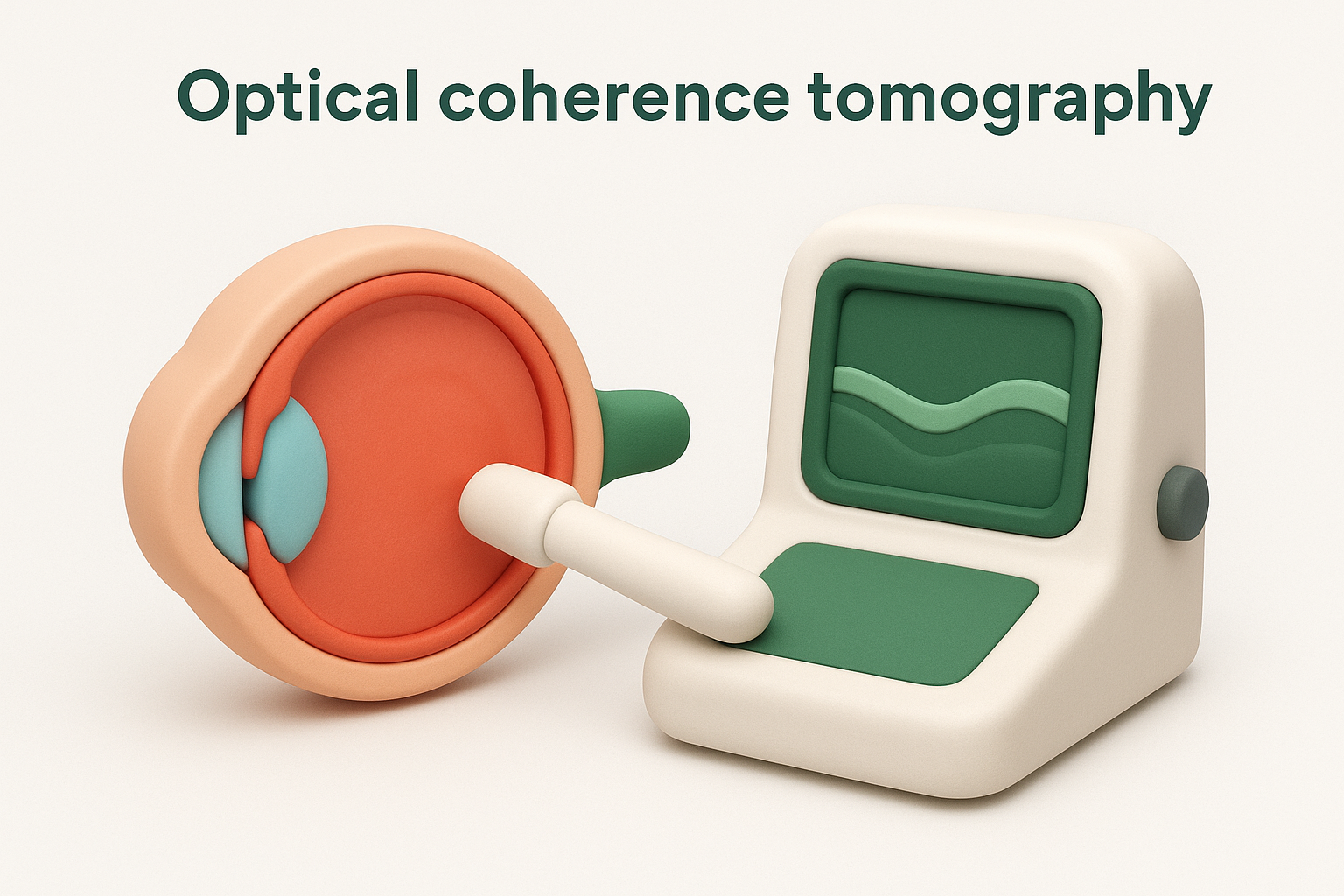
OCT is particularly valuable for detecting vitreomacular traction at an early stage, often before you notice significant changes to your central vision, which gives your specialist more treatment options and a better chance of a straightforward outcome.
Ocular ultrasound
When blood or dense floaters block a direct view of the retina, your specialist turns to ocular ultrasound. This imaging technique uses sound waves to map the structures inside the eye, allowing your ophthalmologist to assess the position of the retina and detect any detachment even when optical methods cannot penetrate the vitreous cavity.
Ultrasound is the primary diagnostic tool used in cases of significant vitreous haemorrhage, where the blood obscures the retina entirely. It can also confirm whether a posterior vitreous detachment has occurred and whether the retina remains in its correct position against the back wall of the eye.
How vitreous problems get treated
Treatment for eye vitreous body conditions depends entirely on what is causing the problem, how severe it is, and whether the retina is involved. Some conditions require nothing more than careful monitoring, while others call for prompt intervention to protect your retinal tissue and preserve your vision. Your ophthalmologist will recommend the most appropriate approach based on a full assessment of your eye.
Laser treatment for retinal tears
When a posterior vitreous detachment pulls a tear in your retina, the priority is sealing that tear before fluid seeps underneath and causes a detachment. Your specialist achieves this with laser photocoagulation, a procedure performed in the consulting room under local anaesthetic drops. The laser creates a series of small burns around the tear, which weld the retina to the underlying tissue and form a barrier that fluid cannot cross.
Laser treatment for a retinal tear is most effective when applied early; the longer a tear remains open, the greater the risk of it progressing to a full retinal detachment that requires more extensive surgery.
The procedure itself takes only a few minutes and causes minimal discomfort. Most patients return to normal activities the same day, though your specialist will schedule follow-up appointments to confirm the seal is holding and that no new tears have formed.
Vitrectomy surgery
A vitrectomy is the main surgical procedure used to treat more complex vitreous problems, including vitreous haemorrhage that does not clear on its own, macular holes, and significant vitreomacular traction. During the procedure, your surgeon removes the vitreous gel entirely through small incisions at the side of the eye, using fine instruments to clear the gel and, where necessary, peel away any membranes from the retinal surface. The vitreous cavity is then filled with a clear fluid or gas to maintain the eye’s shape and support the retina while it heals.
Recovery from vitrectomy varies depending on the underlying condition treated, but most patients notice a gradual improvement in visual clarity over several weeks as the eye settles and any remaining gas bubble absorbs.
Monitoring benign floaters
Not every vitreous change requires active treatment. When your specialist confirms that floaters result from normal age-related changes and that the retina is intact, the standard recommendation is observation. Your brain often adapts to persistent floaters over time, making them less noticeable in daily life. For floaters that remain significantly disruptive, your ophthalmologist can discuss further options such as laser vitreolysis, which uses a targeted laser to break up dense floater clumps.

When to get help
Changes to the eye vitreous body are common, but some require prompt attention rather than a wait-and-see approach. If you notice a sudden increase in floaters, new flashing lights, a shadow or curtain across any part of your vision, or a rapid drop in visual clarity, contact an eye specialist the same day. These symptoms can indicate a retinal tear or detachment, and early treatment significantly improves your outcome.
Not every vitreous change is an emergency, but all of them deserve a professional assessment rather than self-diagnosis. Even floaters that seem minor are worth discussing at your next appointment, as your ophthalmologist can confirm whether your retina is intact and advise on what to watch for going forward. Identifying problems early keeps your treatment options simpler and your prognosis stronger.
If you are experiencing any of these symptoms or want to discuss your eye health, book an appointment with our team at Bright Sight Eye Specialists.