


A tiny bulge in a blood vessel might not sound like much, but an eye microaneurysm is often the earliest detectable sign that diabetes is starting to affect your vision. These microscopic changes in the retinal blood vessels frequently appear before you notice any symptoms at all, which is exactly why understanding them matters. Caught early, they give you and your eye specialist a critical window to act before more serious damage develops.
Microaneurysms form when persistently high blood sugar weakens the walls of the small blood vessels in your retina. They’re the hallmark finding of early diabetic retinopathy, and for many patients, they’re the first clue picked up during a routine eye examination. While a single microaneurysm doesn’t necessarily mean your vision is at risk right now, it does signal that your retinal health needs closer attention and, in many cases, a clear management plan.
At Bright Sight Eye Specialists, Dr. Rahul Dubey and our team in Randwick regularly diagnose and manage diabetic eye disease, from these earliest changes through to advanced treatments including intravitreal injections and laser therapy. This article explains what eye microaneurysms are, why they develop, how they connect to diabetes, and what steps you should take if they’ve been found during your eye check.
What an eye microaneurysm is
An eye microaneurysm is a microscopic outpouching, or tiny balloon-like bulge, that forms in the wall of a small blood vessel in your retina. The retina is the thin, light-sensitive layer lining the back of your eye, and it depends on a dense network of fine capillaries to deliver oxygen and nutrients to the cells responsible for your sight. When those capillary walls weaken at a localised point, blood pressure within the vessel pushes outward, creating this small, sac-like protrusion. The result is a lesion that is invisible to the naked eye but clearly visible to a trained ophthalmologist using the right equipment.
How microaneurysms form in the retina
Your retinal capillaries are built from two key cell types: endothelial cells, which form the vessel’s inner lining, and pericytes, which wrap around the outside and give the wall its structural support. When pericytes are damaged or lost, specific points along the vessel wall lose their integrity. Blood pressure then acts on those weaker spots, gradually pushing them outward until a bulge develops. The microaneurysm sits there, structurally compromised, and prone to leaking fluid, lipids, and small amounts of blood into the surrounding retinal tissue.

Pericyte loss is one of the earliest structural changes in diabetic retinopathy, which is why microaneurysms often appear well before any other visible sign of damage to the retina.
The lesions themselves are tiny, typically between 10 and 100 micrometres in diameter, which is smaller than the width of a human hair. Their size alone is not the concern; the concern is what they signal about the health of the blood vessels feeding your retina, and what can leak from them if left unmonitored.
What microaneurysms look like during an eye examination
During a retinal examination, microaneurysms appear as small, discrete, red dots on the surface of the retina. They can resemble dot haemorrhages, but specific imaging techniques help your specialist tell the difference. On a fundus photograph, they show up as well-defined round lesions clustered near the capillary bed in the inner retinal layers.
Your ophthalmologist will note their number, location, and distribution across different zones of the retina. A handful of microaneurysms in one area tells a very different story from dozens spread across multiple quadrants, and that distribution shapes the entire management plan your specialist will recommend going forward.
Why eye microaneurysms matter
An eye microaneurysm on its own rarely causes symptoms, but that silence is precisely what makes it clinically significant. You can have multiple microaneurysms present in your retina and notice absolutely nothing unusual about your vision. That gap between what is happening at a cellular level and what you actually experience is why regular screening matters so much, particularly if you have diabetes or other vascular risk factors.
The risk of leakage and swelling
The structural weakness in a microaneurysm means the vessel wall cannot reliably contain its contents. Over time, fluid and lipid deposits can leak out into the surrounding retinal tissue. When this leakage accumulates near the macula, the area of your retina responsible for sharp central vision, it causes a condition called diabetic macular oedema, one of the leading causes of vision loss in people with diabetes.
Diabetic macular oedema develops in roughly 1 in 3 people with diabetic retinopathy over a ten-year period, which is why catching microaneurysms early makes a real difference to long-term vision.
Your specialist will pay particular attention to any microaneurysms sitting within the central zone of the macula, as leakage in this area threatens the vision you use for reading, driving, and recognising faces. Even a small amount of fluid accumulation in this region can blur your central sight noticeably.
What happens if they’re left unchecked
Without monitoring, microaneurysms can multiply and spread, signalling worsening vascular damage across your retina. As the number increases, so does the risk of progression to more advanced stages of diabetic retinopathy, including proliferative retinopathy, where abnormal new blood vessels grow on the retinal surface and can bleed suddenly into the eye. Your specialist tracks the number and location of microaneurysms over time specifically to catch this progression before treatment becomes significantly more complicated.
Causes, including the diabetes link
Diabetes is the most common cause of eye microaneurysms, but it is not the only one. The underlying mechanism in every case is the same: sustained damage to the tiny capillaries feeding your retina weakens their structural integrity until small sections of the vessel wall give way and bulge outward.
How diabetes damages retinal vessels
Chronically elevated blood glucose levels are directly toxic to the pericytes that support your retinal capillary walls. Over time, high sugar concentrations trigger inflammation, oxidative stress, and thickening of the basement membrane inside the vessel. These changes progressively strip away the structural reinforcement that keeps capillary walls intact. Once enough pericytes are lost, the vessel wall at those weakened points has no defence against normal blood pressure, and a microaneurysm forms.
The longer blood glucose levels remain poorly controlled, the faster pericyte loss occurs, and the higher your risk of developing multiple microaneurysms across your retina.
The risk increases significantly with the duration of your diabetes. Studies show that after 20 years of diabetes, the majority of people with Type 1 and a large proportion with Type 2 have some degree of retinopathy, with microaneurysms typically appearing first. Poorly controlled blood pressure compounds the damage, because higher intravascular pressure accelerates the outward bulging of already-weakened vessel walls.
Other conditions that raise your risk
While diabetes drives most cases, other conditions that impair your retinal circulation or blood vessel health can also trigger microaneurysm formation. Retinal vein occlusion, hypertension, and hyperlipidaemia each place additional mechanical or metabolic stress on your capillary walls. Anaemia and haematological disorders affecting blood viscosity are also recognised contributors. If microaneurysms appear without a diabetes diagnosis, your specialist will investigate these alternative causes thoroughly.
How doctors find them and what tests show
Detecting an eye microaneurysm requires more than a standard vision test. Your ophthalmologist uses a combination of direct retinal examination and specialist imaging technology to identify these tiny lesions, measure their extent, and track any changes between appointments. Dilation of your pupil is almost always part of this process, as it gives your specialist a much clearer view of the retinal blood vessels at the back of your eye.
Fundus photography and fluorescein angiography
Fundus photography captures a detailed wide-field photograph of your retina through a dilated pupil, giving your specialist a complete map of its blood vessel network. Microaneurysms appear as small, discrete red dots on the retinal surface, and these photographs become a baseline record your specialist compares directly against images from every future visit.
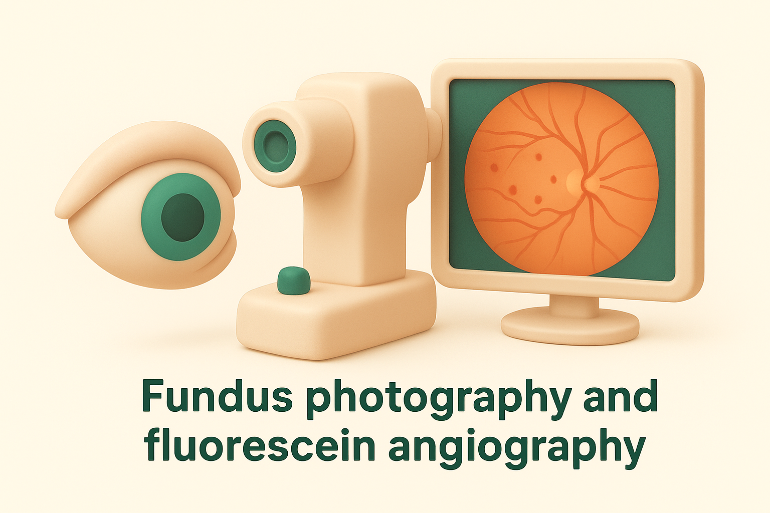
Fluorescein angiography takes the assessment further by injecting a harmless fluorescent dye into a vein in your arm. As the dye moves through your retinal circulation, a rapid sequence of photographs shows exactly where it leaks or pools. Microaneurysms that are actively leaking light up clearly on these images, giving your specialist precise information about which lesions are causing damage right now.
Fluorescein angiography is particularly useful for identifying microaneurysms that are too small or too few to appear clearly on standard fundus photography alone.
Optical coherence tomography
Optical coherence tomography, known as OCT, uses light waves to produce a cross-sectional scan of your individual retinal layers. While it does not visualise microaneurysms as directly as angiography does, it reveals whether fluid or swelling has accumulated in the surrounding tissue. This is critical for catching early diabetic macular oedema before it affects your central vision. Your specialist will typically use OCT alongside fundus photography to build a thorough picture of your retinal health at each monitoring visit.
Next steps and treatment options
Finding an eye microaneurysm during a routine check does not automatically mean you need immediate treatment, but it does mean you need a clear plan. Your specialist will assess the number, location, and any signs of leakage before deciding whether to monitor closely or move to active treatment right away.
Monitoring and blood sugar control
For most patients with early microaneurysms and no signs of macular fluid or swelling, the first step is structured monitoring combined with tight systemic control. Your ophthalmologist will schedule follow-up appointments at regular intervals, typically every six to twelve months, depending on how many microaneurysms are present and how stable they appear on imaging. Between visits, improving blood glucose control and managing blood pressure significantly reduces the rate at which new microaneurysms form and existing ones worsen. Working closely with your GP or endocrinologist during this period makes a measurable difference to your retinal outcomes.
Research consistently shows that achieving stable blood glucose and blood pressure targets slows the progression of diabetic retinopathy more effectively than any other single intervention.
When treatment is needed
Treatment becomes necessary when microaneurysms lead to diabetic macular oedema or progression toward proliferative retinopathy. Intravitreal injections of anti-VEGF medication are currently the first-line treatment for diabetic macular oedema; your specialist injects the medication directly into the eye during a brief, carefully controlled procedure to reduce fluid and protect your central vision. Laser therapy remains an option in specific cases, particularly for focal leakage away from the central macula. In more advanced situations involving abnormal new vessel growth, your specialist may recommend a broader treatment approach combining both modalities. Starting treatment early, before your vision actually deteriorates, gives you the best chance of preserving clear sight long term.

Key takeaways and when to get help
An eye microaneurysm is a small but meaningful warning sign. It tells you that your retinal blood vessels are under stress, and that closer monitoring and tighter systemic control are needed right now, not later. Most people with early microaneurysms retain excellent vision when they attend regular appointments and manage blood glucose and blood pressure consistently. The danger lies in ignoring them, because progression to macular oedema or proliferative retinopathy can happen without any noticeable change in your day-to-day sight.
You should seek a specialist review promptly if you have diabetes and have not had a retinal examination in the past year, if your optometrist has flagged any retinal changes, or if you notice any blurring, distortion, or new floaters. Early detection genuinely changes outcomes. Book an appointment with our team at Bright Sight Eye Specialists to have your retinal health properly assessed and managed.