


Waking up to discover a bright red patch on the white of your eye can be alarming. The sudden appearance of blood, whether a small spot or a larger area, naturally raises questions about what’s happening and whether you need immediate medical attention. An eye hemorrhage refers to bleeding that occurs in or around the eye, and while the term sounds serious, not all cases require urgent intervention.
The challenge lies in distinguishing between minor bleeding that resolves on its own and more significant issues that demand prompt treatment. A broken blood vessel on the eye’s surface often looks dramatic but typically heals without lasting effects. However, bleeding within the deeper structures of the eye can signal conditions affecting your vision and requires specialist assessment.
This guide explains the different types of eye haemorrhage, their causes, symptoms to watch for, and when you should seek professional care. At Bright Sight Eye Specialists, Dr Rahul Dubey and Dr Capucine Odouard diagnose and treat the full spectrum of eye conditions, from routine concerns to complex retinal and macular issues, helping patients across Sydney’s Eastern Suburbs protect their vision.
Why eye haemorrhage matters
Your vision affects nearly every aspect of your daily life, from reading and driving to recognising faces and maintaining independence. When bleeding occurs in or around your eye, the consequences extend beyond the physical appearance of redness. Some forms of eye haemorrhage indicate underlying conditions that can permanently damage your sight if left untreated.
Impact on vision and daily life
Understanding the difference between superficial and deep bleeding determines your response time. A subconjunctival haemorrhage on the eye’s surface typically resolves within two weeks without affecting your vision, though the appearance can concern those around you. Internal bleeding within the vitreous cavity or retina presents a different scenario entirely, as it can blur your central vision, create shadows in your visual field, or signal retinal tears that require immediate surgical intervention.
The timing of treatment directly influences your visual outcome. Conditions like vitreous haemorrhage from diabetic retinopathy or retinal vein occlusion can progress rapidly, and delayed treatment may result in permanent vision loss. Dr Dubey and Dr Odouard regularly see patients who initially dismissed their symptoms as minor, only to discover later that early intervention could have preserved more of their sight.
Bleeding inside your eye requires different management than surface bleeding, and distinguishing between the two often requires specialist equipment and expertise.
Early detection prevents complications
Regular eye examinations allow your specialist to identify risk factors before bleeding occurs. Patients with diabetes, high blood pressure, or blood clotting disorders face higher chances of developing internal eye haemorrhage, and monitoring these conditions helps prevent sight-threatening complications. Your optometrist or GP may detect early warning signs during routine checks and refer you for specialist assessment.
Recognition of symptoms empowers you to act quickly when bleeding does occur. Knowing whether you’re experiencing a harmless broken blood vessel or symptoms suggesting deeper involvement, such as sudden floaters, flashing lights, or vision changes, guides your decision about seeking care. This awareness becomes particularly relevant for patients taking blood-thinning medications or those with existing eye conditions that increase their vulnerability to complications.
Types of eye haemorrhage and what they look like
Eye haemorrhage presents differently depending on where the bleeding occurs within your eye’s structure. The location of the bleeding determines both the appearance and the severity of your condition, with some types visible to you and others requiring specialist equipment to detect. Understanding these distinct categories helps you assess whether you’re dealing with a superficial issue or something requiring immediate attention.
Subconjunctival haemorrhage
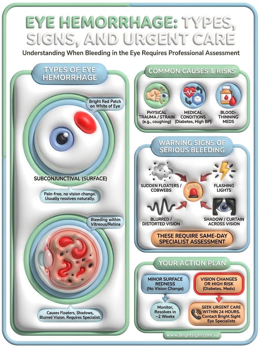
This common form of bleeding occurs beneath the transparent conjunctiva that covers the white part of your eye. You’ll notice a bright red patch that stands out sharply against the white sclera, often appearing suddenly without warning. The blood becomes trapped between the conjunctiva and the underlying tissue, creating a vivid appearance that can spread across part or all of the white surface.
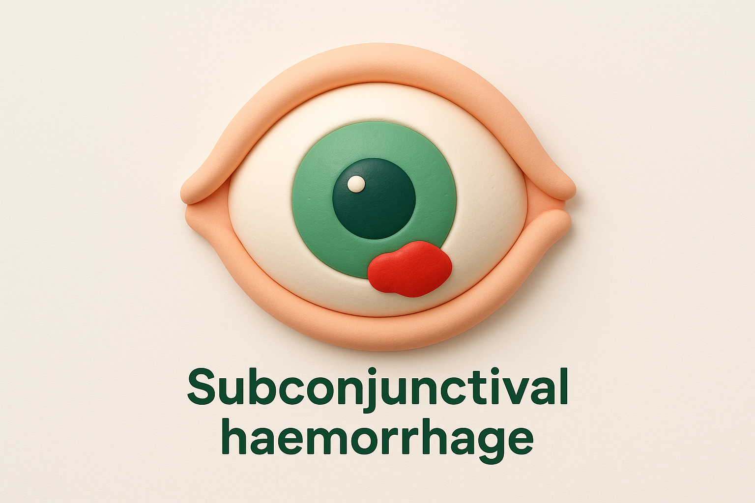
Despite its dramatic look, subconjunctival haemorrhage rarely causes pain or affects your vision. You might feel slight irritation or awareness of the affected area, but the blood remains on the surface rather than interfering with your eye’s internal structures. The patch typically changes colour as it heals, transitioning from bright red to yellow before disappearing completely.
A bright red patch on the white of your eye without vision changes typically indicates surface bleeding that resolves without treatment.
Internal bleeding locations
Bleeding within your eye’s deeper structures creates different symptoms and requires specialist diagnosis. Vitreous haemorrhage occurs when blood enters the gel-like substance filling your eye, causing floaters, shadows, or blurred vision rather than visible redness. Retinal and subretinal bleeding affects the light-sensitive tissue at the back of your eye, potentially creating blind spots or distorted vision that demand urgent assessment by specialists like Dr Dubey or Dr Odouard.
Common causes and risk factors
Eye haemorrhage develops through various pathways, from simple physical triggers to complex medical conditions affecting your blood vessels. Identifying your personal risk factors allows you to take preventive measures and recognise when bleeding requires specialist attention. Your individual circumstances, including existing health conditions and medications, significantly influence both the likelihood of developing eye haemorrhage and the appropriate response.
Physical trauma and medical conditions
Direct injury to your eye from blunt force, rubbing too vigorously, or sudden pressure changes during activities like heavy lifting can rupture delicate blood vessels. Straining during coughing fits, vomiting, or even sneezing forcefully creates enough pressure to cause surface bleeding, though these incidents typically resolve without intervention.
Underlying health conditions increase your vulnerability to internal eye haemorrhage beyond the surface bleeding visible to others. Diabetes damages blood vessels throughout your body, including the tiny capillaries in your retina, whilst high blood pressure weakens vessel walls and makes them prone to leaking. Blood disorders affecting clotting or vessel integrity further elevate your risk of both spontaneous and trauma-related bleeding.
Diabetes and high blood pressure damage the delicate blood vessels in your eye, increasing your risk of serious internal bleeding that can threaten your vision.
Medications and lifestyle factors
Blood-thinning medications like warfarin or aspirin reduce your blood’s ability to clot, making you more susceptible to eye haemorrhage from minor trauma or spontaneous bleeding. These medications serve important purposes for your overall health, but they require careful monitoring and awareness of bleeding risks.
Age naturally weakens blood vessel walls, and alcohol consumption temporarily raises blood pressure, both contributing to increased haemorrhage risk.
Symptoms and when to seek urgent care
Recognizing which symptoms indicate minor surface bleeding versus those suggesting deeper complications determines how quickly you should seek professional care. Surface bleeding typically creates visible redness without affecting your vision, whilst internal haemorrhage produces symptoms that interfere with your ability to see clearly. Your response time becomes critical when symptoms suggest bleeding within the retina or vitreous cavity, as delays can lead to permanent vision loss.
Warning signs of serious bleeding
Sudden onset of floaters (dark spots, cobwebs, or strings drifting across your vision) combined with flashing lights signals potential vitreous haemorrhage or retinal detachment requiring urgent assessment. These symptoms indicate blood or debris floating within your eye’s interior, blocking light pathways and potentially damaging your retina. You might notice a shadow or curtain moving across your peripheral vision, suggesting retinal involvement that demands immediate specialist attention from practitioners like Dr Dubey or Dr Odouard.
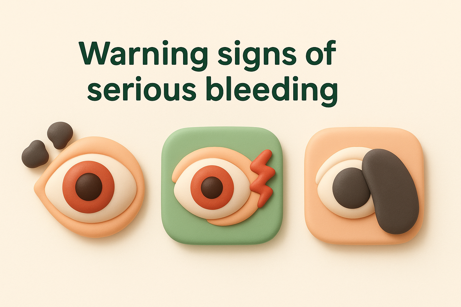
Blurred vision that doesn’t improve with blinking, difficulty reading, or distortion where straight lines appear wavy point to bleeding affecting your central retina or macula. These changes differ from the clear vision you maintain with surface haemorrhage, indicating that blood has entered critical areas responsible for detailed sight.
Sudden floaters, flashing lights, or a shadow crossing your vision require same-day assessment by an eye specialist, as these symptoms suggest internal bleeding that can threaten your sight.
When to seek immediate care
Contact an eye specialist within 24 hours if you experience vision changes accompanying eye hemorrhage, even if the redness appears minor. Patients with diabetes, high blood pressure, or those taking blood-thinning medications should seek prompt evaluation due to their elevated risk of serious complications. Your existing medical conditions increase the likelihood that bleeding extends beyond the surface, making professional assessment essential for protecting your vision.
How doctors diagnose and treat eye haemorrhage
Your specialist begins assessment by examining your medical history and current symptoms to determine the likely source and severity of bleeding. This initial conversation reveals risk factors like diabetes, blood pressure issues, or blood-thinning medications that influence both diagnosis and treatment planning. Dr Dubey or Dr Odouard will ask about vision changes, when the bleeding started, and whether you experienced trauma or physical strain.
Diagnostic examination methods
Specialists use several tools to identify where bleeding occurs within your eye. A slit lamp examination allows detailed viewing of your eye’s surface structures, revealing subconjunctival haemorrhage or inflammation that might accompany deeper bleeding. Your doctor examines your retina and optic nerve through dilated pupils using ophthalmoscopy or fundoscopy, checking for vitreous haemorrhage, retinal tears, or macular involvement that requires intervention.
Advanced imaging through optical coherence tomography (OCT) creates detailed cross-sectional images of your retina, measuring fluid or blood accumulation with precision. Fluorescein angiography highlights blood vessel abnormalities by injecting dye that reveals leaking vessels or areas of poor circulation requiring treatment.
Your specialist uses advanced imaging technology to pinpoint exactly where bleeding occurs, determining whether you need observation or immediate intervention.
Treatment approaches
Treatment depends entirely on the type and location of your eye hemorrhage. Surface bleeding resolves naturally within two weeks without intervention, though artificial tears may improve comfort. Internal bleeding requires targeted approaches: vitreous haemorrhage may clear spontaneously or need vitrectomy surgery, whilst retinal bleeding might require laser treatment, injections, or surgical repair. Your specialist develops a treatment plan addressing both the bleeding and underlying causes like diabetic retinopathy or vascular occlusion.
What to do next
Eye hemorrhage ranges from minor surface bleeding that resolves naturally to serious internal conditions requiring urgent specialist care. You now understand the difference between harmless subconjunctival hemorrhage and symptoms suggesting deeper bleeding that threatens your vision. This knowledge empowers you to respond appropriately when you notice blood in or around your eye.
Surface bleeding typically clears within two weeks without intervention, but vision changes, floaters, or flashing lights demand same-day assessment. Don’t wait to see if symptoms improve, as delayed treatment of internal hemorrhage can result in permanent sight loss. Your specialist uses advanced diagnostic equipment to determine exactly where bleeding occurs and what treatment you require.
If you experience eye hemorrhage with vision changes, or if you have diabetes, high blood pressure, or take blood-thinning medications, contact Dr Dubey or Dr Odouard at Bright Sight Eye Specialists for prompt assessment. They provide comprehensive diagnosis and treatment for all forms of eye hemorrhage, protecting your vision through evidence-based care across Sydney’s Eastern Suburbs.