


You’re reading a book or staring at a clear sky when small, shadowy shapes drift across your vision. These are eye floater dots, and nearly everyone notices them at some point. Most of the time, they’re a normal part of ageing and nothing to worry about. But in some cases, a sudden increase in floaters can signal something that needs urgent attention.
Knowing the difference matters. A harmless floater and one that warns of a retinal tear or detachment can look surprisingly similar, and the window for effective treatment can be narrow. At Bright Sight Eye Specialists in Randwick, Dr Rahul Dubey and Dr Capucine Odouard regularly assess and treat patients with floater-related concerns, from routine monitoring through to vitrectomy surgery for persistent or sight-threatening cases.
This article explains what causes eye floater dots, when they’re serious, and what your treatment options look like, so you can make an informed decision about your next step.
Why eye floater dots happen
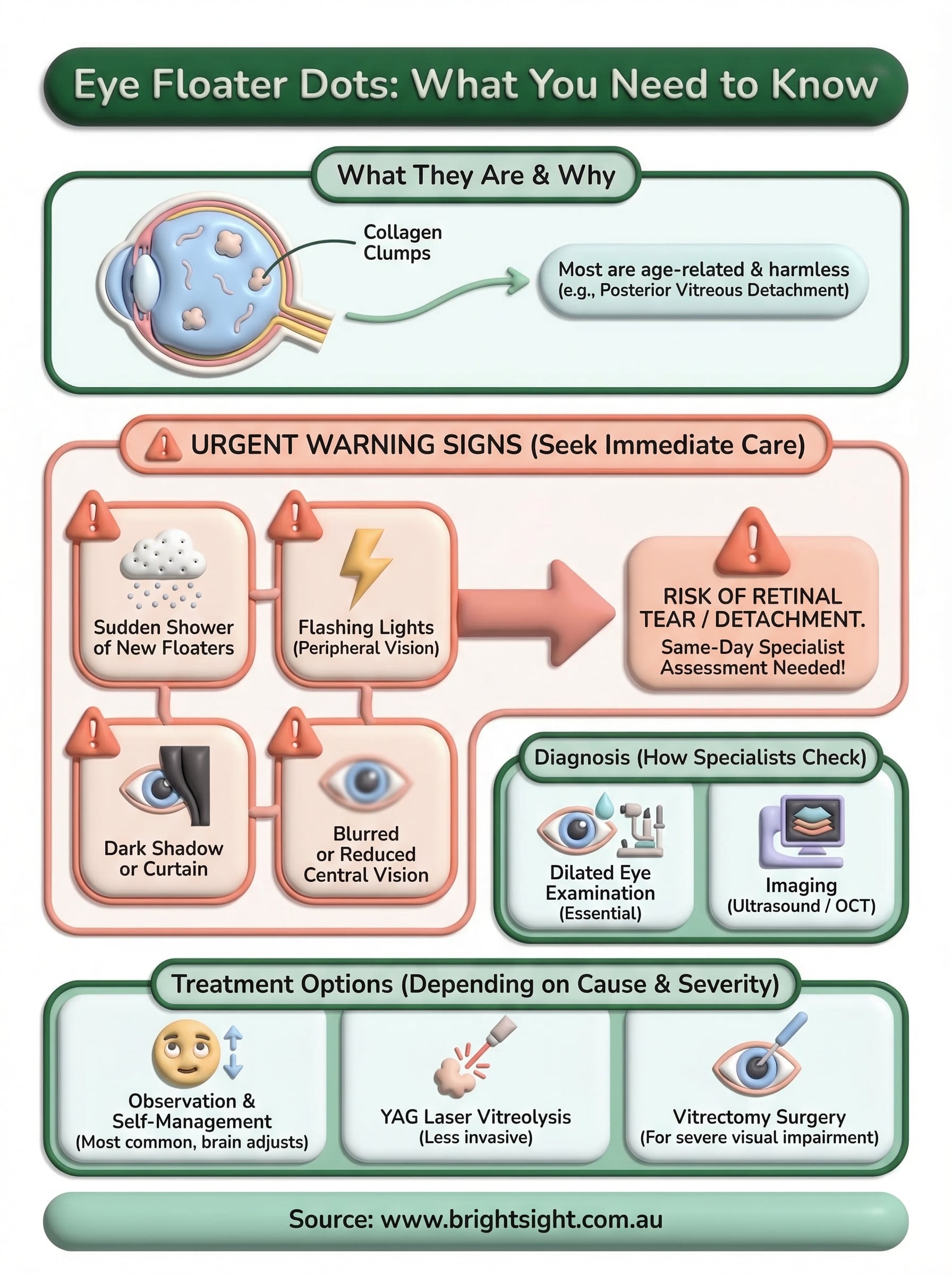
The human eye contains a gel-like substance called the vitreous, which fills the space between the lens and the retina. This gel consists mostly of water, but it also holds microscopic protein fibres called collagen. When these fibres clump together or the vitreous changes in consistency, they cast small shadows onto the retina, and those shadows are what you see as eye floater dots drifting across your field of view.
The vitreous gel and why it changes
Throughout your life, the vitreous gradually loses its uniform, gel-like structure and becomes more fluid. As this happens, the collagen fibres tend to bundle together and drift freely, shifting with every movement your eye makes. That’s why floaters dart away when you try to look directly at them, then slowly drift back into view once your eye settles.
Most floaters result from normal, age-related changes inside the vitreous and pose no threat to your sight.
Posterior vitreous detachment
The most common cause of a sudden increase in floaters is a condition called posterior vitreous detachment (PVD). This occurs when the vitreous gel separates from the inner surface of the retina, a natural process that happens in most people between the ages of 50 and 75. During PVD, a distinct ring-shaped floater often appears alongside brief flashes of light.

Your risk of PVD rises with age, myopia (short-sightedness), and previous eye surgery. Once the gel has fully separated, most people find their floaters gradually become less noticeable as the brain adjusts. However, the period during which PVD develops is the most important time to rule out a retinal tear with an eye specialist.
How to check if they are serious
Most eye floater dots are harmless, but certain changes in your symptoms should prompt you to seek urgent assessment. The key is paying attention to how your floaters behave and whether any new symptoms appear alongside them.
Warning signs that need urgent care
If you notice a sudden large increase in floaters, especially ones you have never seen before, treat it as a reason to contact an eye specialist the same day. The same applies if you see flashes of light in your peripheral vision, which can indicate the vitreous is pulling on the retina.
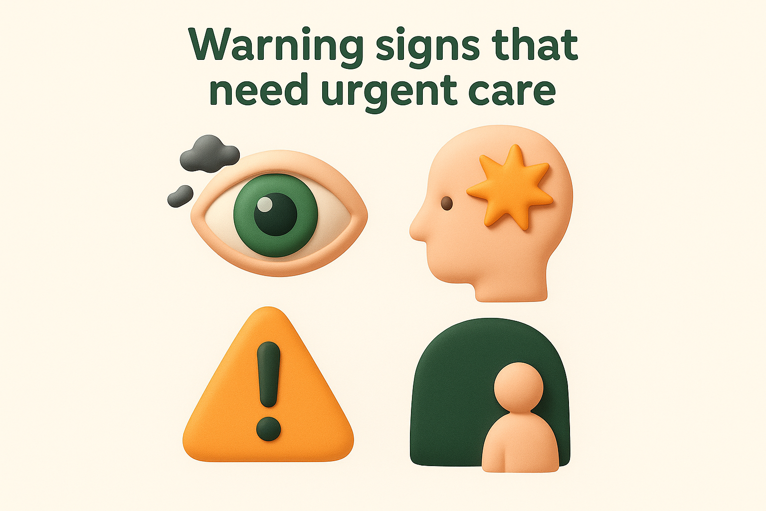
A sudden shower of new floaters, with or without flashes, can be the first sign of a retinal tear, which requires urgent treatment to prevent detachment.
Watch for these specific warning signs:
- A sudden burst of many new floaters appearing at once
- Flashing lights, particularly at the edges of your vision
- A dark shadow or curtain creeping across your visual field
- Blurred or reduced central vision
Any one of these symptoms warrants same-day contact with an eye specialist. Early treatment for a retinal tear can prevent it from progressing to a full retinal detachment, which is far more serious and harder to treat.
Other causes that need treatment
Not all eye floater dots stem from age-related vitreous changes. Some floaters result from conditions that require active medical treatment, and identifying the underlying cause early makes a significant difference to your outcome.
Vitreous haemorrhage
Vitreous haemorrhage occurs when blood leaks into the vitreous cavity, often from damaged or abnormal blood vessels due to diabetic retinopathy, a retinal tear, or trauma. You may notice a sudden dense shower of floaters, red-tinged vision, or significant blurring. It requires prompt assessment because the underlying cause determines the correct treatment.
Diabetic eye disease is one of the most common causes of vitreous haemorrhage and can develop without obvious symptoms until significant damage has already occurred.
Inflammation and infection
Uveitis, or inflammation inside the eye, releases inflammatory cells into the vitreous, producing floater-like symptoms similar to those caused by protein clumps. It may result from infection, autoimmune conditions, or systemic diseases such as rheumatoid arthritis.
Treatment depends on the underlying cause but typically involves anti-inflammatory medication or steroid injections. If your floaters accompany eye pain, redness, or light sensitivity, seek assessment promptly rather than waiting for symptoms to resolve on their own.
How eye specialists diagnose floaters
When you visit an eye specialist about eye floater dots, the assessment goes well beyond a quick look. The goal is to find the cause and confirm whether your retina is intact, so the examination needs to be thorough and use specialised equipment that a standard optometry consultation may not include.
Dilated eye examination
Your specialist will place dilating drops into your eyes to widen the pupil, which allows a clear view of the vitreous and the retina all the way to its edges. Using an instrument called a slit lamp combined with a specialised lens, the doctor can inspect the vitreous for clumps, blood, or inflammation, and scan the retina for any tears or thinning.
A dilated examination is the most reliable way to rule out a retinal tear in someone presenting with new or worsening floaters.
Imaging and additional tests
In some cases, your specialist may use ultrasound imaging (B-scan ultrasonography) to examine the vitreous and retina, particularly if bleeding is present and a direct view is not possible. Optical coherence tomography (OCT) may also be used to assess the macula and the vitreoretinal interface in detail, providing a cross-sectional image of the retinal layers that cannot be seen with the naked eye alone.
Treatment options and self-management
Treatment depends on what is causing your eye floater dots and how much they affect your daily life. For many people, no active treatment is needed, and the right approach is simply to monitor symptoms over time.
Observation and self-management
Most floaters caused by posterior vitreous detachment settle without intervention. Your brain gradually learns to filter them out, and many people find floaters become far less noticeable within a few months. Moving your eyes up and down can briefly shift floaters out of your central vision when they become distracting.
There is no eye drop or supplement proven to reduce floaters, so be cautious of products making those claims.
Surgical and laser options
When floaters significantly impair your vision or quality of life, two clinical options exist. Vitrectomy involves removing the vitreous gel and replacing it with a saline solution, which eliminates the floaters entirely. It carries small but real surgical risks, so your specialist will weigh these carefully against your symptoms. YAG laser vitreolysis uses a laser to break up floater clumps and is a less invasive option, though it suits only certain floater types and locations based on their size and position within the vitreous.
What to do next
If you have new or worsening eye floater dots, the safest step is to book a review with an eye specialist rather than waiting to see if they settle. Most floaters turn out to be harmless, but a dilated examination gives you a definite answer and rules out a retinal tear before any damage can progress.
At Bright Sight Eye Specialists in Randwick, Dr Rahul Dubey and Dr Capucine Odouard assess floater-related concerns as part of a thorough vitreoretinal consultation. Whether you need monitoring, laser treatment, or surgery, they will explain your options clearly and help you decide on the right path for your vision.
Do not delay if you experience sudden floaters, flashes of light, or a shadow creeping across your vision. These symptoms need same-day attention. Book an appointment with Bright Sight Eye Specialists and get a clear answer about what you are seeing.