


An epiretinal membrane is a thin sheet of scar tissue that forms on the retina’s surface, usually over the macula where your sharpest vision happens. Think of it as a piece of cling film stretched across the inside of your eye. Sometimes called macular pucker or cellophane maculopathy, this condition can cause your central vision to blur or distort. Many people have small epiretinal membranes that cause no problems at all. Others notice straight lines appearing wavy or difficulty reading fine print. The membrane develops when cells migrate to the retina’s surface and create a translucent layer.
This article explains how doctors diagnose epiretinal membranes using OCT scans and other tests, what symptoms to watch for, and when surgery might help. You’ll learn about the causes and risk factors, understand when simple monitoring is enough versus when vitrectomy surgery becomes necessary, and discover what recovery looks like. By the end, you’ll know whether your symptoms warrant a specialist assessment and what treatment options are available.
Why epiretinal membranes matter
Your central vision controls nearly every task that requires detail, from reading text messages to recognising faces across a room. When an eye epiretinal membrane forms over your macula, it can interfere with these everyday activities. While many membranes remain mild and stable, others progress gradually and pull on the retina as they contract. This traction causes distortion that makes straight edges appear curved or wavy, affecting your ability to drive safely or read comfortably.

Early detection allows you to track changes and plan treatment before significant vision loss occurs.
Most people adapt to mild distortion without realising their vision has changed. You might hold reading material at different distances or squint more often without connecting these changes to your eye health. Untreated progressive membranes can lead to permanent structural changes in your macula, making surgical outcomes less predictable. The tissue beneath a longstanding membrane may not fully recover its normal architecture even after removal. Regular monitoring helps your ophthalmologist identify when intervention makes sense, typically before the membrane causes irreversible damage to your photoreceptors or disrupts the delicate retinal layers beneath.
How to diagnose an epiretinal membrane
Your eye specialist begins by examining your retina with dilated pupils and specialised lenses that provide a magnified view of your macula. During this clinical examination, they look for signs like a shiny cellophane-like appearance or wrinkling on the retinal surface. This fundus examination reveals whether the membrane affects your fovea (the very centre of your macula) and helps determine its severity. Your ophthalmologist also assesses how much the membrane has distorted your retinal architecture and whether it’s causing any secondary effects like macular oedema or pseudohole formation.
Clinical examination findings
When looking at your retina, your doctor searches for specific visual clues that indicate an eye epiretinal membrane. The membrane typically appears as a translucent sheen over the macula, sometimes with visible striae or folds in the retinal surface. You might not see these changes yourself, but your specialist can spot them using slit-lamp biomicroscopy with special contact lenses. They’ll also check for vascular tortuosity, where blood vessels appear more twisted than normal due to traction from the membrane.
Your specialist evaluates both eyes even if symptoms affect only one, since membranes can develop bilaterally.
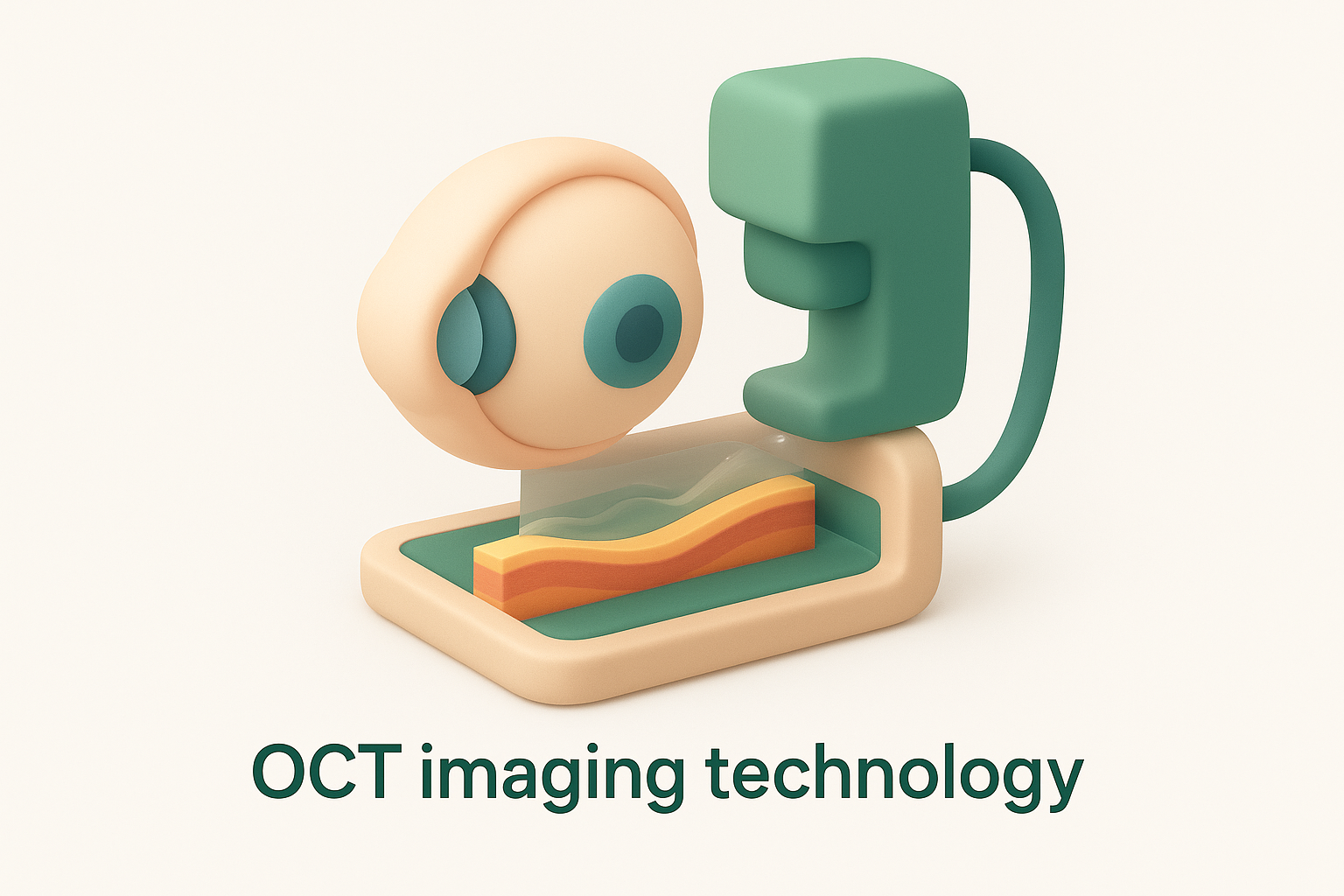
OCT imaging technology
Optical coherence tomography provides the most detailed view of an epiretinal membrane’s structure and its impact on your retina. This non-invasive scan creates cross-sectional images showing the membrane as a bright line on your retina’s inner surface. OCT reveals how much retinal distortion exists, whether the membrane has created foveal irregularities, and if any fluid accumulation has occurred beneath it. Your doctor uses OCT to measure your central retinal thickness and monitor changes over time, making it essential for deciding whether surgery is necessary.
Symptoms, causes and risk factors
Understanding what triggers an eye epiretinal membrane and recognising its symptoms helps you decide when to seek specialist care. Most membranes develop gradually over months or years, making symptoms subtle at first. Your vision changes might seem minor initially, but tracking them becomes important as the membrane evolves. The condition affects each person differently, with some experiencing minimal disruption whilst others notice significant visual distortion that interferes with daily tasks.
Common symptoms you might experience
Blurred central vision ranks as the most frequent complaint, making reading or recognising faces more difficult than usual. You might notice straight lines appearing wavy or bent, particularly when looking at doorframes, window blinds, or text on a page. Some people experience micropsia, where objects appear smaller than they actually are, or develop mild double vision in the affected eye. These symptoms typically worsen gradually rather than appearing suddenly.

Visual distortion often becomes more noticeable when you close your unaffected eye and rely solely on the eye with the membrane.
Difficulty reading fine print or viewing detailed images indicates the membrane has begun affecting your central macula. You may find yourself squinting more often or needing brighter lighting for tasks that previously felt effortless.
What causes epiretinal membranes
The primary trigger involves posterior vitreous detachment, a natural ageing process where the gel-like vitreous pulls away from your retina. This separation can create tiny breaks in the internal limiting membrane, allowing glial cells and other cells to migrate onto the retinal surface. These cells then proliferate and form the translucent membrane you see on examination. Most epiretinal membranes develop without any obvious secondary cause, making them idiopathic in nature.
Secondary membranes form after events like retinal tears, intraocular surgery, eye trauma, or inflammatory conditions. Conditions such as diabetic retinopathy or retinal vein occlusion also increase membrane formation risk.
Key risk factors
Age over 50 years represents the strongest predictor, with prevalence increasing significantly after 75. People who have experienced posterior vitreous detachment face higher risk, as do those with previous retinal surgery or trauma to the eye.
When treatment or surgery is needed
Most eye epiretinal membranes require no immediate treatment and can be safely monitored through regular eye examinations. Your ophthalmologist tracks membrane progression using OCT imaging and visual acuity tests, typically scheduling follow-up appointments every six to twelve months. This observation approach makes sense when your symptoms remain mild and daily activities continue without significant disruption. Many people maintain stable vision for years, with their membrane causing only minor distortion that doesn’t interfere with reading, driving, or recognising faces.
Observation versus intervention
Watchful waiting remains the standard approach when your visual acuity stays at 20/40 or better and metamorphopsia doesn’t significantly affect your quality of life. Your specialist compares OCT scans over time to assess whether the membrane is contracting or pulling on your retina. This monitoring strategy protects you from unnecessary surgical risks whilst ensuring timely intervention if progression occurs. You’ll use an Amsler grid at home between appointments to check for changes in your central vision, reporting any worsening distortion promptly.
Surgery becomes worth considering when your symptoms prevent normal daily functioning. You might struggle to read standard print, experience difficulty with tasks requiring fine detail, or notice substantial visual distortion that affects your independence.
Signs that surgery becomes necessary
Declining visual acuity below 20/60 or progressive metamorphopsia that interferes with reading represents clear indicators that surgical intervention may benefit you. Your ophthalmologist also considers whether the membrane has created macular thickening or structural distortion visible on OCT scans. These anatomical changes suggest the membrane is exerting significant traction on your retina.
Surgery typically provides better outcomes when performed before the membrane causes permanent damage to your photoreceptor layer.
Pseudohole formation or persistent macular oedema despite the membrane’s presence might prompt earlier surgical discussion, as these complications can limit visual recovery potential.
Vitrectomy surgery and recovery
Vitrectomy removes the vitreous gel from your eye and peels away the epiretinal membrane using microsurgical instruments. Your surgeon performs this procedure under local anaesthetic with sedation or general anaesthetic, typically as day surgery lasting 30 to 60 minutes. Three tiny incisions, each smaller than a millimetre, allow instruments to enter your eye. The surgeon removes the vitreous gel to access the retinal surface, then carefully peels the membrane away using fine forceps. In most cases, your ophthalmologist also removes the internal limiting membrane beneath the epiretinal membrane to reduce recurrence risk.
The surgical procedure
Your eye receives numbing drops and injections around the socket to ensure comfort throughout surgery. The surgeon inserts a light source, infusion line, and surgical instruments through the tiny incisions, working whilst viewing the retina through a surgical microscope. Membrane peeling requires meticulous technique, as the tissue adheres tightly to your retina. Your surgeon may use vital dyes like brilliant blue to stain the membrane and make it more visible during removal. The procedure concludes with a check for any retinal tears and the eye is filled with saline or gas, depending on your specific case.

Removing the internal limiting membrane alongside the eye epiretinal membrane significantly lowers the chance of membrane regrowth.
Recovery timeline and expectations
Vision typically worsens immediately after surgery before gradually improving over weeks to months. Your eye remains red and uncomfortable for the first week, requiring regular antibiotic and anti-inflammatory drops. Most patients notice visual improvement within three to six months, though full recovery can take up to a year. You’ll need to avoid heavy lifting, strenuous exercise, and swimming during the initial healing period. If your surgeon uses gas during surgery, you may need to maintain specific head positioning for several days.
Follow-up appointments monitor your healing progress and check for complications like raised eye pressure, infection, or retinal detachment. The visual outcome depends on how long the membrane was present and the extent of retinal damage before surgery. Around 80 to 90% of patients experience improved vision and reduced distortion, with most achieving at least two lines of visual acuity improvement on the eye chart.

Next steps
An eye epiretinal membrane requires specialist assessment to determine whether observation or surgical intervention suits your particular situation. Schedule an appointment with a retinal specialist if you notice visual distortion, blurred central vision, or difficulty reading fine print. Your ophthalmologist will perform a comprehensive examination and OCT imaging to evaluate the membrane’s severity and progression. Early detection allows for better monitoring and treatment planning before permanent retinal damage occurs. Bright Sight Eye Specialists provides expert diagnosis and treatment for epiretinal membranes in Sydney’s Eastern Suburbs, offering advanced surgical solutions when needed.