


Your eyes work hard every day, processing thousands of visual signals from the moment you wake until you close them at night. Yet many people only think about their eye health when something goes wrong. Understanding common eye conditions early can make a real difference in preserving your vision and quality of life.
Whether you’re noticing changes in your sight, have a family history of eye disease, or simply want to stay informed, knowing what to look for matters. Early detection often leads to better treatment outcomes, and many conditions are highly manageable when caught in time.
At Bright Sight Eye Specialists in Randwick, Dr Rahul Dubey and Dr Capucine Odouard treat a wide range of eye conditions across Sydney’s Eastern Suburbs. This guide covers seven of the most common eye conditions we see in practice, including their symptoms, causes, and available treatments, helping you recognise warning signs and understand when to seek specialist care.
1. Cataracts
Cataracts are one of the most common eye conditions affecting older Australians, clouding the lens inside your eye and making everyday tasks like reading or driving increasingly difficult. The natural lens sits behind your pupil and focuses light onto the retina, but when it becomes cloudy, your vision loses clarity and sharpness.
What cataracts are
Your eye’s lens is made mostly of water and protein, arranged in a precise way that keeps the lens clear. Over time, some of these proteins clump together, creating cloudy areas that block and scatter light passing through. This process happens gradually, often over years, and typically affects both eyes at different rates.
Common cataract symptoms
You might notice your vision becoming blurry or hazy, as though you’re looking through a foggy window. Colours often appear faded or yellowed, and you may find yourself needing brighter light for reading. Many people also experience increased glare from car headlights at night or difficulty seeing in low light conditions.
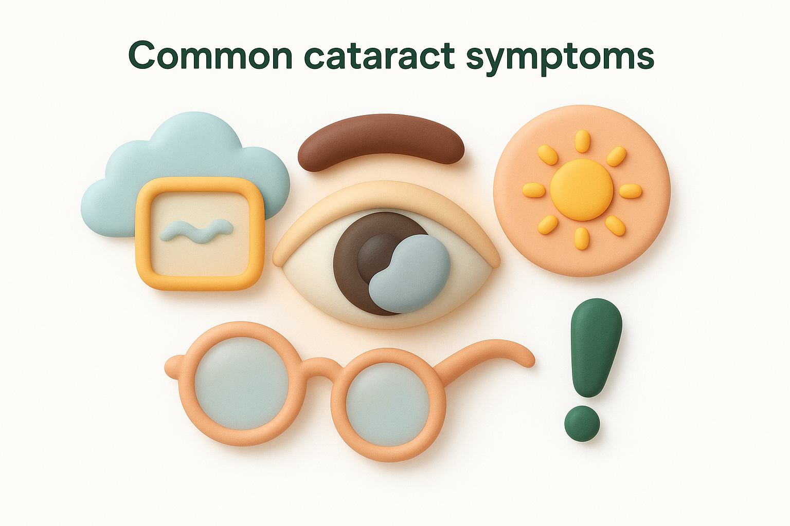
Early cataracts may cause only minor visual disturbances, but as they progress, they can significantly impact your daily activities and quality of life.
Causes and risk factors
Ageing remains the primary cause of cataracts, with most people developing some degree of lens clouding after age 60. Other factors include prolonged UV exposure without eye protection, smoking, diabetes, certain medications like corticosteroids, and previous eye injuries or surgery. Family history also plays a role in determining when and how quickly cataracts develop.
How eye specialists diagnose cataracts
Your eye specialist uses several tests to assess cataracts during a comprehensive examination. They check your visual acuity with an eye chart, examine the lens using a slit lamp microscope, and may dilate your pupils to see the back of your eye. These tests help determine the severity of cataracts and whether they’re affecting your daily life enough to consider surgery.
Treatment options including cataract surgery
Cataract surgery is the only effective treatment for advanced cataracts. During this procedure, your surgeon removes the cloudy lens and replaces it with a clear artificial lens implant. At Bright Sight Eye Specialists, we offer various lens options including standard, multifocal, and extended depth of focus lenses, with many no gap options available for insured patients.
What to expect after surgery and recovery timeline
Most patients notice improved vision within days of surgery, though complete healing takes several weeks. You’ll need to use prescribed eye drops for about a month and avoid strenuous activities initially. Follow-up appointments ensure your eye is healing properly, and many patients report significantly clearer vision and restored colour perception once they’ve recovered.
2. Age-related macular degeneration
Age-related macular degeneration (AMD) affects the central part of your retina called the macula, which you rely on for sharp, detailed vision needed for reading, recognising faces, and driving. This progressive eye condition gradually damages your central vision whilst leaving peripheral vision intact, creating a blind spot in the middle of what you see.
What age-related macular degeneration is
Your macula contains millions of light-sensitive cells that process fine detail and colour. AMD occurs when these cells break down or when abnormal blood vessels grow beneath the retina, distorting your central vision. The condition typically develops after age 50 and represents a leading cause of vision loss in older Australians.
Dry AMD vs wet AMD
Dry AMD accounts for about 85-90% of cases and progresses slowly as small deposits called drusen accumulate under the retina. Wet AMD develops when abnormal blood vessels leak fluid or blood, causing rapid vision loss. You can develop wet AMD in one or both eyes even if you initially had only the dry form.
Symptoms you might notice first
You might notice straight lines appearing wavy or distorted, particularly when reading or looking at door frames. Other signs include difficulty recognising faces from a distance, needing brighter light for close work, and a blurry or dark area in your central vision that gradually enlarges.
Changes in central vision, particularly distortion of straight lines, warrant prompt specialist assessment as early treatment of wet AMD can help preserve your sight.
Risk factors and practical prevention steps
Age over 50 remains the strongest risk factor, along with smoking, family history, high blood pressure, and prolonged UV exposure. You can reduce your risk by quitting smoking, wearing UV-protective sunglasses, maintaining healthy blood pressure, and eating leafy greens rich in lutein and zeaxanthin.
Tests used to diagnose and monitor macular health
Your specialist uses optical coherence tomography (OCT) to create detailed cross-sectional images of your retina, revealing early changes in macular structure. They also perform fluorescein angiography to identify abnormal blood vessels and may ask you to check an Amsler grid at home between appointments to monitor for changes.
Treatment options including injections and lifestyle support
Intravitreal injections of anti-VEGF medication can slow or stop wet AMD progression by reducing abnormal blood vessel growth. Your specialist administers these injections in the consulting room, typically monthly at first then extending intervals based on response. Vitamin supplements containing zinc, vitamins C and E, and specific antioxidants may slow dry AMD progression in certain cases.
3. Glaucoma
Glaucoma represents a group of eye conditions that damage your optic nerve, often through elevated pressure inside your eye. This progressive disease can steal your peripheral vision so gradually that you might not notice changes until significant damage has occurred, making regular eye examinations essential for early detection.
What glaucoma is and why it damages vision
Your eye constantly produces aqueous fluid that normally drains through tiny channels at the front of your eye. When these drainage channels become blocked or don’t work properly, pressure builds inside your eye and damages the optic nerve fibres that carry visual signals to your brain. This damage is irreversible, which makes early treatment crucial.
Early warning signs and why you may not notice symptoms
Most people with open-angle glaucoma experience no early symptoms because the disease affects peripheral vision first whilst leaving central vision intact. You might notice difficulty seeing to the side or trip over objects you didn’t see approaching. Acute angle-closure glaucoma causes sudden eye pain, severe headache, nausea, and blurred vision, requiring emergency care.
Because glaucoma typically causes no pain or obvious symptoms until advanced stages, regular eye examinations with pressure checks and optic nerve assessment remain your best protection against vision loss.
Key risk factors including family history
Your risk increases significantly after age 50, particularly if you have family members with glaucoma, which increases your risk four to nine times. Other factors include high eye pressure, thin corneas, diabetes, high blood pressure, and being of African or Asian ancestry.
How specialists test for glaucoma
Your specialist measures intraocular pressure with a tonometer, examines your optic nerve through dilated pupils, and tests your peripheral vision using automated perimetry. They also use OCT imaging to measure the thickness of nerve fibres around your optic disc, detecting early changes before you notice symptoms.
Treatment options including drops, laser, and surgery
Prescription eye drops form the first line of treatment, lowering eye pressure by reducing fluid production or improving drainage. Your specialist may recommend selective laser trabeculoplasty to improve drainage or surgery to create new drainage channels if drops prove insufficient.
Follow-up schedules and long-term management
You’ll need regular monitoring every three to six months to ensure treatment maintains safe pressure levels and prevents further damage. Your specialist adjusts medications based on pressure readings and visual field tests, ensuring your glaucoma remains stable whilst minimising side effects from treatment.
4. Diabetic retinopathy
Diabetic retinopathy develops when high blood sugar levels damage the tiny blood vessels in your retina, potentially leading to serious vision loss if left untreated. This eye condition affects many people living with diabetes, progressing through stages that range from mild changes to severe complications threatening your sight.
How diabetes affects the retina
High blood sugar weakens and damages the small blood vessels that supply your retina with oxygen and nutrients. These damaged vessels can leak fluid or blood into the retina, causing swelling and blurred vision. As the condition advances, your eye may grow abnormal new blood vessels that bleed easily and lead to scarring, further compromising your vision.
Symptoms and why early disease can feel silent
Early diabetic retinopathy typically causes no noticeable symptoms, which makes regular screening essential. As the condition progresses, you might experience blurred or patchy vision, floaters, difficulty seeing colours, or dark areas in your visual field. Many people only notice problems once significant damage has occurred.
Because diabetic retinopathy often causes no symptoms until advanced stages, regular eye examinations remain crucial for anyone with diabetes, regardless of how well you feel your vision is working.
Screening timing for people with diabetes
You should have a comprehensive eye examination within five years of a Type 1 diagnosis and immediately after a Type 2 diagnosis. Annual screenings become essential for monitoring changes, though your specialist may recommend more frequent examinations if they detect early signs of retinopathy.
How specialists diagnose diabetic eye disease
Your specialist dilates your pupils to examine your retina thoroughly, looking for damaged blood vessels, swelling, or abnormal growth. They use OCT imaging to detect fluid accumulation in your macula and may perform fluorescein angiography to identify leaking vessels or areas of poor blood flow.
Treatment options including laser and injections
Intravitreal injections of anti-VEGF medication help reduce swelling and prevent abnormal blood vessel growth. Your specialist may also recommend laser photocoagulation to seal leaking vessels or destroy abnormal tissue. Advanced cases might require vitrectomy surgery to remove blood or scar tissue from inside your eye.
How to reduce risk through diabetes and blood pressure control
Maintaining tight blood sugar control significantly reduces your risk of developing or worsening diabetic retinopathy. Keeping your blood pressure and cholesterol within target ranges also protects your retinal blood vessels, as does quitting smoking and maintaining a healthy weight through regular exercise and balanced nutrition.
5. Retinal detachment
Retinal detachment occurs when your retina pulls away from the back wall of your eye, cutting off its blood supply and threatening permanent vision loss. This serious eye emergency requires immediate medical attention, as delays in treatment significantly reduce the chances of restoring your sight.
What retinal detachment is
Your retina lines the inside back surface of your eye like wallpaper on a wall, converting light into electrical signals your brain interprets as vision. When tears or holes develop in the retina, fluid can seep underneath and separate it from the supporting tissue beneath, causing it to detach progressively across your visual field.
Symptoms that need urgent care
You might suddenly notice flashes of light in your peripheral vision, a dramatic increase in floaters, or a shadow or curtain moving across your field of view. These warning signs demand immediate specialist assessment, as hours can make the difference between saving and losing your vision.
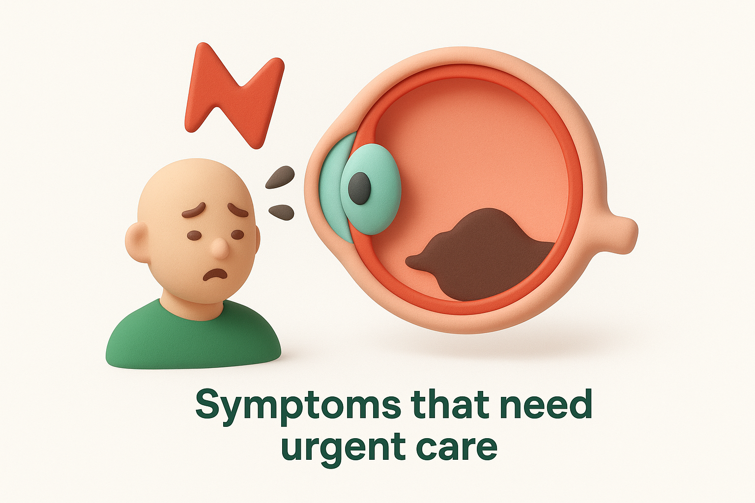
Retinal detachment symptoms require same-day specialist evaluation because treatment becomes less effective as time passes and more retina detaches.
Who has a higher risk
Severe short-sightedness increases your risk significantly, as does previous cataract surgery, eye injuries, or a family history of detachment. You also face higher risk if you’ve had retinal tears or detachment in your other eye, or if you have certain eye conditions like lattice degeneration.
What happens during an emergency eye assessment
Your specialist examines your retina through dilated pupils using specialised lenses and imaging to map the extent of detachment and locate any tears. They assess whether the macula remains attached, which affects your potential visual recovery, and determine the urgency and type of surgery needed.
Treatment options and timing
Most detachments require surgical repair within days to prevent permanent damage. Your surgeon may perform vitrectomy to remove gel pulling on the retina, use laser or freezing to seal tears, or place a silicone band around your eye to support reattachment.
Recovery, follow-up, and vision outcomes
Recovery takes several weeks, during which you may need to maintain specific head positions to help the retina settle. Vision improvement occurs gradually over months, with better outcomes when treatment happens quickly and the macula wasn’t detached.
6. Dry eye and conjunctivitis
Dry eye and conjunctivitis represent two of the most common eye conditions seen in daily practice, affecting people of all ages and causing significant discomfort. Whilst they share some similar symptoms like redness and irritation, these conditions have different causes and require distinct approaches to treatment.
Dry eye overview
Your eyes need a stable tear film to stay comfortable and maintain clear vision. Dry eye occurs when your tears evaporate too quickly or your eyes don’t produce enough quality tears, leaving the surface of your eye inadequately lubricated and protected.
Dry eye symptoms and common triggers
You might experience a gritty or sandy sensation, burning, redness, or paradoxically, watery eyes as your body tries to compensate for dryness. Common triggers include prolonged screen time, air conditioning, contact lens wear, certain medications, and medical conditions like Sjögren’s syndrome or rheumatoid arthritis.
Dry eye treatment options and home care
Artificial tears provide immediate relief for mild cases, whilst your specialist may recommend prescription anti-inflammatory drops or punctal plugs to retain tears for more severe symptoms. Taking regular screen breaks, using a humidifier, and staying well hydrated also help manage symptoms at home.
Conjunctivitis overview
Conjunctivitis, commonly called pink eye, occurs when the clear membrane covering your eye becomes inflamed. This condition can result from bacterial or viral infections, allergies, or irritants, each requiring different management approaches.
How to tell allergic, viral, and bacterial conjunctivitis apart
Allergic conjunctivitis causes intense itching in both eyes with watery discharge, often accompanied by sneezing or nasal symptoms. Viral conjunctivitis produces watery discharge and typically starts in one eye, whilst bacterial forms create thick, yellow-green discharge that may cause your eyelids to stick together overnight.
Bacterial and viral conjunctivitis spread easily through contact, so washing your hands frequently and avoiding touching your eyes helps protect others around you.
When to seek care and how to reduce spread
You should seek specialist assessment if you experience severe pain, light sensitivity, vision changes, or symptoms lasting beyond a week. Avoid sharing towels or pillowcases, wash your hands regularly, and replace eye makeup used whilst infected to prevent recurrence or spreading infection.

Next steps
Understanding these common eye conditions helps you recognise warning signs early and seek appropriate care when needed. Regular comprehensive eye examinations remain your strongest protection against vision loss, particularly as you age or if you have risk factors like diabetes, high blood pressure, or a family history of eye disease.
Your vision affects every aspect of your daily life, from reading and driving to recognising loved ones and enjoying activities you value. Taking prompt action when you notice changes can make a significant difference in preserving your sight and maintaining quality of life.
At Bright Sight Eye Specialists, Dr Rahul Dubey and Dr Capucine Odouard provide comprehensive eye care and surgical solutions across Sydney’s Eastern Suburbs. Book a consultation with our experienced specialists to discuss any concerns about your vision, arrange a routine examination, or explore treatment options for existing conditions.