


Your eyes do an extraordinary amount of work every single day, and most of us don’t give them a second thought until something goes wrong. Understanding eye and eye diseases is one of the most practical steps you can take to protect your vision long-term. The earlier you recognise a problem, the better your chances of effective treatment and preserving your sight.
At Bright Sight Eye Specialists in Randwick, Dr Rahul Dubey and Dr Capucine Odouard diagnose and treat a wide range of eye conditions, from cataracts and macular degeneration to diabetic eye disease and retinal conditions. We see firsthand how much difference early detection makes for our patients across Sydney’s Eastern Suburbs and beyond.
In this guide, we break down seven of the most common eye diseases, covering their symptoms, what causes them, and how they’re treated. Whether you’ve noticed changes in your vision or simply want to stay informed, this is a straightforward, practical resource to help you understand what your eyes might be telling you and when to seek specialist care.
1. Cataract
When it comes to eye and eye diseases, cataracts are among the most common conditions affecting Australians, particularly those over 60. A cataract forms when the natural lens inside your eye gradually becomes cloudy, blocking light from reaching the retina and causing your vision to deteriorate over time.
What it is and why it happens
Your eye’s lens sits just behind the iris and focuses light onto the retina. Over time, proteins within the lens break down and clump together, creating increasing cloudiness. This is largely an age-related process, though injury, certain medications, and systemic diseases can accelerate it.
Symptoms people notice first
Most people notice blurred or hazy vision gradually, often describing it as looking through a fogged window. Other early signs include heightened sensitivity to glare, faded or washed-out colours, difficulty reading in low light, and more frequent changes to your glasses prescription.
Risk factors and who is more likely to get it
Age is the strongest risk factor, with most cataracts developing after 60. Prolonged UV exposure, smoking, diabetes, long-term corticosteroid use, and a family history of cataracts all increase your chances of developing them earlier or more severely.
How an optometrist or ophthalmologist diagnoses it
Your eye specialist will examine your lens using a slit-lamp microscope, which gives a magnified, detailed view of the eye’s internal structures. Visual acuity testing and glare assessment help determine how significantly the cataract is affecting your daily activities and whether surgery is appropriate.
Treatment options and what cataract surgery involves
Surgery is the only effective treatment. The procedure removes the cloudy natural lens and replaces it with a clear artificial intraocular lens (IOL). At Bright Sight, standard and premium lens options are available, including multifocal and EDOF lenses, with no-gap options for eligible privately insured patients.
Cataract surgery is one of the most commonly performed surgical procedures in Australia, with consistently high rates of improved vision outcomes for patients.
Recovery, safety, and what results to expect
Most patients notice clearer vision within days of surgery. Full recovery typically takes four to six weeks, and your surgeon will give you specific aftercare instructions to protect your eye and support healing throughout that period.
When to seek urgent care
Contact an eye specialist promptly if you experience sudden vision loss or significant pain, as these are not typical cataract symptoms and may indicate a separate, more urgent condition requiring immediate assessment.
2. Age-related macular degeneration
Age-related macular degeneration (AMD) is one of the leading causes of permanent vision loss in Australians over 50, and it sits high on the list when discussing eye and eye diseases that need early attention.
What it is and how it affects central vision
Your macula is the small, central part of your retina responsible for sharp, detailed vision needed for reading, recognising faces, and driving. AMD causes the macula to deteriorate over time, leaving a blurred or blank spot in the centre of your visual field while peripheral vision typically stays intact.
Early vs late symptoms
Early AMD often produces no noticeable symptoms, which is why routine testing matters. As it progresses, you may notice straight lines appearing wavy, difficulty reading fine print, or a dark patch developing in the centre of your vision.
AMD can progress slowly over years or rapidly over weeks, depending on the type. Regular monitoring is essential.
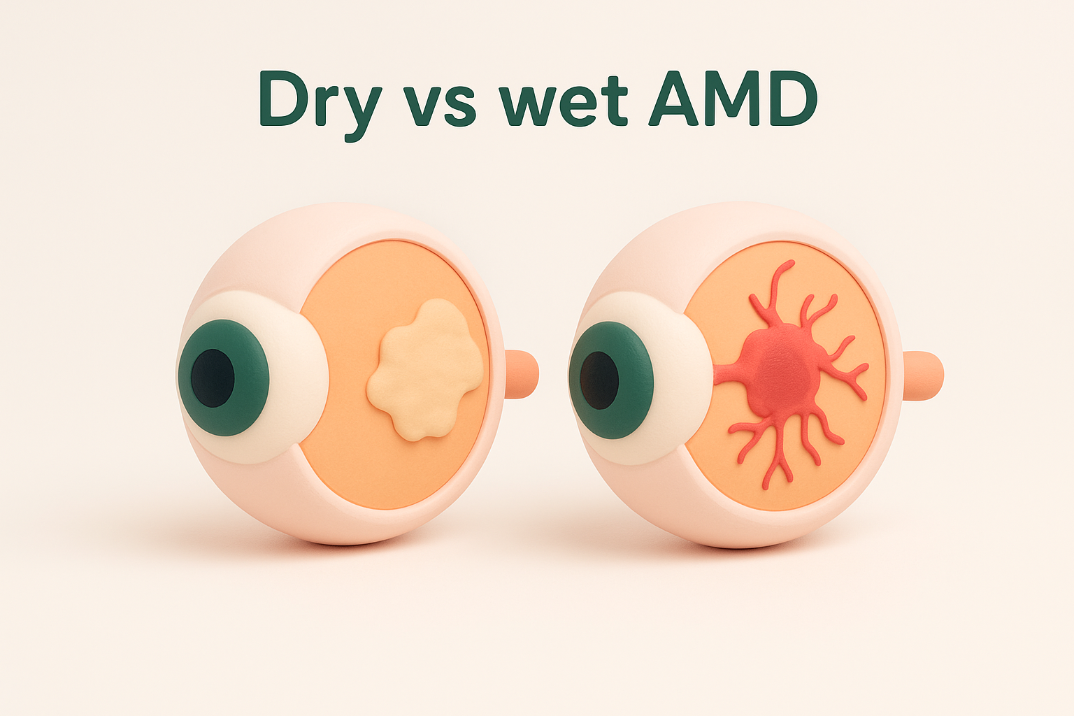
Dry vs wet AMD
Dry AMD accounts for the majority of cases and involves a gradual breakdown of macular tissue. Wet AMD is less common but more aggressive, caused by abnormal blood vessels leaking fluid beneath the retina, leading to faster and more severe vision loss.
Risk factors and prevention steps that help
Age, smoking, and family history are the strongest risk factors. Wearing UV-protective sunglasses, eating leafy green vegetables, and stopping smoking all reduce your risk meaningfully.
How clinicians test for AMD
Your specialist uses an OCT scan to image retinal layers in detail, alongside visual acuity tests and an Amsler grid to detect early central vision distortion.
Treatment options, including injections for wet AMD
Dry AMD has no cure currently, but nutritional supplements (AREDS2 formulation) may slow progression in intermediate cases. Wet AMD is treated with intravitreal injections of anti-VEGF medication, which reduce leakage and can stabilise or improve vision.
When to seek urgent care
If straight lines suddenly look wavy or distorted, or you notice a rapid change in central vision, contact an eye specialist immediately. Wet AMD can cause irreversible damage within days without prompt treatment.
3. Diabetic retinopathy
Diabetic retinopathy is a serious complication of diabetes and one of the most prevalent eye and eye diseases affecting working-age Australians. It develops when high blood sugar damages the small blood vessels inside the retina, and it can cause significant vision loss before you notice anything is wrong.
What it is and why it can progress quietly
The condition advances through stages, and the early stages typically produce no symptoms. Damage accumulates in the retinal blood vessels gradually, which is exactly why routine eye checks matter even when your vision feels unchanged.
Symptoms and warning signs
As retinopathy progresses, you may notice blurred or patchy vision, new floaters, or trouble seeing in low light. Sudden vision loss can occur if a vessel bleeds into the vitreous gel, and this requires same-day assessment.
Who is at risk and how often to check your eyes
Anyone with type 1 or type 2 diabetes carries a risk. Annual dilated eye exams are recommended from diagnosis, or more frequently once retinopathy is detected.
How clinicians diagnose and stage it
Your specialist dilates your pupils and examines your retina directly, supported by OCT imaging and fundus photography to document the precise stage of damage and guide treatment decisions.
Treatment options, including laser and injections
Laser treatment seals leaking vessels and reduces further damage. For diabetic macular oedema, intravitreal anti-VEGF injections reduce swelling and help protect central vision.
Early treatment substantially lowers the risk of severe vision loss from diabetic retinopathy.
How blood sugar, blood pressure, and cholesterol affect outcomes
Keeping your blood sugar well controlled is the most effective way to slow progression. Raised blood pressure and cholesterol also damage retinal vessels, so managing all three together improves your long-term outcome significantly.
When to seek urgent care
Contact an eye specialist immediately if you experience sudden vision loss or a significant new floater, as these symptoms may indicate vitreous haemorrhage or retinal detachment requiring urgent treatment.
4. Glaucoma
Glaucoma is one of the most common eye and eye diseases causing permanent vision loss, yet many people live with it for years without knowing. It develops when the optic nerve is progressively damaged, usually by raised pressure inside the eye.
What it is and why early disease often has no symptoms
The optic nerve carries visual information to your brain, and glaucoma silently damages it over time. Because peripheral vision is lost first, most people notice nothing until significant and irreversible damage has already occurred.
Open-angle vs angle-closure glaucoma
Open-angle glaucoma is the most common form and advances slowly without any pain or obvious symptoms. Angle-closure glaucoma is less common but far more acute, triggered by a sudden narrowing of the eye’s drainage angle and a rapid spike in pressure.
Symptoms that suggest an emergency
Angle-closure glaucoma can produce sudden eye pain, nausea, and blurred vision alongside halos around lights. These symptoms require same-day emergency care, as elevated pressure can permanently damage the optic nerve within hours.
If you develop sudden eye pain with nausea or rapid vision changes, go to an emergency eye hospital immediately.
Risk factors and who should get checked earlier
People over 50, those with a family history of glaucoma, elevated eye pressure, diabetes, or high myopia carry a greater risk and benefit from earlier routine screening.
How clinicians diagnose and monitor glaucoma
Your specialist measures intraocular pressure, inspects the optic nerve, and uses visual field tests and OCT imaging to detect and monitor nerve damage at each visit.
Treatment options, including drops, laser, and surgery
Prescription eye drops that reduce intraocular pressure are typically the first treatment. When drops are insufficient, laser therapy or surgical drainage procedures offer more sustained pressure control.
What ongoing follow-up looks like
Glaucoma requires lifelong management because no treatment reverses existing nerve damage. Regular appointments let your specialist confirm that eye pressure and nerve health are remaining stable over time.
5. Retinal detachment
Retinal detachment sits at the sharp end of eye and eye diseases that require immediate action. When the retina separates from the back wall of your eye, its blood supply cuts off rapidly, and without same-day treatment, permanent vision loss is a real risk.
What it is and why it needs same-day assessment
The retina is a thin, light-sensitive layer lining the inside of your eye. Once it lifts away, retinal cells begin to die quickly without their underlying blood supply, making rapid surgical treatment essential to preserving useful vision.
Classic symptoms: flashes, floaters, curtain or shadow
You may notice sudden flashes of light or a shower of new floaters. A dark curtain or shadow spreading across your visual field is the most urgent warning sign and requires immediate attention.
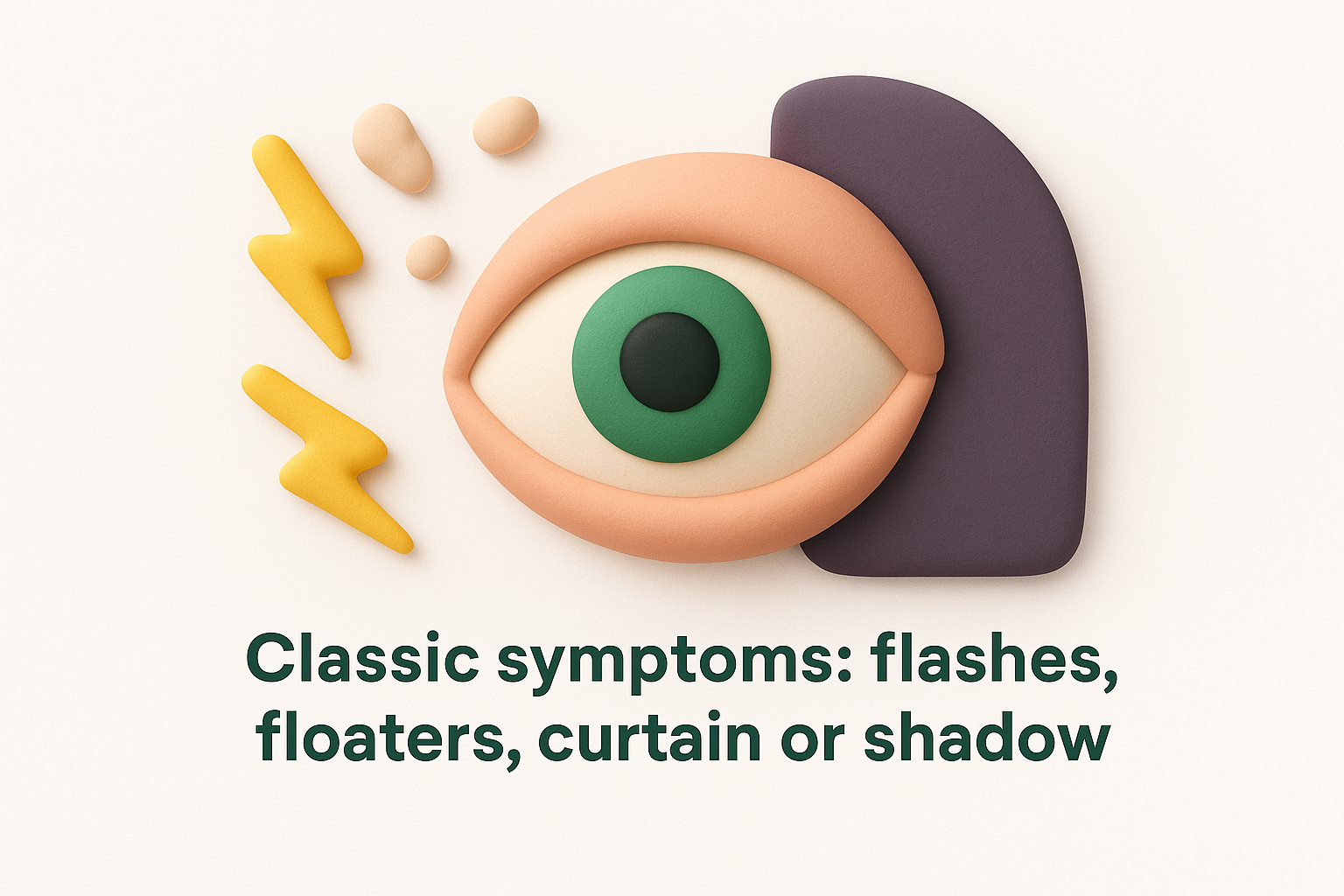
A shadow spreading across your vision is an emergency. Seek same-day eye care without delay.
Common causes and risk factors
High myopia and previous eye surgery are among the strongest risk factors. A prior retinal tear, eye injury, or family history of detachment also raises your risk considerably.
How clinicians confirm a detachment
Your specialist examines your retina through dilated pupils, often combining indirect ophthalmoscopy with ultrasound imaging when the internal view is limited.
Treatment options and timing
Most detachments need surgery, either vitrectomy or scleral buckle, to reattach the retina. The sooner surgery is performed, the stronger your visual outcome is likely to be.
What to do while you wait to be seen
Avoid strenuous activity and follow any positioning advice your specialist provides, as specific positions can slow further detachment spreading in some situations.
When to go straight to the emergency department
Go directly to an emergency eye unit if you experience a spreading shadow or curtain in your vision, sudden central vision loss, or a large burst of new floaters with flashes.
6. Dry eye and conjunctivitis
Not every red or irritated eye signals a serious problem, but knowing the difference between dry eye and conjunctivitis helps you respond appropriately. These are among the most common eye and eye diseases seen in general practice, and both can worsen without the right care.
How to tell irritation from infection or allergy
Dry eye typically produces a gritty, burning sensation that worsens throughout the day. Conjunctivitis usually brings redness, discharge, or marked itching, and often affects one eye before spreading to the other.
Dry eye: symptoms, causes, and common triggers
Your tear film can break down due to screen use, air conditioning, contact lens wear, and ageing. Symptoms include stinging, watery eyes, and fluctuating vision, particularly during extended reading or driving.
Conjunctivitis: viral vs bacterial vs allergic signs
Viral conjunctivitis produces watery discharge and often follows a cold. Bacterial conjunctivitis causes thick, yellow-green discharge, while allergic conjunctivitis brings intense itching and swelling without discharge.
Safe at-home steps that can help and what to avoid
Preservative-free lubricating drops help manage dry eye symptoms effectively. Avoid rubbing your eyes, as this spreads infection and aggravates dry eye further.
For conjunctivitis, avoid sharing towels or pillowcases, as viral and bacterial forms spread quickly through contact.
When you might need prescription treatment
Antibiotic eye drops are needed for confirmed bacterial conjunctivitis. Severe or persistent dry eye may require prescription anti-inflammatory drops or punctal plugs to retain moisture.
Hygiene and prevention to reduce spread and flare-ups
Wash your hands frequently and thoroughly, particularly before touching your eyes. Replace contact lenses and lens cases as directed to reduce infection risk.
When to seek urgent care
Contact an eye specialist promptly if you experience significant pain, light sensitivity, or vision changes, as these suggest a more serious condition than simple irritation or conjunctivitis.

Next steps for your eye health
Understanding eye and eye diseases is only useful if it leads you to take action. Most of the conditions covered in this guide respond far better to early treatment than late intervention, and several cause irreversible damage before you notice any symptoms at all. Knowing what to look out for puts you ahead of that curve.
Your next step is straightforward: book a comprehensive eye examination if you have not had one recently, or if you have noticed any changes in your vision. This applies whether your concern is mild irritation or something more worrying like flashes, floaters, or reduced central vision.
At Bright Sight Eye Specialists, Dr Rahul Dubey and Dr Capucine Odouard provide thorough assessments and personalised care across a wide range of eye conditions in Randwick. Book an appointment with our team and take the first practical step towards protecting your vision.