


ERM eye refers to epiretinal membrane, a condition where scar tissue grows on the surface of your retina. This thin layer sits over your macula, the part of the eye responsible for sharp central vision. You might hear it called macular pucker or cellophane maculopathy. Most people with ERM notice their vision becoming slightly blurred or distorted, though some experience no symptoms at all. The membrane can contract over time, pulling on the retina and affecting how clearly you see fine details like text or faces.
This guide explains everything you need to know about ERM in the eye. You’ll discover how to recognise the early symptoms, understand what causes the condition to develop, and learn about available treatments including surgical options. We’ll walk you through the diagnostic process, explain when treatment becomes necessary, and outline what to expect during recovery. Whether you’ve just been diagnosed or you’re researching symptoms you’ve noticed, you’ll find practical information to help you make informed decisions about your eye health and treatment options.
Why ERM in the eye matters for your vision
Your macula controls the central 10 degrees of your visual field, the area you rely on for reading, driving, and recognising faces. When an epiretinal membrane forms over this critical region, it can contract and wrinkle the underlying retina, distorting the precise arrangement of light-sensitive cells. Even a subtle membrane can affect your ability to see fine details clearly. The condition typically progresses slowly, so you might adapt to gradual changes without realising how much your vision has declined until you compare both eyes.

Your central vision handles all the tasks that require sharp focus and precision throughout your day.
Impact on daily activities
Reading becomes frustrating when straight lines appear wavy and letters seem to jump or blur together on the page. You might struggle to thread a needle, recognise medication labels, or follow subtitles on television. Driving presents particular challenges because distorted vision affects depth perception and makes it harder to judge distances accurately. Many people with ERM eye find themselves holding reading material at arm’s length or tilting their head to find a clearer viewing angle, compensating unconsciously for the membrane’s effects on their vision quality.
How to get ERM in the eye diagnosed and treated
Getting an accurate diagnosis for erm eye starts with a comprehensive eye examination. Your optometrist or ophthalmologist will dilate your pupils to examine your retina and macula closely, looking for the characteristic translucent membrane or wrinkling on the retinal surface. Most cases are discovered during routine eye checks before symptoms become severe, which is why regular eye examinations matter. If your optometrist suspects an epiretinal membrane, they will refer you to a retinal specialist for further assessment and treatment planning.
Initial examination and optical coherence tomography
Your retinal specialist uses optical coherence tomography (OCT) to create detailed cross-sectional images of your macula. This scan shows the membrane’s thickness and how it affects the retinal layers beneath it. The OCT reveals whether the membrane is causing significant distortion or swelling, information that helps determine if treatment is necessary. Your specialist will also assess your visual acuity using standard eye charts and may test for metamorphopsia (distorted vision) using an Amsler grid, a simple chart with intersecting lines that highlights visual distortions you might not notice in everyday tasks.

Early detection through regular eye examinations gives you more treatment options and better outcomes.
Treatment decision based on symptom severity
Treatment depends entirely on how much the membrane affects your daily life. Most people with mild ERM require only monitoring with six-monthly check-ups, as many membranes remain stable for years without causing significant vision loss. Your specialist recommends surgery only when you experience troublesome visual symptoms that interfere with reading, driving, or work tasks. The decision focuses on your functional vision rather than the membrane’s appearance alone, ensuring treatment benefits outweigh surgical risks. Between appointments, you should report any sudden changes in vision distortion or blurriness to your specialist promptly.
Symptoms and stages of ERM in the eye
Most people with erm eye experience gradual changes in their central vision rather than sudden sight loss. The condition typically develops slowly over months or years, allowing your brain to adapt to subtle distortions. Many patients discover their epiretinal membrane during routine eye examinations before noticing any symptoms themselves. Early detection matters because treatment becomes more complex once significant membrane contraction occurs. Your symptoms depend on the membrane’s location, thickness, and how much it pulls on the underlying retinal tissue.
Early symptoms you might notice
The first sign often appears as mild blurriness that doesn’t improve with new glasses or contact lenses. You might notice straight edges like door frames or window blinds looking slightly bent or wavy, a symptom called metamorphopsia. Some people describe seeing a subtle grey patch in their central vision, similar to looking through a slightly foggy window. These early changes can be so gradual that you only notice them when covering your unaffected eye. Reading might take more effort, and you may find yourself struggling with small print that previously posed no difficulty.
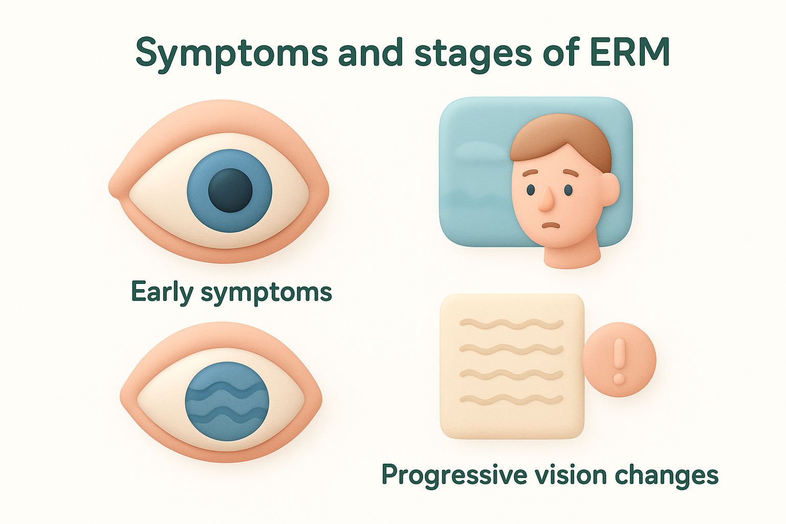
Early symptoms often go unnoticed until you compare your vision between both eyes.
Progressive vision changes
As the membrane thickens and contracts, distortion becomes more pronounced and harder to ignore. Text appears wavy or jumbled, making sustained reading frustrating or impossible. Your vision might seem like you’re looking at reflections in rippled water. Some people notice objects appearing smaller than they should (micropsia), which can affect depth perception and spatial awareness. In advanced cases, central vision can drop to 6/60 (20/200) or worse, though this severe progression affects fewer than 5% of patients. Double vision in one eye occasionally occurs when the membrane pulls unevenly across the macula.
Causes, risk factors and related eye conditions
The primary cause of erm eye relates to posterior vitreous detachment (PVD), a natural ageing process where the gel-like vitreous inside your eye separates from the retina. This separation creates tiny breaks in the retinal surface, allowing specialised cells called glial cells to migrate through and form scar tissue on the macula. In most cases, no specific trigger causes the membrane to develop beyond normal age-related changes. Secondary causes include previous retinal tears or detachment, diabetic retinopathy, retinal vein occlusion, eye trauma, or inflammation inside the eye.
Age and hereditary factors
Your risk of developing an epiretinal membrane increases significantly after age 50, with studies showing that approximately 20% of people over 75 have detectable membranes on retinal examination. Both eyes develop ERMs in roughly 10 to 20% of cases, though one eye typically shows more advanced changes than the other. If you have already developed an epiretinal membrane in one eye, your annual risk of developing one in the fellow eye reaches 2.7%. No genetic link has been confirmed, though having a family history of retinal conditions may warrant more frequent monitoring.
Your risk increases substantially with each decade after age 50.
Associated retinal conditions
Diabetic retinopathy commonly triggers secondary epiretinal membrane formation through chronic inflammation and vascular changes in the retina. Retinal vein occlusion creates similar inflammatory conditions that promote scar tissue development. Previous vitrectomy surgery or retinal detachment repair increases your membrane risk because these procedures alter the vitreoretinal interface. Uveitis, an inflammatory condition affecting the eye’s interior, can also lead to membrane formation as part of the healing response.
Treatment options, surgery and recovery
Treatment for erm eye follows a watch-and-wait approach for most patients because many membranes remain stable without causing significant functional problems. Your specialist monitors the condition through regular OCT scans and visual acuity tests, typically scheduled every six months. If your vision remains adequate for daily tasks and distortion doesn’t worsen, surgery carries more risk than benefit. Only 20 to 30% of patients eventually require surgical intervention, with the decision based entirely on how symptoms affect your quality of life rather than the membrane’s appearance on scans.
Conservative monitoring versus surgical intervention
Your specialist recommends vitrectomy surgery when metamorphopsia interferes with essential activities like reading, driving, or work tasks that demand clear central vision. Surgery becomes appropriate if your vision drops below 6/12 (20/40) or if distortion causes significant frustration despite reasonable acuity. Some patients opt for surgery earlier when bilateral membranes affect both eyes, as operating on the worse eye can restore balance and improve overall visual function. Your specialist will discuss realistic outcome expectations based on your membrane’s characteristics, duration of symptoms, and pre-operative vision level.
Surgery focuses on improving your functional vision and reducing distortion, not achieving perfect sight.
The vitrectomy procedure and what to expect
During vitrectomy, your surgeon makes three tiny incisions in the white part of your eye and removes the vitreous gel to access the retinal surface. Using microscopic forceps, they carefully peel away the epiretinal membrane and often remove the internal limiting membrane beneath it to reduce recurrence risk. The procedure takes 30 to 45 minutes under local anaesthetic with sedation, performed as day surgery. Most surgeons use brilliant blue dye to enhance membrane visibility during removal, ensuring complete peeling. You’ll need someone to drive you home afterwards, and your eye may feel gritty or uncomfortable for a few days.
Recovery timeline and visual outcomes
Visual improvement occurs gradually over three to six months, with some patients noticing continued enhancement up to 12 months post-surgery. Most people gain two to three lines on the eye chart, though results vary depending on membrane duration and pre-existing retinal damage. Distortion typically improves more reliably than visual acuity itself. You’ll use antibiotic and anti-inflammatory drops for four weeks and attend regular check-ups to monitor healing. If you still have your natural lens, cataract progression accelerates after vitrectomy, often requiring cataract surgery within two years.

Next steps and support
Regular eye examinations remain your most important preventative measure for detecting erm eye early, before symptoms become troublesome. Schedule comprehensive eye checks every two years if you’re over 50, or annually if you have diabetes, previous retinal problems, or a history of epiretinal membrane in one eye. Your optometrist can spot early membrane formation during routine dilated examinations and refer you promptly to a retinal specialist when necessary.
If you’ve noticed distorted or blurred central vision that doesn’t improve with glasses, don’t wait for your next scheduled appointment. Seek assessment promptly to understand whether an epiretinal membrane or another condition requires attention. At Bright Sight Eye Specialists, Dr. Rahul Dubey provides comprehensive assessment and surgical treatment for epiretinal membranes using advanced techniques and technology. Early evaluation gives you more treatment options and better long-term outcomes, allowing you to maintain the clear vision you need for daily activities.