


When the inner layer of your cornea stops working properly, vision becomes cloudy and daily tasks grow difficult. For years, full-thickness corneal transplants were the standard fix, but they came with long recovery times and higher rejection rates. The DMEK procedure (Descemet Membrane Endothelial Keratoplasty) changed that. It replaces only the thin, damaged layer of cells rather than the entire cornea, resulting in faster visual recovery and better outcomes.
At Bright Sight Eye Specialists in Randwick, Dr Capucine Odouard performs DMEK surgery for patients with conditions such as Fuchs’ endothelial dystrophy and bullous keratopathy. Her training at Sydney Eye Hospital and subspecialty focus on corneal conditions means you’re in experienced hands, whether you’re from the Eastern Suburbs or regional Australia.
This article covers how the DMEK procedure works step by step, what success rates look like, the risks involved, and what to expect during recovery.
What the DMEK procedure involves
The DMEK procedure focuses on replacing only the Descemet membrane and the endothelial cell layer, which is roughly 10 to 15 micrometres thick. Because the surgery targets such a thin, precise layer, it requires careful preparation of donor tissue and a controlled surgical technique.
Preparing the donor tissue
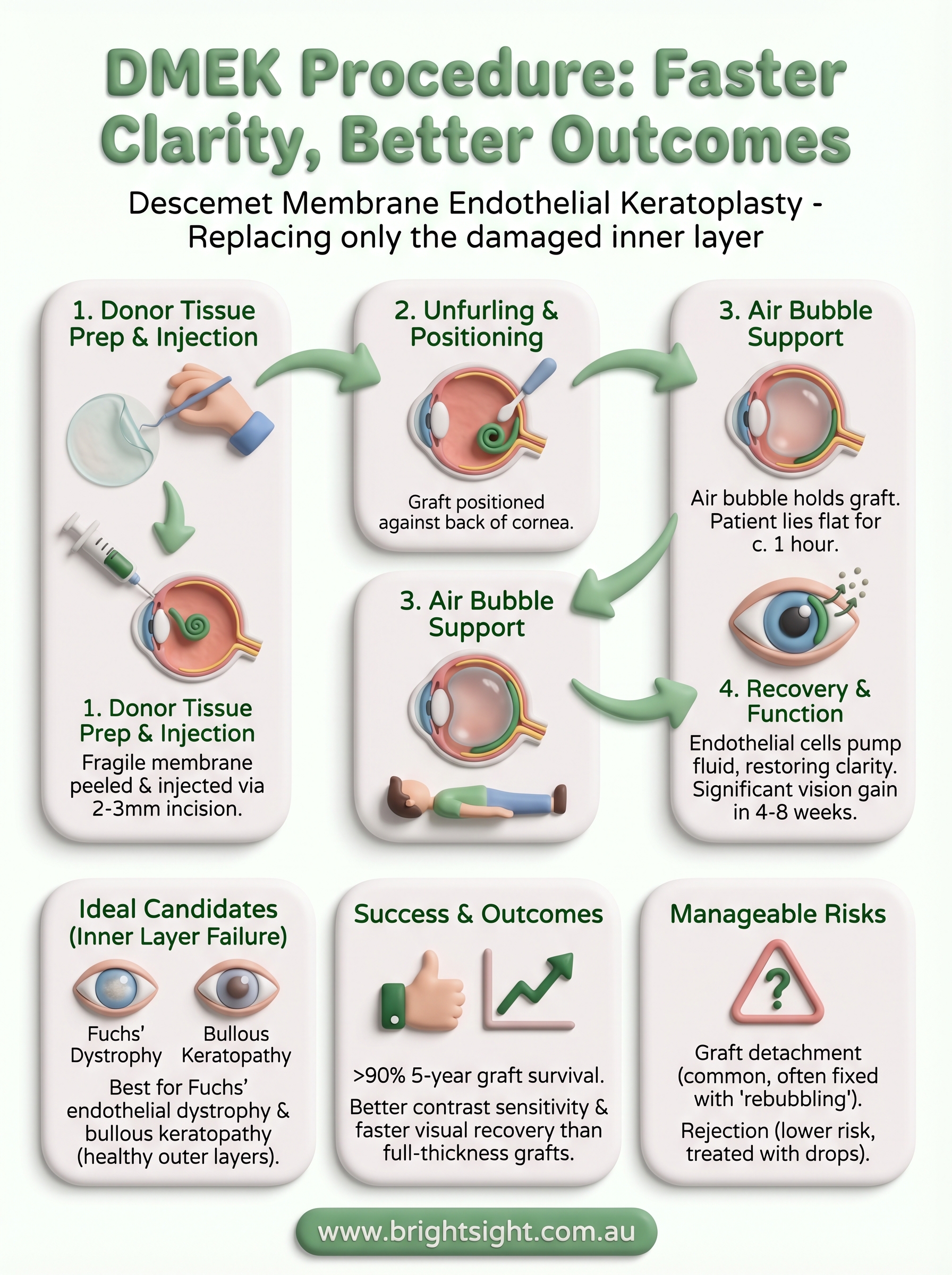
Before surgery begins, your surgeon carefully peels the Descemet membrane and endothelial cells from a donor cornea. This step demands significant technical skill because the tissue is extremely fragile. The prepared graft is then rolled up, ready to be inserted through a small incision of around 2 to 3 millimetres in your cornea.
The donor tissue must be prepared and handled with precision; any damage at this stage directly affects the success of the graft.
The surgical steps on the day
Your surgeon makes a small incision and removes the diseased endothelial layer from the back of your cornea. The rolled donor graft is then injected into your eye using a specialised inserter. Once inside the anterior chamber, your surgeon carefully unfurls and positions the graft against the back surface of your cornea.
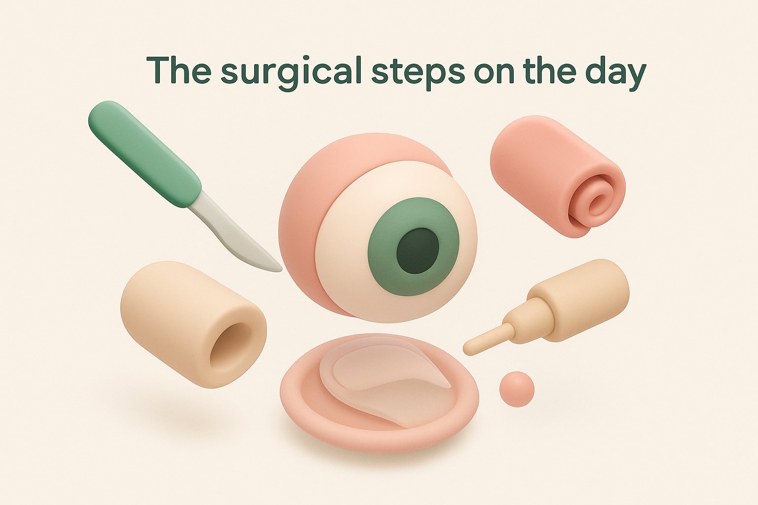
After correct positioning is confirmed, an air bubble is injected to press the donor tissue into place and hold it there while it adheres. You will need to lie flat on your back for around one hour post-surgery so the air bubble maintains contact between the graft and your cornea.
What happens after the graft adheres
Over the following days, your endothelial cells begin to function, pumping fluid out of the corneal tissue and restoring clarity. Most patients notice a meaningful improvement in vision within four to eight weeks, with continued gains over several months.
Who benefits and who should avoid DMEK
The dmek procedure works best for patients whose corneal problems stem specifically from endothelial cell failure rather than damage to the outer layers of the cornea. If your stroma and epithelium are healthy but your endothelial cells are no longer clearing fluid effectively, DMEK is likely a strong option for you.
Conditions suited to DMEK
Fuchs’ endothelial dystrophy is the most common reason surgeons recommend DMEK. In this condition, endothelial cells gradually deteriorate, causing corneal swelling and progressively blurred vision. Bullous keratopathy, where cells fail following cataract surgery or other intraocular procedures, is another key indication. Your surgeon will assess your corneal thickness, cell count, and overall eye health before confirming this approach.
If your vision is worsening due to corneal swelling that no longer responds to drops or ointments, DMEK is worth discussing with your specialist.
When DMEK is not the right choice
Significant stromal scarring or a history of previous failed grafts with substantially altered anatomy may rule out DMEK for some patients. Advanced glaucoma or other structural problems inside the eye can also reduce how well the donor tissue adheres and functions over time.
Your surgeon will review your full eye history and recommend the most suitable alternative, such as DSAEK or a full-thickness graft, if DMEK is not appropriate for your situation.
DMEK vs DSAEK and full-thickness grafts
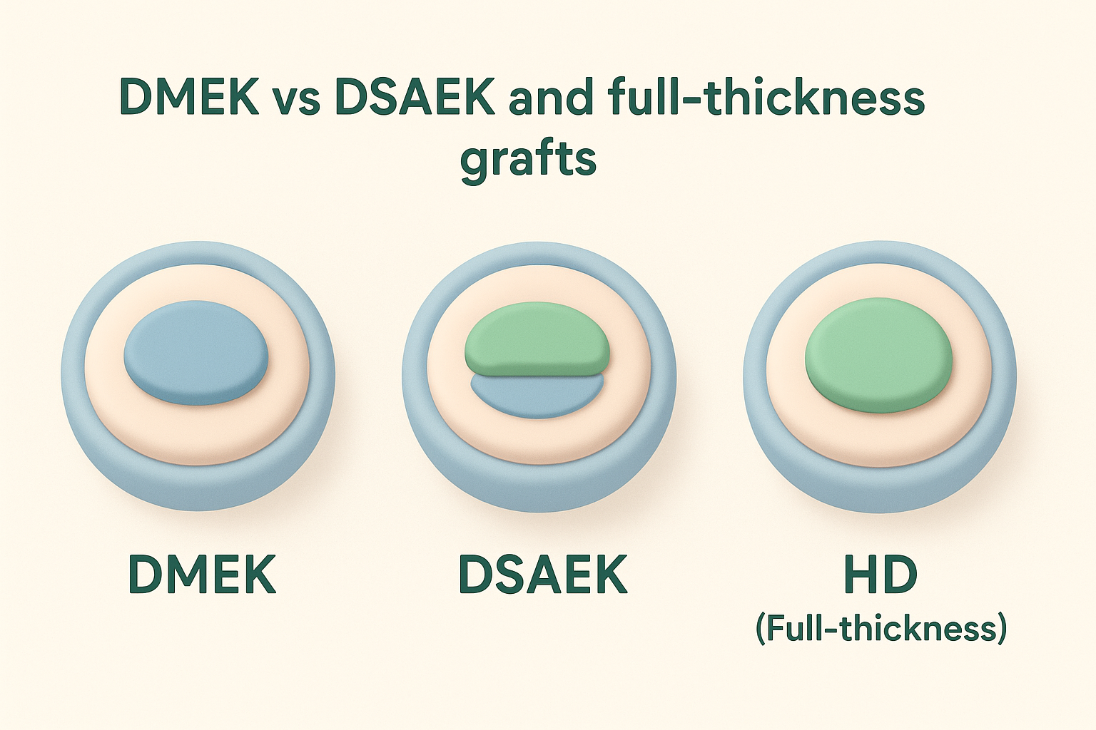
Understanding how the dmek procedure compares to other corneal transplant options helps you have a more informed discussion with your surgeon. All three approaches differ significantly in how much of the cornea is replaced and the speed of visual recovery.
DSAEK: the thicker alternative
DSAEK also targets the endothelial layer, but the graft includes a thin layer of donor stroma alongside the Descemet membrane. This extra thickness makes the tissue easier to handle, which some surgeons prefer for complex cases.
The trade-off is that final visual acuity is generally not as sharp as with DMEK. The thinner DMEK graft consistently produces better contrast sensitivity and faster clarity in the months following surgery.
If sharp vision is your main goal and your cornea is otherwise undamaged, DMEK is usually the stronger option.
Full-thickness transplant (PKP)
A penetrating keratoplasty (PKP) replaces every corneal layer and is reserved for cases involving stromal scarring or disease affecting multiple layers. When only your endothelial cells have failed, PKP is rarely the first recommendation.
Recovery after PKP can extend to one to two years, and the risk of graft rejection is considerably higher than with DMEK. Your surgeon will only suggest PKP when less invasive approaches are not suitable.
Success rates and what affects outcomes
The dmek procedure carries strong outcomes when performed by an experienced surgeon on suitable candidates. Published data consistently show graft survival rates above 90% at five years, with many patients achieving vision of 6/6 or better. These figures are notably higher than those reported for full-thickness transplants, making DMEK the preferred approach for isolated endothelial failure in most ophthalmology centres.
Patient selection and surgeon experience are the two most significant factors in determining how well your graft performs long term.
What the research shows
Studies report that 80 to 90% of DMEK patients reach visual acuity of 6/9 or better within 12 months. A small proportion require a graft rebubbling procedure, where additional air is injected to reattach partially detached tissue. When addressed promptly, this rarely affects long-term visual outcomes and remains a manageable complication.
Factors that affect your result
Your pre-operative endothelial cell count and the overall health of your cornea play a direct role in how quickly and completely vision improves after surgery. Underlying glaucoma, a history of uveitis, or prior intraocular surgery can each reduce graft adherence and slow recovery. Your surgeon will factor all of these variables into your individual assessment before confirming that DMEK is the right approach for you.
Risks, complications, and follow-up care
Like any intraocular surgery, the dmek procedure carries risks that your surgeon will discuss with you during your consultation. Understanding these upfront helps you prepare and recognise any warning signs early.
Contact your surgeon immediately if you notice a sudden drop in vision, increased redness, or eye pain in the weeks after surgery.
Complications you may encounter
Graft detachment is the most common complication, occurring in roughly 30 to 40% of cases to some degree. Most partial detachments are managed with a rebubbling procedure performed in the clinic or operating room, and vision outcomes remain strong when this is caught early. Graft rejection, while less frequent than with full-thickness transplants, is still possible and presents as sudden blurring, redness, or light sensitivity.
Your follow-up schedule
Regular post-operative appointments are essential in the first three months, typically at one day, one week, one month, and three months after surgery. Your surgeon will monitor your endothelial cell count and corneal thickness at each visit to confirm the graft is functioning well. Most patients use steroid eye drops for at least 12 months to reduce rejection risk, and your surgeon will taper this schedule based on your individual progress.
Next steps if you are considering DMEK
If your vision has been declining due to corneal swelling and drops are no longer helping, the dmek procedure is worth exploring with a specialist who performs it regularly. The best starting point is a comprehensive corneal assessment, which gives your surgeon accurate data on your endothelial cell count, corneal thickness, and overall eye health before any decisions are made.
At Bright Sight Eye Specialists in Randwick, Dr Capucine Odouard assesses each patient individually and explains all available treatment options clearly, including whether DMEK, DSAEK, or another approach suits your specific condition. Her training at Sydney Eye Hospital and subspecialty focus on corneal surgery means you receive advice grounded in current evidence and real surgical experience.
Telehealth appointments are also available for patients based outside Sydney. To book your consultation and take the first step toward clearer vision, contact Bright Sight Eye Specialists today.