


You’re reading a book or gazing at a clear sky when something drifts across your vision, a shadowy shape, a tiny speck, or what looks like dark spots in front of eyes. These visual disturbances, commonly called floaters, are one of the most frequent concerns patients bring to eye specialists.
For most people, floaters are completely harmless and simply part of the natural ageing process. But in some cases, they can signal something more urgent, like a retinal tear or detachment that requires prompt medical attention.
At Bright Sight Eye Specialists in Sydney’s Eastern Suburbs, Dr Rahul Dubey and the team regularly help patients understand and manage these symptoms. This guide explains what causes dark spots in your vision, when you should seek help, and the treatment options available if you need them.
Why dark spots can appear in your vision
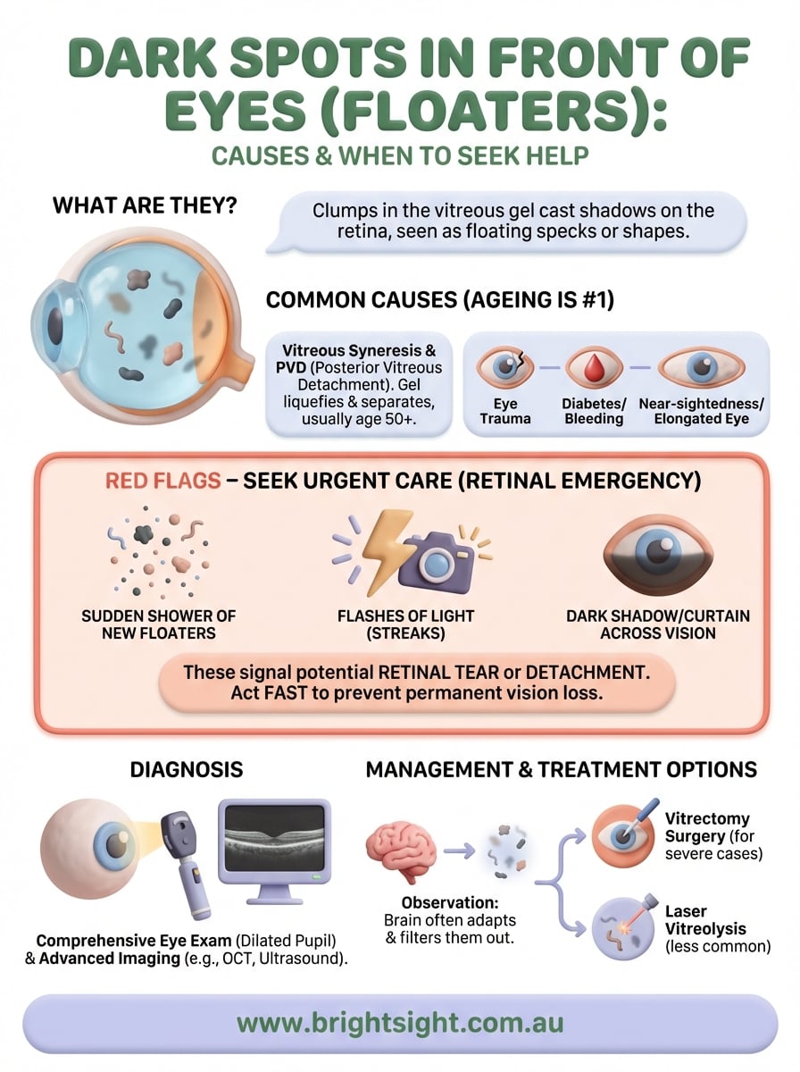
The dark spots you see moving across your field of vision come from inside your eye, not from objects in the outside world. Your eye contains a gel-like substance called the vitreous that fills the space between your lens and retina. When this gel develops small clumps or strands, they cast shadows on your retina, and your brain interprets these shadows as floating spots or shapes.
The vitreous gel and ageing
As you age, the vitreous gel naturally begins to break down and liquefy. This process, called vitreous syneresis, usually starts around age 50 but can occur earlier in people who are short-sighted or have experienced eye trauma. The gel starts to pull away from the back wall of your eye, creating pockets where tiny fibres clump together.

These clumps drift through the liquid portions of the vitreous, moving when your eyes move and seeming to dart away when you try to look directly at them. Most people notice them more when looking at bright, plain backgrounds like a white wall or blue sky because the contrast makes the shadows more visible.
While floaters become more common with age, a sudden increase in their number or size needs immediate medical evaluation.
Other factors that contribute
Several conditions beyond normal ageing can cause dark spots in front of eyes to appear. Your risk increases if you have diabetes, as high blood sugar can damage the blood vessels in your retina and cause bleeding into the vitreous. Inflammation inside the eye, eye injuries, and certain eye surgeries can also trigger floater formation. Even severe near-sightedness stretches your eyeball slightly, which accelerates vitreous changes and makes floaters more likely to develop at a younger age.
Common causes of dark spots and floaters
Understanding what triggers dark spots in front of eyes helps you know whether your symptoms need immediate attention or simply careful monitoring. While the natural ageing process accounts for most floaters, several other conditions can produce similar visual disturbances that range from completely benign to potentially serious.
Age-related vitreous changes
The most common cause of floaters is posterior vitreous detachment (PVD), which occurs when the vitreous gel separates from your retina. This separation happens to roughly 75% of people over age 65 and usually produces a sudden increase in floaters, sometimes accompanied by brief flashes of light. Your vitreous continues to shrink and condense over time, creating more visible strands and clumps that cast shadows on your retina.
Most age-related floaters settle below your line of sight within a few months and become less noticeable with time.
Eye conditions and injuries
Bleeding inside your eye from diabetic retinopathy or torn blood vessels creates red or dark floaters that can temporarily cloud your vision. Inflammation in the middle layer of your eye, called uveitis, releases inflammatory debris that appears as small dots or hazy spots. Direct trauma to your eye, recent cataract surgery, or laser procedures can also introduce particles into the vitreous. If you have severe near-sightedness (usually stronger than minus 6 dioptres), your elongated eyeball stretches the vitreous gel more than normal, making you three times more likely to develop floaters at a younger age.
When dark spots signal an emergency
While most floaters are harmless, certain patterns of dark spots in front of eyes indicate a retinal emergency that needs same-day evaluation. Your retina can tear or detach when the vitreous pulls away too forcefully, and without prompt treatment, you risk permanent vision loss. Learning to recognise the warning signs helps you know when to seek immediate medical attention rather than waiting for a routine appointment.
Signs that require immediate attention
You need urgent eye care if you notice a sudden shower of new floaters, especially if they appear alongside flashes of light that look like lightning streaks or camera flashes. A dark curtain or shadow spreading across your peripheral vision signals that your retina may be detaching from the back wall of your eye. Any rapid decrease in vision, particularly if it seems like a veil is dropping over part of your visual field, requires emergency evaluation.
If you experience a sudden increase in floaters with flashes of light, contact an eye specialist immediately, even outside regular business hours.
What happens during a retinal emergency
When your retina tears, fluid can seep through the gap and lift the retina away from its underlying support tissue. This separation, called retinal detachment, cuts off your retina’s blood supply and causes permanent damage within 24 to 48 hours if left untreated. Prompt surgical repair can often restore your vision, but delays reduce the chance of a good outcome.
How eye specialists diagnose the cause
Your eye specialist uses several diagnostic techniques to determine what’s causing your dark spots in front of eyes and whether they pose any risk to your vision. The examination focuses on examining your retina and vitreous in detail, looking for tears, detachment, bleeding, or inflammation that might need treatment.
The diagnostic examination
Your appointment begins with a comprehensive eye examination that includes checking your visual acuity and eye pressure. Your specialist then dilates your pupils with eye drops, which takes about 20 to 30 minutes to take full effect. This dilation allows complete access to view your entire retina and vitreous. Using a specialised lens and bright light, your doctor examines the back of your eye thoroughly, looking for any areas where the retina might be torn or pulled away from its normal position.
Dilating your eyes is essential for a thorough examination, so plan to have someone drive you home afterwards as your vision will be temporarily blurred.
Advanced imaging when needed
In some cases, your specialist orders additional tests like optical coherence tomography (OCT), which creates detailed cross-sectional images of your retina, or ultrasound scanning if bleeding or severe floaters prevent a clear view. These imaging techniques help identify subtle changes that might not be visible during a standard examination.
How to manage and treat troublesome floaters
Most people with dark spots in front of eyes don’t need any treatment because the floaters gradually become less noticeable as your brain learns to ignore them. Your vitreous gel continues to liquefy over time, allowing the clumps to settle below your line of sight where they cause minimal disruption. However, when floaters significantly interfere with your daily activities or quality of life, several treatment options can help.
Conservative observation
Your eye specialist typically recommends watchful waiting for the first few months after floaters develop. During this period, most floaters settle naturally and become less bothersome without any intervention. You can help manage the symptoms by moving your eyes up and down, which shifts the vitreous fluid and temporarily moves floaters out of your central vision. Wearing sunglasses in bright conditions also reduces the contrast that makes floaters more visible against light backgrounds.
Most floaters fade into the background within three to six months as your brain adapts and filters them out of your conscious awareness.
Surgical treatment options
When floaters remain severely disruptive, your specialist may recommend vitrectomy surgery, which removes the vitreous gel and replaces it with a clear saline solution. This procedure eliminates floaters completely but carries surgical risks including cataract formation and retinal detachment. Laser vitreolysis offers a less invasive alternative that uses targeted laser pulses to break up large floaters, though it works best for specific types of floaters in certain locations within your eye.
What to do next
If you’ve recently noticed dark spots in front of eyes that don’t interfere with your vision, you can safely monitor them for a few months to see if they settle naturally. Most floaters become less noticeable as your brain adapts and filters them from your conscious awareness. Keep track of any changes in their number or size, and maintain your regular eye examination schedule.
However, you should seek immediate medical attention if you experience a sudden increase in floaters, flashes of light, or a shadow spreading across your vision. These symptoms can indicate a retinal tear or detachment that needs urgent treatment to prevent permanent vision loss.
At Bright Sight Eye Specialists, Dr Rahul Dubey provides comprehensive retinal examinations and treatment for patients experiencing concerning visual symptoms. Contact our team in Sydney’s Eastern Suburbs for a thorough assessment if you’re worried about floaters or any other changes in your vision.