


When the clear, dome-shaped window at the front of your eye becomes damaged or diseased, vision can deteriorate significantly. A cornea transplant, also called keratoplasty, replaces damaged corneal tissue with healthy donor tissue, restoring clarity and function. For many Australians facing conditions like keratoconus, Fuchs’ dystrophy, or corneal scarring, this procedure offers a path back to clear, comfortable vision.
At Bright Sight Eye Specialists in Randwick, Dr. Capucine Odouard brings extensive expertise in corneal conditions and advanced surgical techniques, including DSEK and DMEK procedures. Her patient-centred approach ensures you receive tailored treatment based on your specific condition and lifestyle goals. Understanding what a cornea transplant involves, from the procedure itself to recovery expectations, helps you make informed decisions about your eye health.
This guide covers everything you need to know: the types of corneal transplant surgery available, what conditions they treat, the recovery timeline, potential risks, and realistic success rates based on current evidence.
Who needs a cornea transplant
Your cornea might need replacement when disease, injury, or degeneration causes vision loss that cannot be corrected with glasses, contact lenses, or medication. A cornea transplant becomes necessary when the corneal tissue loses its transparency or structural integrity, preventing light from entering your eye properly. Conditions affecting the cornea range from inherited disorders to infections, each requiring careful evaluation to determine if surgical intervention offers the best outcome.
Conditions that damage the cornea
Keratoconus ranks among the most common reasons for transplant in younger Australians. This progressive condition causes your cornea to thin and bulge into a cone shape, distorting vision significantly. When rigid contact lenses no longer provide adequate correction, transplant surgery may restore normal corneal contour and visual function.
Fuchs’ dystrophy affects the inner corneal layer responsible for maintaining clarity. As this endothelial layer deteriorates, fluid accumulates in the cornea, causing swelling, cloudiness, and painful blisters on the surface. This condition typically progresses slowly in both eyes, eventually requiring surgical treatment when vision deteriorates.
Corneal scarring from severe infections (bacterial, viral, or fungal keratitis) can permanently damage the tissue. Previous eye injuries, chemical burns, or complications from earlier surgeries may also leave scars that obstruct vision. These opaque areas prevent light transmission, making transplant the only option for vision restoration.
Other conditions include corneal dystrophies (inherited disorders affecting corneal structure), chronic oedema (swelling) after cataract surgery, and corneal perforations requiring urgent tissue replacement.
A thorough assessment by a corneal specialist determines whether your specific condition will benefit from transplant surgery and which technique offers the best prognosis for your situation.
When conservative treatment isn’t enough
You might be considered for surgery when non-surgical approaches fail to maintain functional vision for daily activities. This includes driving, reading, working, or performing tasks requiring clear sight. Your ophthalmologist will first explore alternatives like prescription lenses, corneal collagen cross-linking (for early keratoconus), or medications to manage inflammation and infection.
Transplant becomes the recommended option when corneal damage progresses beyond the point where these treatments provide adequate improvement. Reduced visual acuity, persistent discomfort, light sensitivity, and inability to tolerate contact lenses all signal that conservative management may no longer suffice.
The decision also considers your overall eye health, age, and ability to comply with post-operative care requirements. Conditions like uncontrolled glaucoma, severe dry eye, or active infection must be addressed before proceeding with transplant surgery. Your specialist evaluates these factors comprehensively to ensure you’re a suitable candidate for the procedure.
Types of cornea transplant procedures
Your surgeon selects from several cornea transplant techniques based on which layers of your cornea require replacement. Modern approaches aim to replace only the damaged tissue rather than the entire cornea, leading to faster recovery and better outcomes. Understanding these options helps you discuss treatment choices with your specialist and know what to expect from your specific procedure.
Full-thickness transplant
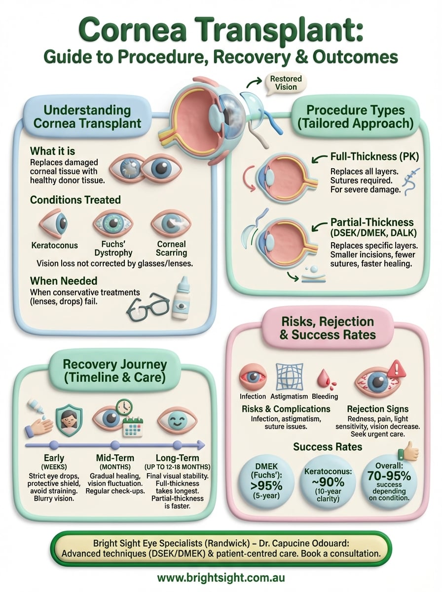
Penetrating keratoplasty (PK) removes your entire cornea and replaces it with donor tissue measuring approximately 7.5 to 8.5 millimetres in diameter. Your surgeon uses tiny sutures to secure the new cornea in place, which typically remain for 12 to 18 months. This approach suits cases where all corneal layers are damaged, such as advanced keratoconus with scarring, severe infections affecting multiple layers, or significant corneal thinning.
Recovery takes longer with full-thickness procedures because the eye needs time to heal completely around the entire circumference. You’ll experience vision fluctuation during the first year as your cornea settles and sutures are gradually removed. However, this technique provides excellent structural support and remains the gold standard for conditions affecting all corneal layers.
Partial-thickness procedures
Endothelial keratoplasty replaces only the inner corneal layer, leaving healthy outer tissue intact. DSEK (Descemet’s Stripping Endothelial Keratoplasty) and DMEK (Descemet’s Membrane Endothelial Keratoplasty) treat conditions like Fuchs’ dystrophy where the endothelium fails but outer layers remain clear. DMEK transplants an ultra-thin layer (15 micrometres), offering faster visual recovery and lower rejection rates than DSEK.
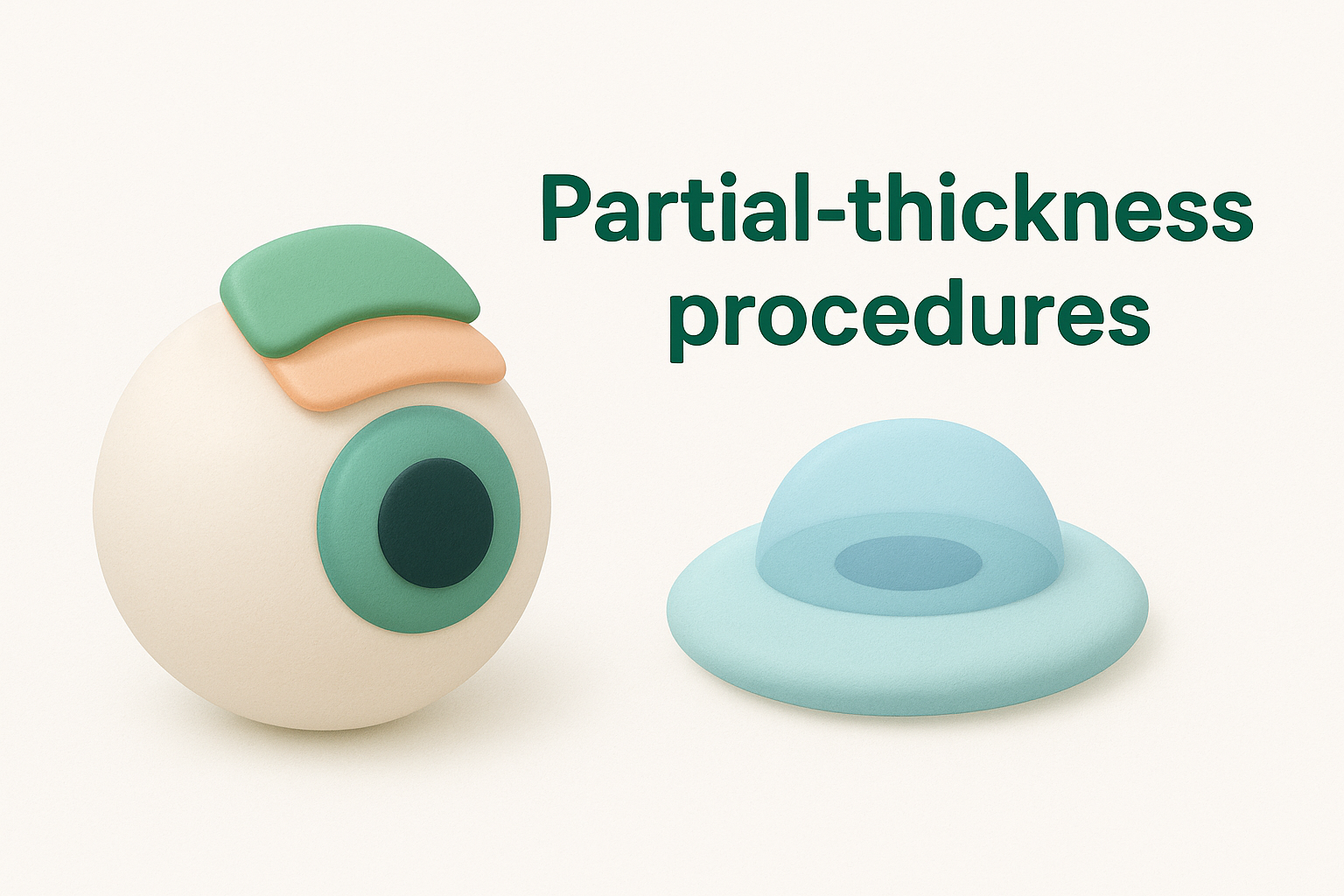
Deep Anterior Lamellar Keratoplasty (DALK) replaces the front and middle corneal layers while preserving your natural endothelium. This technique benefits patients with scarring or keratoconus affecting only the outer layers, reducing rejection risk since your own endothelial cells remain.
Partial-thickness procedures offer significant advantages: smaller incisions, fewer sutures, faster healing, and reduced risk of complications compared to full-thickness replacement.
Your corneal specialist determines which approach matches your specific condition, considering factors like disease location, corneal thickness, and your overall eye health.
What happens before, during and after surgery
Your cornea transplant journey involves several stages, each designed to ensure the best possible outcome. Understanding what to expect at each phase helps you prepare physically and mentally, reducing anxiety and improving cooperation with your surgical team. The process follows a structured pathway from initial assessment through surgery to the critical first hours of recovery.
Pre-operative preparation
Your surgeon conducts a comprehensive eye examination several weeks before surgery, measuring corneal thickness, curvature, and overall eye health. Blood tests and medical history review identify any conditions that might affect healing, such as diabetes or autoimmune disorders. You’ll receive detailed instructions about stopping certain medications (particularly blood thinners) and using antibiotic eye drops to prevent infection.
On surgery day, you arrive at the surgical centre having fasted for several hours as instructed. The surgical team administers anaesthetic (typically local numbing drops with sedation, though general anaesthesia suits some patients), ensuring you remain comfortable throughout the procedure.
The surgical procedure
Surgery typically takes 45 to 90 minutes, depending on the technique used. Your surgeon removes the damaged corneal tissue using precise instruments, then carefully positions the donor tissue in place. For full-thickness transplants, sutures secure the new cornea; partial-thickness procedures may use an air bubble to hold the tissue against your eye.
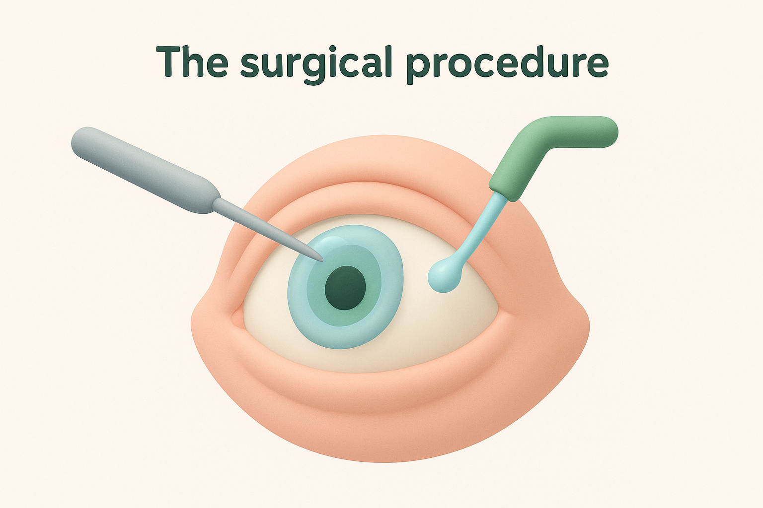
You remain awake but relaxed during most corneal procedures, experiencing no pain due to effective anaesthesia and the surgeon’s gentle approach.
Immediate post-operative care
After surgery, nursing staff place a protective eye shield over your eye and monitor you in the recovery area. You’ll receive specific instructions about sleeping position (typically on your back), applying prescribed eye drops, and avoiding activities that increase eye pressure. Most patients return home the same day with a companion, as driving isn’t permitted immediately post-surgery.
Recovery timeline and aftercare
Your recovery from cornea transplant surgery progresses through distinct phases, each requiring specific attention to protect your healing eye. Following your surgeon’s aftercare instructions precisely determines how well your new cornea integrates and functions. The timeline varies depending on the transplant technique used, with partial-thickness procedures typically healing faster than full-thickness replacements.
Early recovery phase (first few weeks)
The initial fortnight after surgery demands the most vigilant care. You’ll apply antibiotic and steroid eye drops multiple times daily to prevent infection and control inflammation. Your vision remains blurry during this period as your cornea begins healing, though partial-thickness procedures often show improvement within days. Expect frequent follow-up appointments, typically at one day, one week, and two weeks post-surgery, where your surgeon monitors healing progress and adjusts medications as needed.
Strict adherence to your eye drop schedule during the first month significantly reduces rejection risk and promotes optimal healing.
Protecting your eye from accidental trauma becomes essential. You’ll wear a protective shield while sleeping for several weeks and avoid rubbing or pressing on your eye. Activities like heavy lifting, bending over, or straining increase intraocular pressure and risk dislodging the transplanted tissue, so you’ll need to modify these behaviours temporarily.
Long-term healing and care
Complete healing takes considerably longer than you might expect. Full-thickness transplants require 12 to 18 months for final visual stability, while DSEK and DMEK procedures stabilise within three to six months. Your surgeon gradually tapers steroid drops over many months, balancing inflammation control against rejection risk. Some patients continue low-dose drops indefinitely to maintain graft health.
Vision fluctuation occurs as sutures are removed (for full-thickness procedures) and your cornea settles into its final shape. You’ll attend regular check-ups for at least a year, allowing your specialist to detect early complications and optimise your outcome.
Risks, rejection signs and success rates
Every cornea transplant carries risks like any surgical procedure, though modern techniques and careful post-operative management have significantly improved safety profiles. Understanding potential complications, recognising rejection symptoms early, and knowing realistic success rates helps you approach surgery with informed expectations. Your surgical team monitors you closely throughout recovery, but your ability to identify warning signs plays a vital role in protecting your transplanted cornea.
Common risks and complications
Infection represents the most serious early complication, though antibiotic drops and sterile surgical protocols keep incidence rates below 2%. You’ll notice increased pain, redness, discharge, or sudden vision loss if infection develops, requiring immediate medical attention. Bleeding, elevated eye pressure, and swelling also occur occasionally, typically managed with medication adjustments.
Later complications include astigmatism (irregular corneal curvature causing distorted vision), which may require glasses, contact lenses, or additional surgery to correct. Suture-related issues in full-thickness transplants can cause irritation or loosening, though your surgeon monitors and removes problematic stitches during follow-ups.
Warning signs of rejection
Graft rejection occurs when your immune system attacks the donor tissue, affecting approximately 10-30% of transplant patients depending on the technique used. DMEK procedures show the lowest rejection rates (around 1-2%), while full-thickness transplants carry higher risk. Rejection can happen months or years after surgery, making lifelong vigilance essential.
Contact your specialist immediately if you experience sudden vision decrease, increased light sensitivity, eye redness, or pain, these symptoms suggest possible rejection requiring urgent treatment.
Your surgeon treats rejection with intensive steroid drops, often reversing the process if caught early. Most rejection episodes respond well to prompt intervention, preserving graft function.
Success rates and outcomes
Corneal transplant surgery demonstrates excellent long-term outcomes, with overall success rates ranging from 70% to 95% depending on the underlying condition. DMEK procedures achieve the highest success rates (over 95% at five years) for Fuchs’ dystrophy, while keratoconus transplants maintain approximately 90% clarity at ten years. Conditions involving extensive scarring, previous surgeries, or active inflammation show lower success rates, though many patients still achieve functional vision improvement.
Next steps
Cornea transplant surgery offers proven restoration of vision and quality of life for Australians living with corneal disease. Armed with knowledge about the procedure, recovery expectations, and realistic outcomes, you can approach your consultation with informed questions and clear understanding of what lies ahead.
If you’re experiencing vision deterioration from corneal conditions like keratoconus, Fuchs’ dystrophy, or scarring, seeking specialist evaluation determines whether transplant surgery suits your situation. Early assessment allows your ophthalmologist to explore all treatment options and plan the most appropriate approach for your specific needs.
Dr. Capucine Odouard at Bright Sight Eye Specialists in Randwick brings extensive experience in advanced corneal procedures, including DSEK and DMEK techniques. Her patient-centred approach ensures you receive tailored treatment based on your condition and lifestyle goals. Book a consultation with Dr. Odouard to discuss your corneal health and explore whether transplant surgery offers the best path forward for your vision.