


A conjunctival haemorrhage in eye occurs when a tiny blood vessel breaks beneath the clear surface of your eye, creating a bright red patch on the white part. This happens in the conjunctiva, the thin transparent membrane covering your sclera. The trapped blood can’t be wiped away and often appears suddenly, sometimes overnight or after coughing, sneezing or rubbing your eyes. Despite its alarming appearance, this condition is usually harmless and rarely causes pain or affects your vision.
This guide explains everything you need to know about conjunctival haemorrhages, from recognising the symptoms to understanding when you should seek urgent care. You’ll learn what causes these blood spots, how long they typically take to heal, and what treatment options exist. We’ll also cover the warning signs that indicate a more serious problem requiring immediate medical attention. Whether you’ve just noticed a red patch on your eye or you’re experiencing repeated episodes, this article provides the practical information you need to respond appropriately.
Why conjunctival haemorrhage in the eye matters
Understanding conjunctival haemorrhage in eye helps you distinguish between a benign condition and a genuine medical emergency. The sudden appearance of blood on your eye’s surface naturally causes concern, and knowing whether you need immediate care or simply reassurance can save you unnecessary worry and costly emergency visits. This knowledge becomes especially important if you take blood-thinning medications, have high blood pressure, or experience repeated episodes that might signal an underlying health issue.
Understanding the visual impact
The dramatic appearance of a conjunctival haemorrhage can create significant social anxiety and affect your daily interactions. You might notice people staring or feel self-conscious during work meetings, video calls, or social gatherings. Many people worry that others will assume they have a contagious infection or have been in a fight. This visible change to your appearance can last one to two weeks, during which you may feel compelled to repeatedly explain your condition to concerned friends, family members, and colleagues.
While a conjunctival haemorrhage looks alarming, it typically causes no pain and doesn’t affect your ability to see clearly.
What it reveals about your health
A single conjunctival haemorrhage rarely indicates serious disease, but recurrent episodes can serve as an early warning sign for conditions requiring medical attention. Your eye’s small blood vessels are particularly sensitive to changes in blood pressure, clotting function, and vascular health. When these vessels rupture repeatedly, they may signal uncontrolled hypertension, diabetes complications, or bleeding disorders that need proper diagnosis and management. People over 60 face higher risks due to age-related changes in blood vessel walls and increased likelihood of taking medications that affect blood clotting. Recognising when your conjunctival haemorrhage represents more than a cosmetic concern allows you to seek appropriate medical evaluation and potentially prevent more serious complications in other parts of your body.
How to respond when you notice a red eye
Discovering a bright red patch on your eye can trigger immediate alarm, but your first response should be calm observation rather than panic. Take a moment to assess whether you’re experiencing pain, vision changes, or discharge, as these symptoms help determine if you need urgent care. Most conjunctival haemorrhages appear suddenly and cause no discomfort beyond perhaps a slight scratchy feeling. Your initial reaction matters because rushing to the emergency department for a benign condition wastes time and resources, while delaying care for a serious injury can risk permanent damage.
Initial assessment steps
Check your visual clarity by covering one eye at a time and confirming you can read text at normal distances. Gently touch the area around your eye to assess for tenderness or swelling, which shouldn’t accompany a simple conjunctival hemorrhage in eye. Look at the red patch in good lighting to determine if it’s a flat, well-defined blood spot confined to one area, or if the redness spreads diffusely across the entire white of your eye. Note whether both eyes are affected, as bilateral haemorrhages may suggest a more serious underlying problem.
A simple conjunctival haemorrhage appears as a flat red patch with sharp borders, causes no pain, and doesn’t affect your ability to see clearly.
Document when you first noticed the red patch and what you were doing beforehand. Recent activities like heavy lifting, coughing fits, or forceful sneezing often trigger these haemorrhages and help explain their appearance. If you wear contact lenses, remove them immediately and switch to glasses until the haemorrhage resolves completely.
Managing the first 24 hours
Avoid rubbing or touching your eye, as this can worsen the bleeding or introduce bacteria that lead to infection. Apply cool compresses for comfort if you experience mild irritation, but understand this won’t speed healing. Continue your normal activities unless they involve significant physical strain or environments that could irritate your eye, such as dusty workshops or chlorinated swimming pools.
Resist the urge to use over-the-counter eye drops that claim to reduce redness, as these products constrict blood vessels and may cause rebound redness when you stop using them. Simple lubricating drops (artificial tears) provide safe relief if your eye feels dry or gritty. Keep track of any changes in the haemorrhage’s size or appearance over the first day, as rapid expansion or new symptoms warrant prompt medical evaluation.
Symptoms and warning signs to watch for
Most conjunctival hemorrhages produce no symptoms beyond the visible red patch, but understanding what’s normal versus concerning helps you respond appropriately. The typical presentation includes a bright red area on the white of your eye that appears suddenly and remains stable over the first few days. You won’t experience pain, vision loss, or discharge in straightforward cases, and the hemorrhage shouldn’t spread beyond the initial affected area. Recognising these benign features allows you to distinguish between a simple blood vessel rupture and conditions that need immediate medical attention.
Common symptoms of uncomplicated hemorrhages
An uncomplicated conjunctival hemorrhage in eye appears as a flat, well-defined red patch confined to one section of the white part of your eye. The blood sits beneath the clear conjunctiva and creates a uniform appearance without bubbles, elevation, or irregular edges. Your vision remains completely normal because the blood doesn’t cross into the cornea or affect any internal eye structures. You might notice a mild scratchy sensation or feeling of fullness, similar to having a small grain of sand in your eye, but these sensations shouldn’t intensify over time.
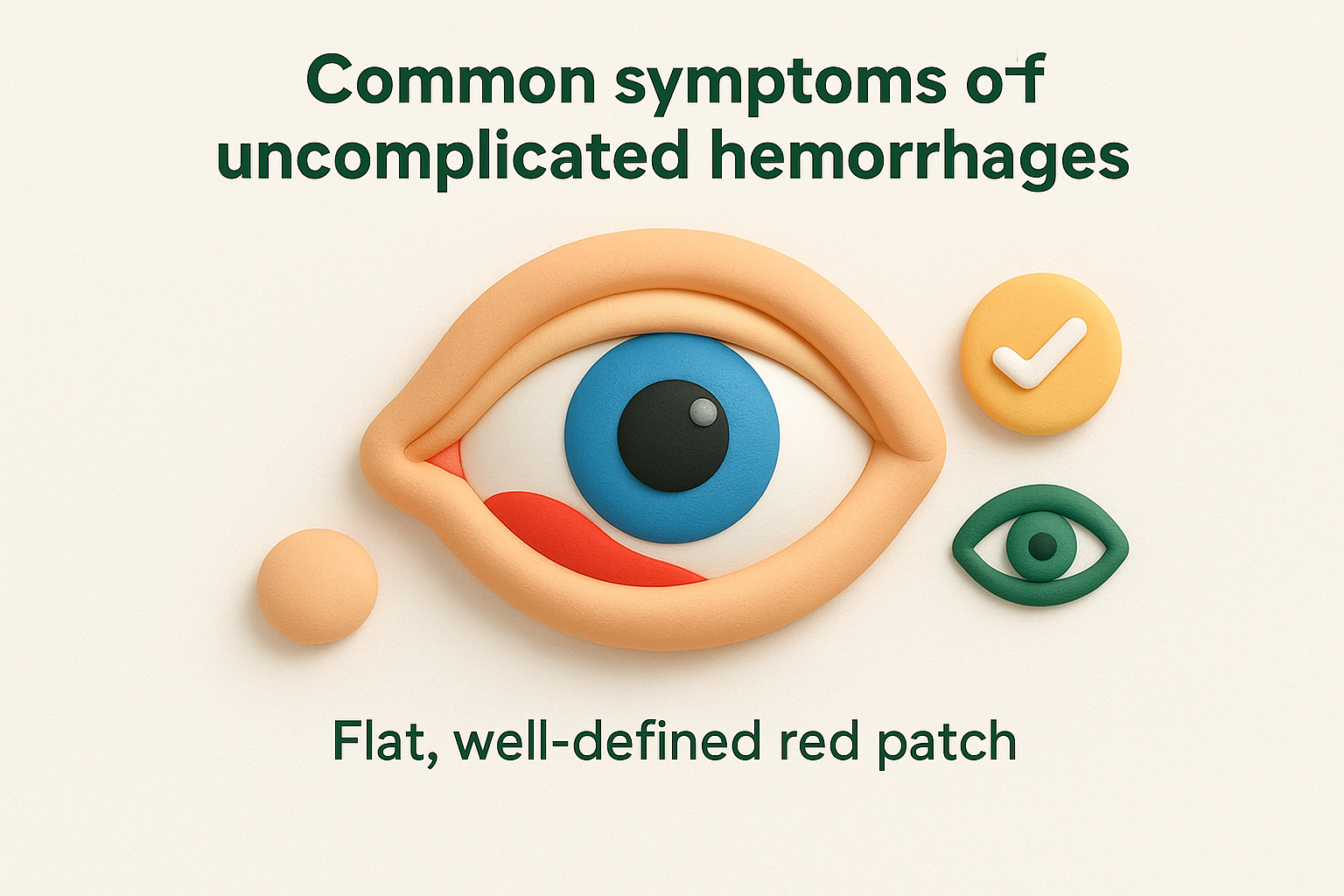
The red patch typically changes colour as your body reabsorbs the blood, progressing from bright red to darker red, then to yellowish-brown before disappearing completely. This colour transformation mirrors the healing process of a skin bruise and confirms normal resolution. You may also notice the hemorrhage spreading slightly in the first 24 hours before stabilising, which occurs naturally as the trapped blood settles across the conjunctival space. These changes are expected and don’t indicate worsening or complications.
A simple conjunctival hemorrhage causes no pain, maintains clear vision, and shows predictable colour changes during the natural healing process.
Red flag symptoms requiring urgent care
Seek immediate medical attention if you experience vision changes alongside the hemorrhage, including blurred vision, double vision, or blind spots in your visual field. These symptoms suggest involvement of deeper eye structures rather than just surface bleeding. Severe pain or a feeling of pressure inside your eye indicates potential complications like increased intraocular pressure or internal bleeding that needs urgent assessment. If you notice both eyes affected simultaneously, especially after head trauma, this pattern warrants immediate evaluation for skull fracture or bleeding disorders.
Watch for signs of globe rupture if the hemorrhage followed direct eye trauma, including a misshapen pupil, dark tissue visible through the hemorrhage, or a soft feeling when you gently touch the closed eyelid. Any discharge (pus, thick mucus, or excessive watering) combined with redness suggests infection rather than simple hemorrhage and requires prompt treatment to prevent complications. Seek care within 24 hours if you develop increasing swelling of your eyelids, inability to move your eye normally, or protrusion of the eyeball, as these signs indicate orbital involvement beyond the conjunctiva.
Main causes and risk factors
Understanding what triggers a conjunctival hemorrhage in eye helps you identify your personal risk factors and take preventive action when possible. These blood vessel ruptures occur when fragile capillaries in your conjunctiva break under pressure or strain, allowing blood to leak into the space beneath the clear membrane covering your eye. The causes range from momentary physical stress to chronic medical conditions that weaken blood vessels over time. While many hemorrhages appear without an obvious trigger, recognising common patterns helps you assess whether your episode represents a random event or signals an underlying health concern that needs attention.
Mechanical triggers and physical strain
Sudden pressure increases in your head and neck create the most common triggers for conjunctival hemorrhage. When you cough forcefully, sneeze, vomit, or strain during bowel movements, the veins in your upper body compress rapidly and blood pressure spikes in the small vessels of your eyes. This Valsalva effect explains why people often wake up with a red eye after a night of coughing from a cold or flu. Heavy lifting, intense exercise, childbirth, and even bending forward whilst picking up objects can produce similar pressure changes that rupture delicate conjunctival vessels.
Direct trauma to your eye represents another frequent mechanical cause, particularly in younger people. Rubbing your eyes vigorously, accidentally poking yourself whilst applying makeup, or receiving a blow during sports can tear blood vessels instantly. Contact lens wearers face additional risk because improper insertion or removal techniques, wearing lenses too long, or using damaged lenses creates friction that stresses the conjunctiva. Foreign particles trapped under a lens can scratch and damage blood vessels, whilst poor hygiene practices increase inflammation that makes vessels more prone to breaking.
Medical conditions and medications
High blood pressure ranks as the leading systemic risk factor for spontaneous conjunctival hemorrhages, particularly in adults over 50. Your persistently elevated blood pressure weakens vessel walls throughout your body, and the tiny capillaries in your conjunctiva become especially vulnerable to spontaneous rupture. Diabetes also compromises blood vessel integrity through chronic inflammation and structural changes that affect capillary walls. People with bleeding disorders such as haemophilia or von Willebrand disease experience hemorrhages more frequently because their blood doesn’t clot properly after even minor vessel damage.
Blood-thinning medications increase your risk of conjunctival hemorrhage because they prevent the rapid clotting that normally stops bleeding from damaged vessels.
Medications that affect blood clotting create significant risk for eye hemorrhages. Anticoagulants like warfarin, apixaban, and rivaroxaban, along with antiplatelet drugs such as aspirin and clopidogrel, prevent the normal clotting process that seals broken blood vessels. You might experience larger hemorrhages that take longer to resolve whilst taking these medications, though this doesn’t necessarily mean you should stop them. Your prescribing doctor balances the cardiovascular benefits against the cosmetic inconvenience of occasional eye bleeding.
Age and lifestyle factors
Advancing age naturally increases your susceptibility to conjunctival hemorrhages as blood vessel walls lose elasticity and structural support. The conjunctiva’s tiny capillaries become more fragile after age 60, making them prone to rupture from minimal stress. Your body’s ability to maintain healthy connective tissue decreases with time, further weakening the support structure around these delicate vessels. Older adults also face higher likelihood of taking blood pressure medications or anticoagulants that compound their baseline risk.
Chronic alcohol consumption and vitamin C deficiency weaken blood vessel walls throughout your body, including the eyes. Regular heavy drinking damages capillary integrity whilst vitamin C plays essential roles in collagen production that maintains vessel strength. Smoking contributes to vascular disease that affects all blood vessels, though its connection to conjunctival hemorrhage remains less direct than other risk factors.
When conjunctival haemorrhage is an emergency
Most conjunctival haemorrhages resolve without medical intervention, but certain circumstances demand immediate attention to prevent permanent vision loss or identify life-threatening conditions. Recognising the difference between a benign blood spot and a serious emergency protects your eyesight and overall health. You need to assess specific warning signs that indicate damage beyond the conjunctiva, particularly when the haemorrhage follows head trauma or appears alongside other concerning symptoms. Understanding when to seek urgent care versus waiting for your regular eye specialist appointment helps you make informed decisions during potentially stressful situations.
Distinguishing routine cases from true emergencies
A simple conjunctival haemorrhage in eye never affects your visual acuity or causes significant pain, so any departure from this pattern requires urgent evaluation. Visit an emergency department immediately if you experience sudden vision loss, blurred vision, seeing flashes of light, or developing blind spots in your visual field. These symptoms suggest retinal detachment, vitreous haemorrhage, or other internal eye problems that need emergency treatment to preserve sight. Sharp, severe pain rather than mild irritation indicates possible acute glaucoma or corneal involvement that demands urgent care.
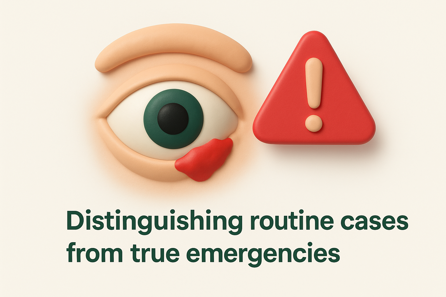
Seek immediate attention if you notice blood filling the anterior chamber (the space between your cornea and iris), creating a visible red layer that settles at the bottom of your eye. This condition, called hyphema, differs significantly from surface conjunctival bleeding and can cause permanent damage through elevated eye pressure or corneal staining. Any misshapen pupil or visible dark tissue beneath the conjunctiva after trauma suggests globe rupture, a surgical emergency requiring immediate specialist intervention.
Trauma-related warning signs
Eye injuries that produce conjunctival haemorrhage warrant emergency assessment when accompanied by specific danger signs. If the haemorrhage appeared after a forceful blow, penetrating injury, or explosion, you need immediate evaluation even if your vision seems normal initially. Watch for restricted eye movement, double vision, or an eyeball that protrudes forward or sits deeper than usual, as these signs indicate orbital fracture or retrobulbar haematoma requiring urgent imaging and treatment.
Any conjunctival haemorrhage that follows significant head trauma requires emergency assessment to rule out skull fracture or intracranial bleeding.
Blood vessels in your conjunctiva connect to deeper structures, and a posterior haemorrhage that appears to originate from behind your eye rather than the surface suggests more serious injury. You should also seek urgent care if both eyes develop haemorrhages simultaneously after trauma, particularly when combined with facial bruising or bleeding from your nose or ears.
Same-day versus emergency department visits
Contact your eye specialist for same-day evaluation if you develop increasing pain, progressive swelling, or discharge over several hours following an initial haemorrhage. These evolving symptoms may indicate developing infection or inflammation that needs prompt treatment but doesn’t constitute an immediate threat to vision. Schedule urgent assessment if you take blood-thinning medication and notice the haemorrhage expanding significantly or if you develop multiple haemorrhages across different areas of your eye, as these patterns might signal excessive anticoagulation requiring dose adjustment.
Diagnosis and what to expect at the eye specialist
Most cases of conjunctival hemorrhage in eye don’t require specialist evaluation, but your general practitioner or optometrist may refer you if the hemorrhage is extensive, recurrent, or accompanied by concerning symptoms. The diagnostic process focuses on confirming the hemorrhage is purely superficial and identifying any underlying conditions that might have triggered it. Your eye specialist uses clinical examination rather than complex testing to make the diagnosis, as the characteristic appearance of blood beneath the conjunctiva is unmistakable. The appointment typically takes 15 to 30 minutes and aims to rule out serious complications whilst addressing your concerns about the hemorrhage’s appearance and healing timeline.
The clinical examination process
Your eye specialist begins by taking a detailed history covering when you first noticed the hemorrhage, any recent trauma or illness, medications you take, and whether you’ve experienced similar episodes previously. They’ll ask about straining activities like heavy lifting, coughing fits, or vomiting that might have triggered the bleed. Your medical history receives particular attention, especially conditions like high blood pressure, diabetes, or bleeding disorders that increase hemorrhage risk.
During the physical examination, your specialist uses a slit lamp microscope to examine your eye’s surface structures in magnified detail. This instrument allows them to assess the hemorrhage’s extent, check for signs of trauma or foreign bodies, and examine your cornea, anterior chamber, and lens for additional problems. They’ll measure your visual acuity with a standard eye chart and check your eye movements to ensure no restriction exists. Your specialist will also measure your intraocular pressure using tonometry to exclude glaucoma, particularly if you’re experiencing any discomfort.
A thorough eye examination confirms whether your hemorrhage is limited to the conjunctiva or involves deeper structures that need different treatment.
Tests and investigations
Blood pressure measurement forms an essential component of your evaluation, especially if you have no known hypertension history or your hemorrhage occurred spontaneously. Elevated readings during your visit may prompt your specialist to recommend follow-up monitoring with your general practitioner. Your specialist might order blood tests if you experience recurrent hemorrhages without obvious cause, checking your clotting function, platelet count, and overall blood health. These investigations help identify bleeding disorders or medication effects that need management.
Additional testing remains uncommon for straightforward cases. Your specialist may request imaging studies only if they suspect orbital involvement or if your hemorrhage followed significant trauma. Most patients leave the appointment with reassurance, advice about expected healing time, and guidance about when to return if new symptoms develop.
Treatment, home care and recovery time
Most cases of conjunctival hemorrhage in eye require no medical treatment whatsoever, as your body naturally reabsorbs the trapped blood over time. Your eye specialist or general practitioner will typically recommend watchful waiting rather than active intervention, since the hemorrhage poses no threat to your vision or eye health. The main treatment goal focuses on managing any underlying conditions that might have triggered the bleeding, such as high blood pressure or medication adjustments, rather than treating the hemorrhage itself. Understanding what to expect during the healing process helps you manage the cosmetic concerns whilst avoiding unnecessary worry or ineffective remedies.
Medical treatment options
Your doctor rarely prescribes specific medications for simple conjunctival hemorrhages because they resolve naturally without intervention. If you experience significant irritation or dryness, your eye specialist might recommend preservative-free artificial tears to keep your eye surface comfortable, though these drops don’t accelerate healing. Avoid over-the-counter redness-relieving drops, as these products constrict blood vessels and can cause rebound redness when discontinued.
Treatment becomes necessary only when an underlying condition requires attention. Your doctor may adjust your blood pressure medication if poorly controlled hypertension triggered the hemorrhage, or consult with your cardiologist about anticoagulation levels if you experienced unusually large bleeding whilst taking blood thinners. These management decisions focus on preventing future episodes rather than treating the current hemorrhage. Doctors never drain or aspirate the blood from your conjunctiva, as this intervention risks infection and doesn’t speed recovery.
Home care strategies
You can safely manage your hemorrhage at home using simple comfort measures that reduce irritation without interfering with natural healing. Apply cool compresses to your closed eyelid for 10 to 15 minutes several times daily if you notice mild discomfort, though this provides symptom relief rather than faster resolution. Avoid rubbing or touching your eye, as this mechanical stress can worsen the bleeding or introduce bacteria that lead to infection.
Keep your eye clean and protected whilst allowing your body’s natural healing process to reabsorb the blood without interference.
Continue your normal daily activities unless they involve heavy lifting, intense exercise, or environments that irritate your eyes. You can read, watch television, use computers, and drive safely, since the hemorrhage doesn’t affect your vision. Remove contact lenses immediately and wear glasses until the blood completely disappears and your eye specialist confirms you can resume lens wear. Maintain proper hand hygiene before applying any eye drops or touching the area around your eye.
Expected recovery timeline
Your conjunctival hemorrhage typically takes one to two weeks to resolve completely, though larger hemorrhages may persist for three weeks or slightly longer. The blood doesn’t drain externally but instead undergoes gradual reabsorption by your body’s natural processes, similar to how a skin bruise fades over time. You’ll notice distinct color changes during healing, starting with bright red that darkens to deep crimson, then transitions through purple and brown shades before finally turning yellowish-green just before disappearing.
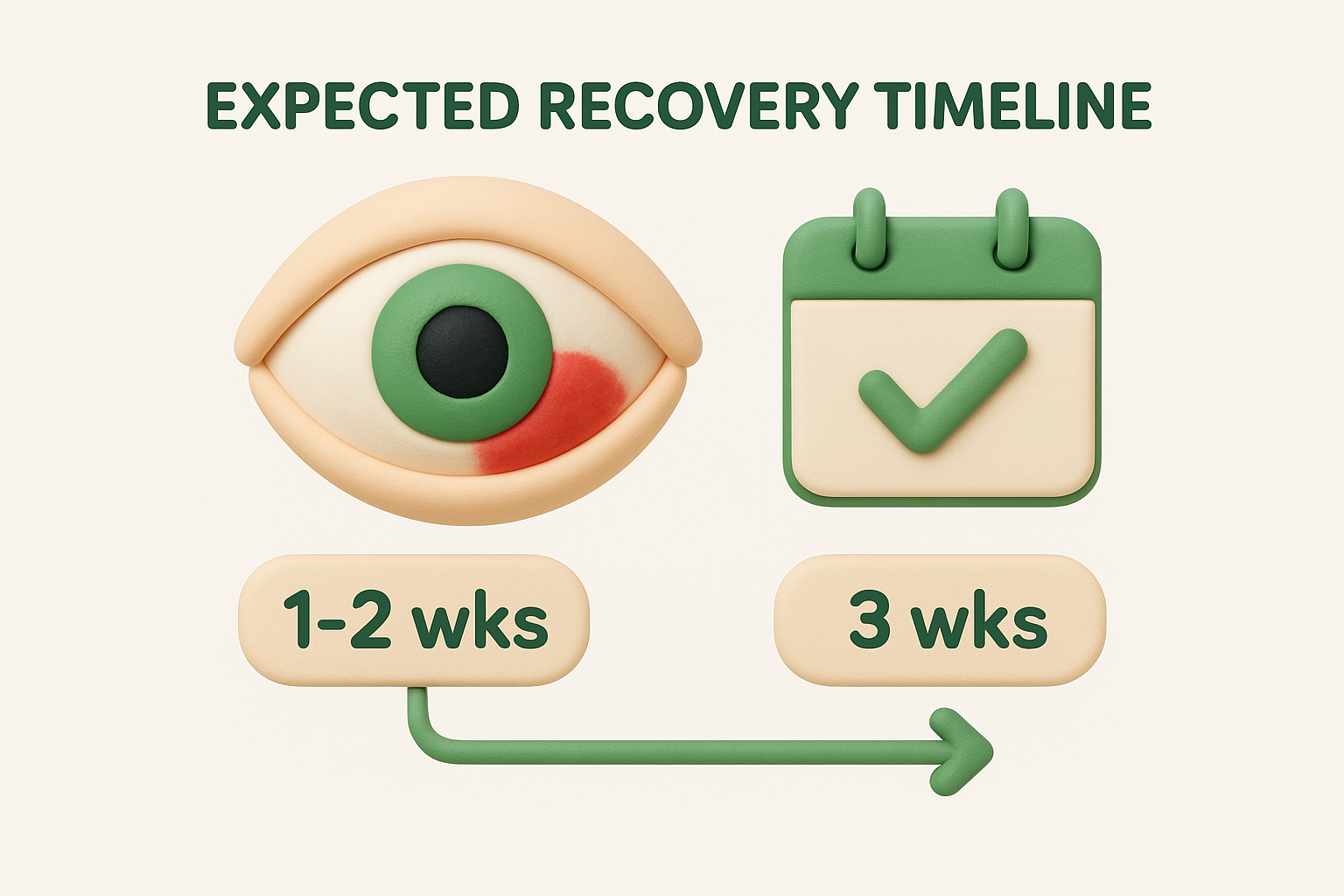
The hemorrhage usually appears largest within the first 24 to 48 hours as the blood spreads across the conjunctival space, then remains stable before slowly shrinking from the edges inward. Factors affecting your recovery speed include the hemorrhage size, your age, overall vascular health, and whether you take blood-thinning medications. Older adults and people on anticoagulants typically experience slower reabsorption but still achieve complete resolution without complications. Contact your eye specialist if the hemorrhage expands significantly after the first two days, fails to show any fading after two weeks, or you develop new symptoms during the healing period.
Preventing future conjunctival haemorrhages
While you can’t eliminate all risk of future conjunctival hemorrhage in eye episodes, you can significantly reduce your chances through targeted lifestyle modifications and health management. Most prevention strategies focus on protecting fragile blood vessels and avoiding activities that spike blood pressure suddenly in your head and neck. Your approach depends on whether previous hemorrhages resulted from identifiable triggers like trauma or physical strain, or whether they appeared spontaneously without obvious cause. Understanding your personal risk factors allows you to implement practical measures that address the most likely causes whilst maintaining your normal quality of life.
Managing controllable risk factors
Your blood pressure control represents the single most effective prevention strategy if you experience recurrent hemorrhages. Work with your general practitioner to achieve consistent readings within the recommended range through medication adherence, dietary sodium reduction, and regular monitoring. Uncontrolled hypertension weakens blood vessel walls throughout your body, making spontaneous ruptures more likely even during routine activities. Schedule regular check-ups to ensure your treatment remains effective, particularly if you’ve had multiple unexplained hemorrhages.
If you take anticoagulant or antiplatelet medications, maintain regular communication with your prescribing doctor about any bleeding episodes. Your specialist may need to adjust dosages or switch medications if you develop frequent hemorrhages, though never stop these drugs without medical guidance as they protect against serious cardiovascular events. People with diabetes should maintain good glycemic control through diet, exercise, and medication compliance, as elevated blood sugar damages blood vessels over time and increases hemorrhage risk.
Consistent management of underlying health conditions like hypertension and diabetes provides the most reliable protection against recurrent conjunctival hemorrhages.
Protective measures and lifestyle adjustments
Modify your physical activities if strenuous exercise or heavy lifting repeatedly triggers hemorrhages. You don’t need to avoid these activities entirely, but proper breathing techniques prevent the Valsalva effect that causes sudden pressure spikes. Exhale during the exertion phase of lifting rather than holding your breath, and take breaks during intense workouts to prevent sustained pressure elevation. Treat chronic coughing promptly with appropriate medical care rather than allowing persistent symptoms that repeatedly stress your eye’s blood vessels.
Contact lens wearers should follow strict hygiene protocols and replacement schedules to minimise conjunctival irritation and inflammation. Replace your lenses according to manufacturer recommendations, never sleep in lenses unless specifically designed for extended wear, and remove them immediately if you experience redness or discomfort. Protective eyewear during sports or activities with eye injury risk provides essential prevention if trauma has caused previous hemorrhages.

Key takeaways
A conjunctival hemorrhage in eye typically appears alarming but rarely signals serious disease, resolving naturally within one to two weeks without treatment. You can manage most episodes at home using simple comfort measures whilst your body reabsorbs the blood through its natural healing process. The condition becomes concerning only when accompanied by vision changes, severe pain, or trauma, which warrant immediate medical evaluation to exclude deeper eye injuries.
Prevention focuses on controlling blood pressure, maintaining medication compliance, and avoiding activities that create sudden pressure spikes in your head and neck. Understanding your personal risk factors allows you to implement targeted strategies that reduce recurrence whilst maintaining your normal lifestyle. Most people experience only occasional episodes throughout their lifetime, with complete resolution leaving no lasting effects on vision or eye health.
If you’re experiencing recurrent conjunctival hemorrhages or have concerns about your eye health, schedule a consultation with Dr Rahul Dubey at Bright Sight Eye Specialists for comprehensive evaluation and personalised management strategies.