


Waking up to a bright red patch on the white of your eye can be alarming. The sight of blood where it shouldn’t be naturally triggers concern, and your first instinct might be to search for bleeding eyeball treatment options. The good news is that in most cases, that dramatic-looking red blotch is a subconjunctival haemorrhage, a condition that typically looks far worse than it actually is.
However, not every instance of blood in or around the eye is harmless. Certain symptoms warrant immediate medical attention, particularly when bleeding occurs inside the eye rather than on its surface, or when it accompanies vision changes, pain, or recent trauma. Understanding the difference can save your sight.
At Bright Sight Eye Specialists, Dr Rahul Dubey and the team regularly assess patients concerned about bleeding in their eyes. This article explains what causes these bleeds, how to manage minor cases at home, and the warning signs that indicate you should see a specialist promptly. Whether you’re dealing with a simple burst blood vessel or something that needs professional evaluation, you’ll find clear guidance on your next steps.
What bleeding in the eye can mean
The term "bleeding in the eye" covers several distinct conditions that vary dramatically in severity. Blood can appear on the surface of your eye, inside the eye itself, or in the tissues surrounding it. The location and cause of the bleeding determine whether you’re dealing with a harmless cosmetic issue or a medical emergency requiring immediate intervention.
Subconjunctival haemorrhage: blood on the eye’s surface
When you see a bright red patch on the white part of your eye, you’re typically looking at a subconjunctival haemorrhage. This occurs when a tiny blood vessel breaks beneath the conjunctiva, the transparent membrane covering the sclera (the white of your eye). The blood becomes trapped between these two layers, creating that vivid red appearance that can spread across part or all of the white surface.

Common triggers include straining during bowel movements, coughing fits, heavy lifting, vigorous rubbing, or even sneezing. Sometimes you won’t identify any cause at all. The condition usually doesn’t hurt, doesn’t affect your vision, and resolves on its own within one to two weeks. Blood thinning medications like aspirin or warfarin can make these bleeds more likely to occur.
Subconjunctival haemorrhages look alarming but rarely indicate serious disease when they appear alone without other symptoms.
Internal eye bleeding: hyphema and vitreous haemorrhage
Blood inside the eye presents a completely different situation from surface bleeding. A hyphema occurs when blood collects in the anterior chamber, the space between your cornea and iris. You’ll notice a visible layer of blood pooling at the bottom of your coloured iris, often following blunt trauma to the eye. This condition can increase eye pressure dangerously and requires prompt medical assessment.
Vitreous haemorrhage involves bleeding into the gel-like substance that fills the centre of your eyeball. Rather than seeing a red patch, you’ll experience floaters, shadows, or a sudden decrease in vision. Causes include diabetic retinopathy, retinal tears, retinal detachment, or age-related macular degeneration. Unlike surface bleeding, vitreous haemorrhage demands urgent evaluation because it often signals serious retinal problems.
Bleeding linked to trauma or underlying conditions
Eye bleeding can accompany injuries ranging from minor scratches to severe trauma. A direct blow to your eye or face might cause multiple types of bleeding simultaneously. You might see blood in the white of your eye alongside internal bleeding, and orbital fractures can produce bleeding in the tissues around your eyeball, leading to bruising and swelling of the eyelids.
Certain medical conditions make eye bleeding more common. Diabetes weakens retinal blood vessels, increasing your risk of both vitreous haemorrhage and other bleeding complications. High blood pressure can cause retinal blood vessels to rupture. Bleeding disorders or blood-thinning medications raise the likelihood of bleeds from minor triggers. Inflammatory conditions affecting blood vessels, such as vasculitis, can also produce bleeding within or around the eye.
Bleeding eyeball treatment varies dramatically based on which type you’re experiencing. Surface bleeding requires minimal intervention, whilst internal bleeding often needs specialist examination, imaging, and potentially surgical treatment. Recognising these differences helps you respond appropriately when blood appears where it shouldn’t be.
Why a bleeding eyeball can look worse than it is
The dramatic appearance of blood on your eyeball creates an immediate sense of panic that’s rarely justified by the actual medical situation. Your eye’s unique anatomy makes even tiny amounts of blood appear far more serious than similar bleeding elsewhere on your body. Understanding why this visual alarm doesn’t match the typical severity helps you respond calmly rather than rushing to emergency departments for conditions that will resolve naturally.
The stark contrast between appearance and actual harm
A subconjunctival haemorrhage occupies a thin layer between two transparent membranes, spreading the blood across a wide surface area without depth. This creates the illusion of massive bleeding when the actual volume might equal just a few drops. The same amount of blood beneath your skin would produce barely noticeable bruising, but on your eye it appears shocking because it spreads thinly across the white sclera rather than pooling in tissue.
Your body’s response compounds the visual drama. The conjunctiva lacks pain receptors in most areas, so a broken blood vessel typically doesn’t hurt. This absence of pain might increase your anxiety because you expect serious-looking injuries to cause discomfort. The bleeding also develops instantly, meaning you discover it suddenly rather than watching it gradually appear, which can feel more alarming than conditions that develop slowly over time.
Surface bleeding on your eye typically involves less blood volume than a minor graze on your knee, despite looking far more severe.
Why blood displays so prominently against the eye’s white background
The sclera provides a stark white backdrop that makes red blood cells intensely visible. Unlike skin, which has varied pigmentation and texture that diffuses the appearance of bruising, the smooth white surface of your eye offers maximum contrast. Photographers use white backgrounds to make subjects stand out, and your eye creates this same effect with any blood present.
Lighting conditions intensify the appearance further. Bright overhead lights or sunlight illuminate the blood through the transparent conjunctiva, making it appear even more vivid. The curved surface of your eyeball also catches light from multiple angles, ensuring the haemorrhage remains visible regardless of where someone looks from. This visibility persists whether your eyes are open or partially closed, keeping the bleeding constantly noticeable.
When bleeding eyeball treatment becomes necessary despite benign appearance
Not every case of eye bleeding matches this reassuring pattern. Internal bleeding doesn’t always show dramatic external signs but requires urgent intervention. You might experience floaters, vision loss, or pain without seeing obvious blood on the surface. Conversely, multiple recurrent surface bleeds can indicate underlying conditions like hypertension or clotting disorders that need treatment even though each individual episode looks worse than it is. The appearance alone never tells the complete story.
What to do now at home
Most surface eye bleeding requires no active bleeding eyeball treatment beyond patience and basic comfort measures. Your body will naturally absorb the blood over the next week or two without any intervention. The challenge lies in resisting the urge to touch or manipulate your eye whilst it heals, and knowing which simple steps actually help versus those that waste your time or potentially cause harm.
Leave the bleed alone without touching or rubbing
Your natural instinct might push you to touch the red area or rub your eye, but this represents the worst action you can take. Rubbing increases pressure on fragile blood vessels, potentially causing additional bleeding or extending the affected area. The conjunctiva heals best when left completely undisturbed, allowing your body’s natural absorption processes to work without interference.
Avoid applying pressure to your eye or pressing on the area around it. Keep your hands away from your face, particularly if you wear contact lenses. Remove your contacts immediately if you have a subconjunctival haemorrhage, and switch to glasses until the bleeding completely resolves. Contact lenses can irritate the healing tissue and potentially trap bacteria against the damaged area.
The fastest healing occurs when you resist touching your eye entirely, despite the strong urge to investigate the red patch.
Use artificial tears to soothe any irritation
Lubricating eye drops provide genuine comfort without interfering with healing. You might feel a scratchy sensation or mild irritation, particularly if you have dry eyes alongside the haemorrhage. Preservative-free artificial tears work best because they avoid adding chemicals that might irritate your already-compromised eye surface. Apply drops three to four times daily, or more frequently if you experience persistent discomfort.
Choose simple lubricating drops rather than products marketed to "reduce redness." Redness-relieving drops constrict blood vessels, which won’t help your existing bleed and might complicate healing. Stick to basic saline or hypromellose-based tears that simply add moisture and create a protective layer over your eye’s surface.
Apply gentle cool compresses if your eye feels uncomfortable
A clean, cool cloth placed over your closed eyelid can reduce minor swelling and provide psychological comfort during the first 24 hours after you notice the bleeding. Soak a clean flannel in cool (not ice-cold) water, wring it out thoroughly, and rest it gently against your closed eye for five to ten minutes. Never apply ice directly to your eye or use frozen items that might cause cold injury to the delicate tissues.
Repeat this cooling process up to four times daily if it helps you feel better, but understand that compresses don’t speed healing or remove blood faster. They simply offer temporary symptom relief whilst your body naturally absorbs the haemorrhage at its own pace.
When to seek urgent care or emergency help
Whilst most surface bleeding resolves without intervention, certain symptoms indicate serious conditions that require immediate professional evaluation. You need to distinguish between harmless subconjunctival haemorrhage and potentially sight-threatening problems that demand urgent bleeding eyeball treatment. Recognising these warning signs can prevent permanent vision loss and catch serious eye conditions before they cause irreversible damage.
Red flags that demand immediate attention
Several symptoms accompanying eye bleeding require emergency department assessment within hours, not days. You should seek immediate help if you experience any of the following alongside the bleeding:
- Sudden, significant vision loss or blurring that doesn’t clear with blinking
- Severe eye pain that persists or worsens over time
- Sensitivity to light that makes it difficult to keep your eyes open
- Double vision or seeing two images of the same object
- Pupils that appear different sizes or don’t react normally to light
- Blood visible inside the coloured part of your eye (hyphema)
- Headache, nausea, or vomiting occurring with the eye bleeding
- The bleeding followed a direct blow to your eye or head
Vision changes that signal internal bleeding
Floaters that appear suddenly in large numbers suggest vitreous haemorrhage, where blood leaks into the gel filling your eye. You might describe these as dots, cobwebs, or shadows drifting across your field of view. A sudden shower of floaters accompanied by flashing lights indicates possible retinal tear or detachment, conditions that require intervention within 24 to 48 hours to preserve your sight.
Progressive vision loss developing over hours rather than staying stable points to ongoing bleeding or pressure buildup inside your eye. This differs from the stable vision you experience with simple surface bleeding. If your vision deteriorates noticeably whilst you watch, you need immediate specialist assessment regardless of how the outside of your eye appears.
Any vision change beyond mild irritation warrants professional evaluation, even when surface bleeding looks like a harmless subconjunctival haemorrhage.
When recent trauma requires professional assessment
Direct impact to your eye demands urgent evaluation regardless of symptom severity. Blunt force can cause hidden damage including orbital fractures, globe rupture, or retinal injuries that don’t produce immediate obvious symptoms. Chemical splashes or foreign objects embedded in your eye constitute medical emergencies requiring immediate irrigation and specialist care.
Multiple recurrent bleeds without clear triggers suggest underlying conditions like hypertension, diabetes, or clotting disorders that need investigation. Contact your GP or eye specialist if you experience repeated episodes, particularly if they occur more than twice within several months.
How doctors diagnose a bleeding eyeball
Your doctor begins diagnosis by examining your eye’s external appearance and asking specific questions about when you first noticed the bleeding, whether you experienced trauma, and what symptoms accompany the visible blood. This initial assessment helps distinguish between simple surface bleeding and conditions requiring more detailed investigation. The examination typically progresses from basic visual inspection to increasingly sophisticated tests based on what your doctor discovers at each stage.
Visual examination with slit lamp microscopy
Your eye specialist uses a slit lamp, a specialised microscope with bright illumination, to examine your eye’s surface and internal structures in magnified detail. This instrument allows them to see exactly where bleeding originated and whether blood has entered spaces inside your eye. The bright, focused beam reveals subtle details invisible to the naked eye, including tiny tears in blood vessels, inflammation, or foreign bodies that might have triggered the haemorrhage.
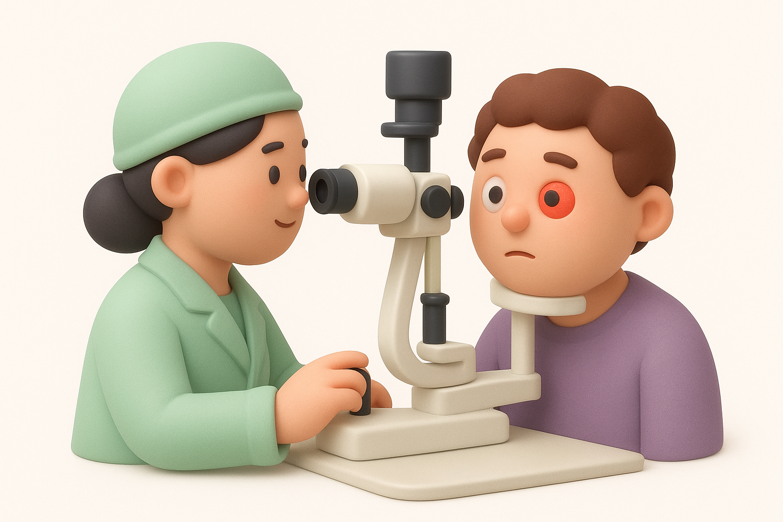
During this examination, your doctor inspects the conjunctiva, cornea, anterior chamber, iris, and lens systematically. They look for blood layering in the anterior chamber (hyphema), signs of trauma, or abnormal blood vessel growth that might explain recurrent bleeding. The slit lamp examination takes just a few minutes but provides crucial information about whether you need bleeding eyeball treatment beyond simple observation.
Eye pressure measurement and internal assessment
Measuring your intraocular pressure becomes essential when blood appears inside your eye rather than just on the surface. Your doctor uses a tonometer to check whether bleeding has increased pressure dangerously, which can occur with hyphema or certain types of internal haemorrhage. Elevated pressure requires immediate intervention to prevent optic nerve damage and permanent vision loss.
Your specialist also performs ophthalmoscopy, using specialised lenses and lights to examine your retina, vitreous cavity, and optic nerve. This reveals vitreous haemorrhage, retinal tears, diabetic changes, or other conditions that might have caused internal bleeding. They dilate your pupils with drops to gain the clearest possible view of these structures.
Internal bleeding often requires dilated pupil examination to reveal the full extent of damage and guide treatment decisions.
When imaging or blood tests become necessary
Your doctor orders additional tests when initial examination suggests trauma, recurrent bleeding, or underlying systemic disease. Ultrasound imaging helps visualise internal structures when blood obscures the view during standard examination. CT or MRI scans become necessary if your doctor suspects orbital fractures, foreign bodies, or injuries extending beyond your eyeball itself.
Blood tests check for clotting disorders, diabetes, or inflammatory conditions that might explain repeated episodes of eye bleeding. Your doctor typically orders these when you experience multiple haemorrhages without obvious triggers, or when bleeding accompanies other symptoms suggesting systemic disease.
Recovery time and how to prevent it coming back
Surface bleeding on your eye typically resolves without any active bleeding eyeball treatment, with your body absorbing the blood naturally over one to two weeks. The timeline varies based on how much blood leaked from the broken vessel and your individual healing capacity. The appearance changes gradually as your body breaks down and reabsorbs the red blood cells, often shifting from bright red to orange, yellow, or greenish-brown before disappearing completely.
How long surface bleeding takes to heal
Most subconjunctival haemorrhages clear within seven to fourteen days for small bleeds, whilst larger areas of bleeding might take three weeks to resolve fully. You’ll notice the red patch shrinking and fading rather than disappearing all at once. This gradual absorption pattern reassures you that healing progresses normally. Your vision should remain completely normal throughout this period unless you have separate underlying conditions affecting your sight.
Internal bleeding follows different timelines entirely. Vitreous haemorrhage might take weeks to months to clear, depending on severity and underlying cause. Your specialist monitors these cases closely and may recommend treatment to address the source of bleeding rather than waiting for natural resolution alone.
Your body absorbs surface bleeding naturally without intervention, but the process takes time that you cannot rush.
Steps to reduce your risk of future bleeds
You can lower your chances of recurrent bleeding by managing underlying health conditions that weaken blood vessels. Keep your blood pressure controlled through medication, diet, and regular exercise if you have hypertension. Maintain stable blood sugar levels if you have diabetes, as uncontrolled glucose damages retinal vessels over time. Your GP or endocrinologist can help optimise these chronic conditions.
Avoid activities that spike pressure suddenly, including heavy lifting with breath-holding, straining during bowel movements, or violent coughing. If constipation causes regular straining, increase your fibre intake and discuss stool softeners with your doctor. Treat persistent coughs promptly rather than allowing them to continue for weeks.
When you need ongoing monitoring or treatment
Your eye specialist schedules regular follow-up appointments if you experience repeated bleeds or if initial examination revealed diabetic retinopathy, age-related macular degeneration, or other conditions requiring monitoring. These check-ups track changes in your retinal blood vessels and allow early intervention before serious bleeding occurs. You might need laser treatment, injections, or other therapies to stabilise fragile vessels and prevent vision-threatening haemorrhage.
Blood-thinning medications create a balancing act between preventing dangerous clots and increasing bleeding risk. Never stop these medications without consulting your prescribing doctor, even if you experience eye bleeds. Your cardiologist or GP can assess whether the benefits outweigh the bleeding risk in your specific situation.

Next steps if you feel unsure
Most cases of surface bleeding require no specific bleeding eyeball treatment and resolve naturally within two weeks. You can manage minor discomfort at home with artificial tears whilst your body absorbs the blood. However, trust your instincts when something feels wrong beyond the alarming appearance.
Contact your eye specialist promptly if you experience vision changes, pain, or symptoms that concern you. Professional assessment provides peace of mind and catches serious conditions early when treatment works best. Your GP can also evaluate whether underlying health issues like hypertension or diabetes need better management to prevent future episodes.
Dr Rahul Dubey and the team at Bright Sight Eye Specialists assess patients concerned about eye bleeding regularly in our Randwick practice. We provide thorough examinations to determine whether your symptoms require treatment or simple observation. Early evaluation prevents complications and ensures you receive appropriate care tailored to your specific situation.