


Noticing black spots in my eyeball is one of the most common concerns patients bring to our clinic at Bright Sight Eye Specialists in Randwick. These spots, often described as tiny dots, cobwebs, or shadowy shapes drifting across your vision, can appear suddenly or develop gradually over time. Understandably, they raise questions: are they harmless, or a warning sign of something more serious?
In most cases, these spots are eye floaters caused by age-related changes inside the eye. But in some situations, they can signal conditions like retinal detachment or vitreous haemorrhage, which require urgent attention. Dr. Rahul Dubey, our retinal and macular surgery specialist, regularly diagnoses and treats the full range of conditions behind these visual disturbances, from benign floaters through to sight-threatening emergencies.
This article explains what causes black spots in your vision, when you should be concerned, and what treatment options are available to you.
What black spots in your vision usually mean
When you notice black spots in your eyeball, what you are seeing is not a problem on the surface of the eye itself. The spots appear because of changes happening inside the eye, specifically within the vitreous humour, the clear gel that fills the space between your lens and retina. Most people experience this at some point in their lives, and in the majority of cases the cause is benign.
The vitreous gel and what happens to it
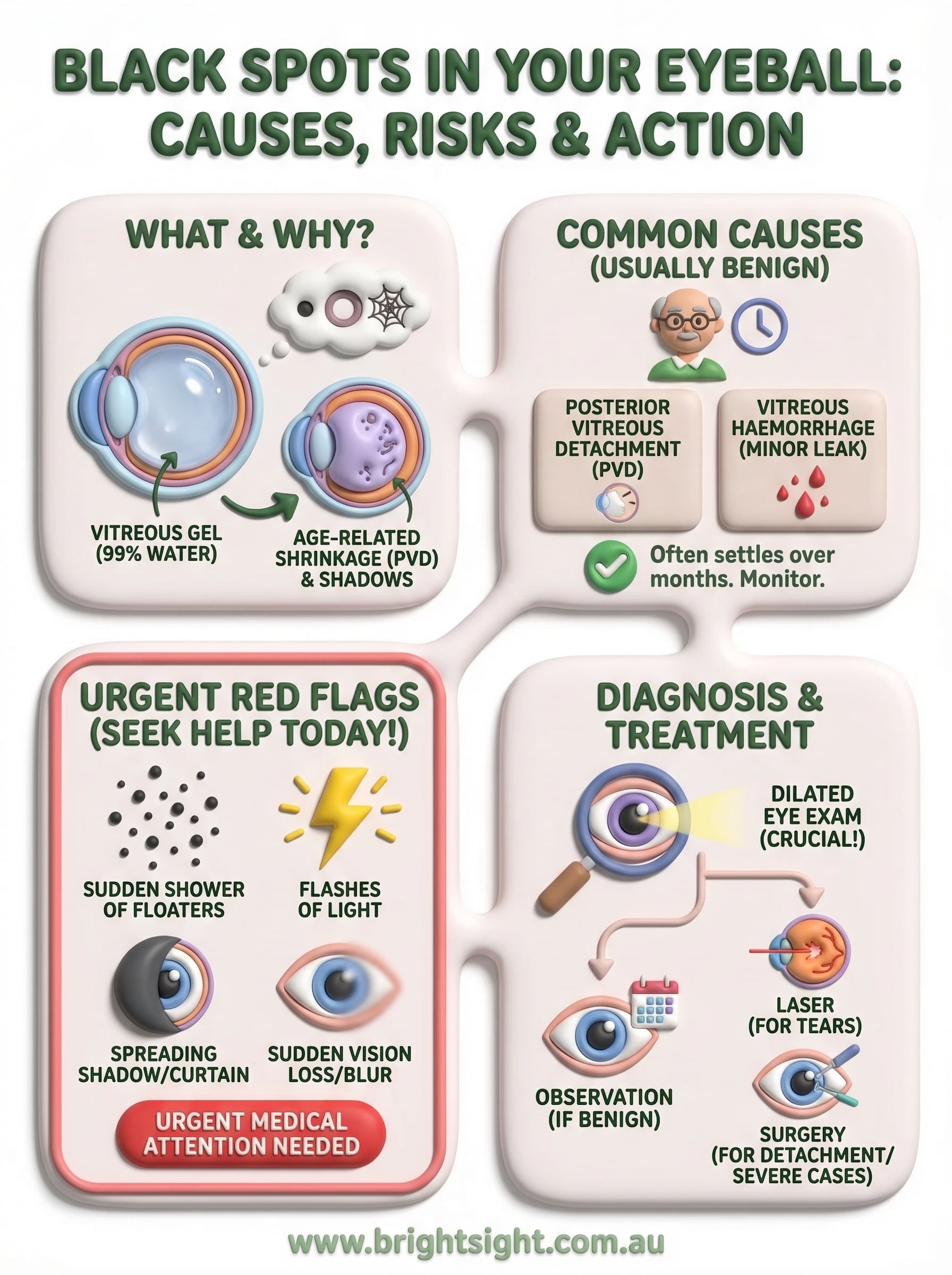
The vitreous gel is roughly 99% water, with the remainder made up of collagen fibres and hyaluronic acid. When you are young, this gel is firm and relatively uniform. As you age, the gel begins to liquefy and shrink, a process called vitreous syneresis. The collagen fibres clump together and cast shadows on your retina as light passes through the eye. Those shadows are what you perceive as spots, threads, or cobweb shapes moving across your vision.

Most floaters are a direct result of the vitreous gel pulling away from the retina, a process called posterior vitreous detachment (PVD), which affects the majority of people over the age of 60.
What these spots look and feel like
The appearance of floaters varies from person to person. You might notice small dark dots, curved lines, rings, or irregular clumps that shift as your eye moves. They tend to be most visible when you look at a plain, bright surface, such as a clear sky or a white wall.
Common descriptions patients use include:
- Tiny black or grey dots that drift when you move your eye
- Cobweb or thread-like shapes in your central vision
- A shadowy ring or curved line that follows your gaze
- A cluster of spots that appears suddenly after a bright flash of light
Common causes and risk factors in Australia
While black spots in my eyeball is a phrase we hear regularly at our Randwick clinic, the underlying cause is not always the same. Several distinct conditions can produce floaters, and understanding the difference helps you know what to expect.
Age-related vitreous changes
The most common cause in Australian adults is posterior vitreous detachment (PVD), where the vitreous gel shrinks and pulls away from the retina. This process accelerates after the age of 50 and is largely unavoidable. Another contributing cause is vitreous haemorrhage, where small amounts of blood leak into the gel from fragile or damaged retinal blood vessels, casting dark shadows across your vision.
Floaters caused by a simple PVD often settle or become less noticeable within a few months, but they rarely disappear entirely.
Who is most at risk
Your risk of developing floaters increases if you are short-sighted (myopic), as longer eyeballs place greater mechanical stress on the vitreous. People with diabetes are also at higher risk due to retinal vessel fragility. Previous eye surgery, eye trauma, and a family history of retinal conditions further raise your likelihood of experiencing significant visual disturbances.
When black spots are urgent red flags
Most floaters are harmless, but certain changes in your vision demand immediate attention. If you experience any of the symptoms described below, treat them as a medical emergency and contact an eye specialist or emergency department the same day rather than waiting for a routine appointment.
Warning signs that need same-day assessment
A sudden increase in black spots in my eyeball, particularly when combined with flashes of light or a spreading shadow at the edge of your vision, may indicate that your retina is tearing or detaching. This is a sight-threatening emergency that requires urgent surgical intervention.
A dark curtain or veil creeping across your field of vision is a retinal detachment until proven otherwise. Acting within hours can be the difference between saving and losing your sight.
Watch specifically for these warning signs:
- A sudden shower of new floaters appearing all at once
- Flashes of light, particularly in dim or dark conditions
- A grey or dark shadow expanding from the side of your vision
- Sudden blurred or lost vision in one eye
Conditions behind urgent symptoms
Retinal detachment and vitreous haemorrhage are the two most serious causes of sudden, dramatic floaters. Both conditions respond well to treatment when caught early, which is why same-day assessment genuinely matters.
Diabetic retinopathy can also trigger abrupt floaters when fragile retinal blood vessels rupture. If you have diabetes and notice a rapid visual change, contact your eye specialist promptly rather than waiting for your next scheduled review.
How an eye specialist diagnoses the cause
When you report black spots in my eyeball, your specialist begins with a detailed history, asking about the onset, duration, and any associated symptoms such as flashes or visual loss. This initial conversation helps determine whether further urgent testing is needed.
The dilated eye examination
Your specialist will use dilating eye drops to widen your pupil, then examine the vitreous and retina using a slit lamp and indirect ophthalmoscope. This allows a clear assessment of the retina’s condition, including any tears, detachment, or signs of bleeding.
A dilated examination remains the most reliable way to distinguish harmless floaters from a sight-threatening retinal problem.
Imaging and additional tests
When the internal view is unclear, your specialist may request additional imaging to confirm a diagnosis. Ocular ultrasound (B-scan) maps structures at the back of the eye when blood or opacity blocks a direct view. Optical coherence tomography (OCT) produces detailed cross-sectional images of the retina, revealing subtle changes that may not appear during a standard examination.

Common imaging tools used include:
- B-scan ultrasound for vitreous haemorrhage or opaque media
- OCT for macular and retinal detail
- Fundus photography to document and monitor retinal changes over time
Treatment options and what you can do today
Treatment for black spots in my eyeball depends entirely on the underlying cause. If your specialist confirms a simple PVD with no retinal complications, no immediate treatment is usually required, and most patients adapt to mild floaters over time.
When observation is the right approach
For benign floaters, your specialist will monitor your eye at scheduled review appointments to confirm no retinal changes develop. Many patients find that floaters become less intrusive within three to six months as the brain learns to filter them out. Keeping a note of any sudden changes in the meantime is worthwhile.
If your floaters worsen, new ones appear, or flashes begin, contact your specialist immediately rather than waiting for your next review.
Active treatments for significant floaters or serious conditions
When floaters are severe enough to affect your daily activities, your specialist may recommend vitrectomy, a surgical procedure to remove the vitreous gel and replace it with a clear saline solution. For retinal tears, laser photocoagulation seals the tear before detachment occurs. Retinal detachment itself requires prompt surgical repair, either through vitrectomy or scleral buckling. Diabetic or haemorrhage-related floaters are treated by addressing the underlying disease process directly.
Next steps
Black spots in my eyeball are something many people notice and then put off investigating, often assuming the spots will resolve on their own. In most cases, benign floaters do settle and become less noticeable over time. But the key risk is missing an early retinal tear or detachment, both of which are treatable when caught quickly and far more difficult to manage once they progress.
If your floaters appeared suddenly, came with flashes of light, or are accompanied by any shadow in your peripheral vision, do not wait. Same-day assessment is the right course of action. Even if your symptoms seem mild, a thorough examination by a specialist gives you certainty and rules out anything serious.
At Bright Sight Eye Specialists in Randwick, Dr. Rahul Dubey provides expert diagnosis and treatment for the full range of conditions behind visual disturbances. Book an appointment today and get a clear answer about what you are seeing.