


You’re reading, driving, or simply looking at a clear sky when you notice it, a black spot in vision that moves across your field of view, drifting away when you try to focus on it. These fleeting shadows, commonly known as eye floaters, are something most people will experience at some point in their lives. While they’re often harmless, understanding what causes them, and recognising when they might signal something more serious, is essential for protecting your sight.
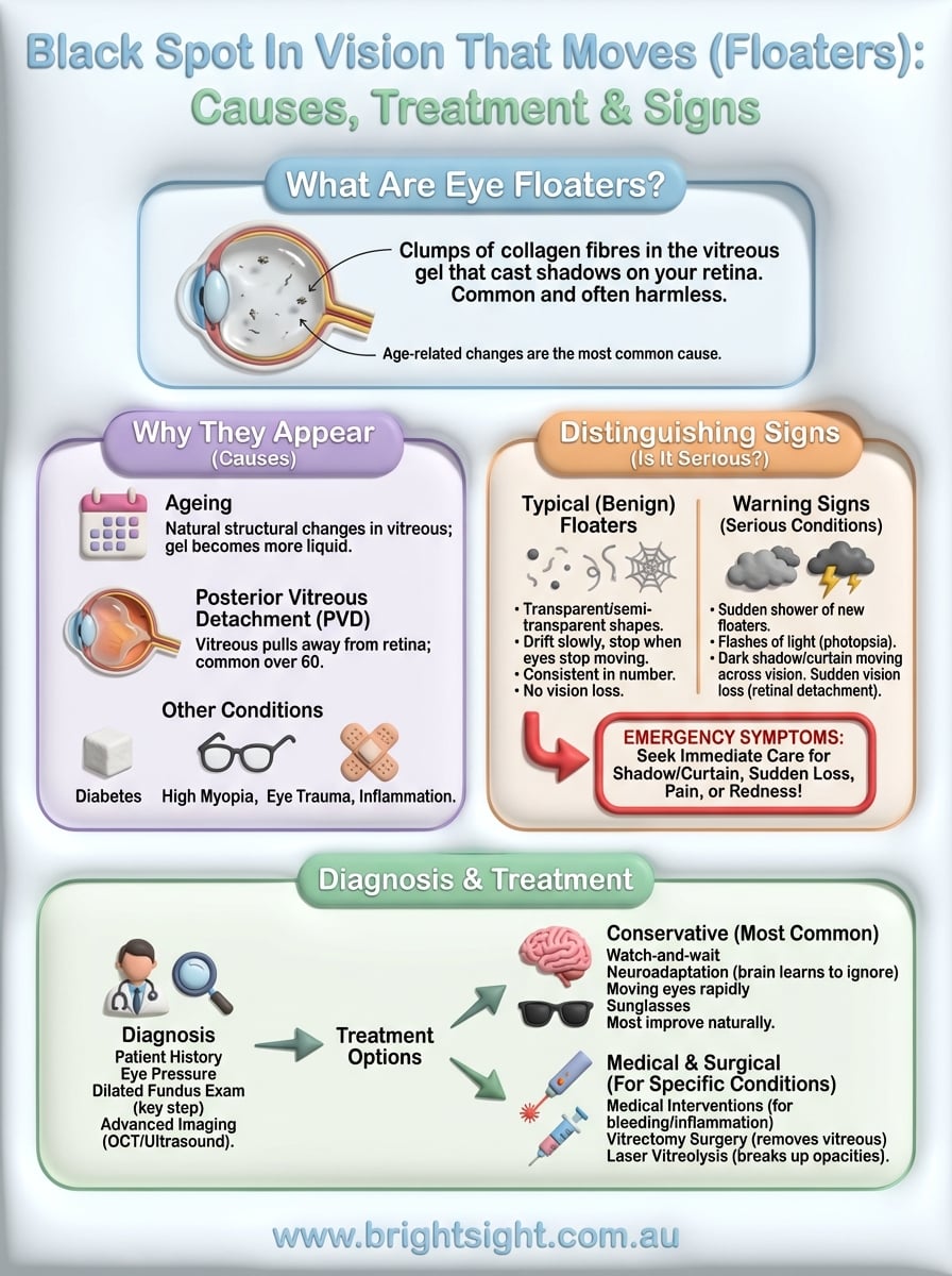
Floaters typically appear when tiny fibres within the vitreous (the gel-like substance filling your eye) clump together and cast shadows on your retina. Age-related changes are the most common cause, but sudden increases in floaters, especially when accompanied by flashes of light, can indicate conditions like retinal detachment that require urgent attention.
At Bright Sight Eye Specialists, Dr Rahul Dubey and our team regularly help patients in Sydney’s Eastern Suburbs understand and manage floaters and the underlying conditions behind them. This article explains what causes moving black spots in your vision, when to seek professional assessment, and the treatment options available, giving you the knowledge to make informed decisions about your eye health.
Why a moving black spot appears
Understanding why a black spot in vision that moves appears starts with knowing what’s happening inside your eye. Your vitreous humour (the clear, gel-like substance filling about 80% of your eyeball) plays a central role in this phenomenon. As you age or experience certain eye conditions, changes to this gel create the shadows you perceive as floaters, spots, or cobweb-like shapes drifting across your field of view.
The vitreous and age-related changes
Your vitreous begins life as a perfectly clear, thick gel that helps maintain your eye’s shape and allows light to pass cleanly to your retina. Over time, this gel naturally undergoes structural changes, becoming more liquid in some areas while developing tiny clumps of collagen fibres in others. These clumps cast shadows on your retina, which your brain interprets as moving spots or strings floating in your vision.
Most people start noticing floaters in their 40s or 50s, though they can appear earlier in people who are short-sighted (myopic) or have had eye trauma. The process is gradual, and while it affects nearly everyone eventually, you might never notice minor floaters unless you’re looking at a bright, uniform background like a blue sky or white wall. The spots move because the vitreous fibres drift within the liquid portion of the gel, following your eye movements but with a slight delay that creates that characteristic drifting sensation.
Posterior vitreous detachment
A more significant cause of sudden floaters is posterior vitreous detachment (PVD), which occurs when the vitreous gel pulls away from its attachment to your retina. This is a normal part of ageing for most people over 60, but it can happen earlier in short-sighted individuals or following eye surgery. When PVD occurs, you typically notice a sudden increase in floaters, often accompanied by flashes of light (photopsia) as the vitreous tugs on your retina.

PVD itself isn’t dangerous, but the tugging motion during detachment can sometimes create a tear in your retina, which requires immediate treatment to prevent vision loss.
The detachment process usually completes over several weeks, during which you might notice floaters are most prominent. Once the vitreous has fully separated, the intense tugging stops, though the floaters themselves may persist for months or permanently. Your brain typically learns to ignore them over time, a process called neuroadaptation, making them less noticeable in daily life.
Other causes of moving black spots
Several less common conditions can produce similar symptoms to age-related floaters. Vitreous haemorrhage (bleeding into the vitreous cavity) can occur from diabetic retinopathy, retinal tears, or blood vessel abnormalities, creating sudden showers of dark spots that obscure your vision more significantly than typical floaters. Inflammation inside your eye (uveitis or vitritis) can cause white blood cells to float in the vitreous, appearing as moving spots or haziness.
Certain systemic conditions also contribute to floater development. Diabetes affects the blood vessels in your retina, potentially leading to bleeding or inflammatory changes that create new floaters. High myopia (severe short-sightedness) causes your eyeball to elongate, stretching the vitreous and making it more prone to early liquefaction and detachment. Even eye injuries can accelerate vitreous changes, introducing blood cells or inflammatory debris that appear as moving shadows in your visual field.
How to tell floaters from other causes
Distinguishing between harmless floaters and symptoms of serious eye conditions requires you to understand specific visual characteristics and accompanying signs. While most people who notice a black spot in vision that moves are experiencing benign age-related changes, certain patterns should prompt immediate professional assessment. Recognising these differences helps you respond appropriately and protects your sight from preventable damage.
Characteristics of typical floaters
Normal floaters follow predictable patterns that help you identify them as non-threatening vitreous changes. They appear as transparent or semi-transparent shapes (dots, threads, cobwebs, or rings) that drift slowly across your field of view, moving in the direction your eye moves but continuing to float after you stop moving your eyes. You notice them most clearly against bright, uniform backgrounds like white walls, blue skies, or computer screens, while they seem to disappear in dim lighting or against patterned surfaces.
Benign floaters remain relatively consistent in number and appearance from day to day, though you might notice them more on some days depending on lighting conditions and what you’re looking at. They don’t obstruct your central vision significantly, and while they can be annoying, they don’t create blind spots or cause pain, redness, or changes in your peripheral vision. Most importantly, typical floaters develop gradually over months or years, giving your brain time to adapt and making them less noticeable over time.
Warning signs of serious conditions
Several characteristics distinguish dangerous symptoms from routine floaters and require urgent evaluation. A sudden shower of new floaters appearing within hours or days, especially if accompanied by flashes of light (like lightning bolts or sparkles in your peripheral vision), suggests your vitreous may be pulling on your retina and potentially causing tears. This pattern occurs in approximately 15% of people experiencing posterior vitreous detachment and needs same-day assessment.

If you notice a shadow or curtain moving across your field of view, or experience sudden vision loss in any part of your visual field, you’re likely experiencing retinal detachment that requires emergency treatment within hours to prevent permanent vision loss.
Other red flags include floaters that appear as dense clouds or veils obscuring your vision rather than transparent shapes, which may indicate vitreous haemorrhage from diabetic retinopathy or torn blood vessels. You should also seek immediate evaluation if floaters develop after eye trauma, during an active infection, or alongside eye pain, redness, or rapid vision changes, as these combinations suggest inflammation, bleeding, or structural damage requiring prompt intervention.
When to treat it as an emergency
Not every black spot in vision that moves requires urgent medical attention, but certain combinations of symptoms demand immediate evaluation to prevent permanent vision loss. The distinction between symptoms that can wait for a routine appointment and those requiring same-day or emergency assessment often determines whether you preserve full vision or experience irreversible damage. Understanding these time-sensitive warning signs helps you respond appropriately when new visual symptoms appear.
Symptoms requiring same-day assessment
You need professional evaluation within 24 hours if you notice a sudden increase in floaters (particularly if dozens appear within hours), even without other symptoms. This pattern suggests your vitreous may have recently detached, creating a period of increased risk for retinal tears that typically lasts several weeks. Contact your ophthalmologist or visit an eye casualty service the same day if you experience persistent flashes of light in one eye, especially if they occur in your peripheral vision or continue when your eyes are closed, as this indicates ongoing traction on your retina.
New floaters appearing alongside reduced visual clarity or a sense of heaviness in your eye also warrant urgent assessment, even if you don’t notice other alarming symptoms. Your eye specialist can perform dilated retinal examination to check for tears, bleeding, or inflammation that requires prompt treatment. Most practices, including Bright Sight Eye Specialists, accommodate emergency appointments for these presentations because early detection significantly improves treatment outcomes.
If you develop sudden visual symptoms during weekends or after hours when your regular specialist isn’t available, visit your nearest hospital’s emergency department or eye casualty unit rather than waiting for business hours.
True emergencies requiring immediate action
You need emergency department assessment within hours if you notice a dark curtain, shadow, or veil moving across any part of your visual field, as this indicates active retinal detachment requiring surgical repair. Similarly, sudden vision loss in one eye (whether complete blackness or significant blurring that doesn’t improve with blinking) demands immediate attention, particularly if accompanied by floaters or flashes. Dense clouds of floaters that appear suddenly and obscure your vision suggest vitreous haemorrhage, which requires urgent evaluation to identify and treat the underlying bleeding source.
Seek immediate care if visual symptoms appear alongside eye pain, redness, or headache, as these combinations may indicate inflammation, infection, or elevated eye pressure requiring emergency treatment to prevent complications.
How eye doctors diagnose the cause
When you report a black spot in vision that moves, your eye specialist follows a systematic diagnostic process to determine whether you’re experiencing benign floaters or a condition requiring intervention. Accurate diagnosis requires specialised equipment and expertise, which is why your regular optometrist might refer you to an ophthalmologist if they detect concerning signs during their initial screening. The examination typically progresses from simple observation to detailed imaging, depending on what your specialist finds at each stage.
Initial examination and patient history
Your appointment begins with a detailed discussion about when symptoms started, how they’ve progressed, and any accompanying signs like flashes or vision changes. Your specialist asks about your medical history, particularly diabetes, high myopia, or previous eye trauma, as these factors significantly influence the likely cause. You’ll undergo visual acuity testing to establish whether the spots affect your ability to see clearly, followed by eye pressure measurement to rule out complications like glaucoma or intraocular inflammation that might contribute to your symptoms.
Your specialist pays particular attention to the pattern of floaters, as sudden showers of spots appearing within hours suggest different underlying causes than gradual development over months or years.
Dilated fundus examination
The most important diagnostic step involves dilating your pupils with eye drops to allow comprehensive examination of your vitreous and retina. Once dilated (which takes 15 to 20 minutes), your specialist uses specialised lenses and bright lights to examine the back of your eye in detail, looking for vitreous changes, retinal tears, bleeding, or signs of detachment. This examination identifies whether your floaters result from normal posterior vitreous detachment, inflammation, haemorrhage, or retinal complications requiring treatment.
Advanced imaging when needed
Your specialist may use optical coherence tomography (OCT) to create detailed cross-sectional images of your retina and vitreous, particularly if they suspect macular involvement or need to assess the extent of vitreous changes. Ultrasound examination helps when dense floaters or bleeding obscure the view of your retina, allowing your specialist to detect retinal detachment or other structural problems that aren’t visible through standard examination. These imaging techniques provide precise information about your eye’s internal structures, guiding treatment decisions and establishing baseline measurements for monitoring future changes.
Treatment options and what to expect
Most people who notice a black spot in vision that moves receive reassurance rather than active treatment, as benign floaters typically improve with time as your brain adapts to their presence. Your treatment pathway depends entirely on what’s causing your symptoms, ranging from watchful observation for harmless age-related changes to urgent surgical intervention for sight-threatening conditions. Understanding the available options helps you set realistic expectations about your recovery timeline and what improvements you can anticipate.
Conservative management for benign floaters
Your specialist recommends a watch-and-wait approach if examination confirms your floaters result from uncomplicated posterior vitreous detachment without retinal tears or other complications. This strategy recognises that your brain naturally learns to ignore floaters over weeks to months through neuroadaptation, making them less noticeable in daily activities. You’ll receive advice about returning immediately if symptoms worsen (such as sudden increases in floaters, new flashes, or vision loss), but otherwise no active treatment is needed.
Simple adjustments help you manage persistent floaters while neuroadaptation occurs. Moving your eyes rapidly from side to side or up and down shifts the vitreous gel and temporarily relocates floaters out of your central vision. Wearing sunglasses in bright conditions reduces contrast between floaters and your background view, making them less prominent. Most patients find floaters become significantly less bothersome within three to six months, though the physical spots themselves remain present in your vitreous gel.
Around 85% of people with new floaters from posterior vitreous detachment experience substantial improvement in symptoms without any medical intervention within the first year.
Medical interventions for specific conditions
Your specialist prescribes targeted treatment when floaters indicate underlying disease rather than simple age-related changes. Vitreous haemorrhage from diabetic retinopathy often responds to improved blood sugar control and sometimes clears spontaneously over weeks, though you might need laser treatment or injections to address the damaged blood vessels causing the bleeding. Inflammation inside your eye requires steroid drops or other anti-inflammatory medications to reduce white blood cell activity and prevent permanent damage to your vision.
When surgery becomes necessary
Your ophthalmologist recommends vitrectomy surgery only for severe cases where floaters significantly impair your daily function or when treating complications like retinal detachment. This procedure involves removing the vitreous gel and replacing it with a clear saline solution, eliminating floaters but carrying risks including cataract development, retinal detachment, and infection. Laser vitreolysis offers a less invasive alternative for specific types of floaters, using focused laser energy to break up larger opacities, though its effectiveness varies depending on floater location and characteristics.
Next steps
Noticing a black spot in vision that moves doesn’t automatically signal an emergency, but understanding when to act quickly protects your sight from preventable damage. Most floaters result from normal age-related changes that improve naturally over time as your brain adapts, requiring no treatment beyond monitoring for warning signs like sudden increases, flashes of light, or vision loss. Your response should match your symptoms: schedule a routine appointment for gradual floaters that don’t affect your daily activities, arrange same-day assessment for sudden increases or persistent flashes, and seek emergency care immediately if you notice shadows, curtains, or sudden vision loss in any part of your visual field.
Book an appointment with Dr Rahul Dubey at Bright Sight Eye Specialists if you’ve developed new floaters or have concerns about existing ones. Our practice in Sydney’s Eastern Suburbs provides comprehensive retinal examination and transparent advice about your treatment options, helping you maintain clear, healthy vision throughout your life.