


A black spot in the vision of one eye refers to a dark area that blocks or dims part of what you can see. This spot might appear as a tiny dot, a larger shadow, or a fixed dark patch that stays in place when you move your eye. Unlike the drifting floaters most people experience, a single black spot in one eye can feel more concerning because it may signal a problem within the eye itself, from a harmless clump of debris in the vitreous gel to a more serious issue like a retinal tear or macular disorder.
This article explains the most common causes of a black spot in one eye, from age related changes and floaters to retinal detachment and macular degeneration. You will learn which symptoms require immediate attention, how your eye specialist diagnoses the underlying cause, and what treatment options are available. Understanding when a black spot is harmless and when it demands urgent care can help you protect your vision and respond quickly if something goes wrong. By the end, you will know exactly what steps to take if you notice a dark spot affecting just one eye.
Why a black spot in one eye needs attention
A black spot affecting just one eye deserves prompt evaluation because it can indicate problems confined to that specific eye, rather than a temporary visual disturbance affecting both eyes. When you notice a dark spot that appears only when you cover the unaffected eye, it suggests the issue originates within the eye’s internal structures such as the vitreous, retina, or macula. Unlike bilateral symptoms that might point to migraine or neurological causes, a black spot in vision of one eye often signals localised damage, bleeding, or detachment that requires an ophthalmologist’s assessment. Your brain cannot always compensate for monocular vision loss, so even a small spot may grow larger if left untreated.
The earlier you seek care for a black spot in one eye, the better your chances of preserving full vision.
When normal floaters become concerning
You probably experience occasional floaters that drift across your field of view and disappear within seconds. These harmless specks form when collagen fibres clump together in the vitreous gel as you age. However, a persistent black spot that remains fixed or appears suddenly with flashes of light signals a more serious problem. If the spot darkens over hours or days, or if you notice a curtain-like shadow spreading from the periphery, your retina may have torn or detached. Age-related floaters move when you shift your gaze, but a stationary or expanding dark patch suggests structural damage that needs urgent intervention.
One eye vs both eyes: what the difference means
Symptoms limited to one eye point to local pathology within that eye, such as a retinal tear, vitreous haemorrhage, or macular defect. Bilateral symptoms often stem from systemic issues like migraine aura or neurological conditions affecting the visual pathways. When you cover each eye in turn and confirm the black spot appears only on one side, you help your specialist narrow the diagnostic possibilities immediately. This distinction also affects treatment urgency, as monocular retinal detachment progresses faster than some bilateral conditions and can cause permanent vision loss within days if ignored.
How to respond when you notice a black spot
Your first response to a black spot in vision of one eye should be calm observation rather than panic. Most dark spots result from harmless age-related changes, but some signal urgent problems that need immediate treatment. Stop what you are doing and take a few moments to assess the situation carefully. Note the spot’s size, shape, and position, and check whether it moves when you shift your gaze or stays fixed in place. This initial assessment helps you decide whether to book a routine appointment or seek emergency eye care within hours.
Document what you observe
Write down the exact time you first noticed the spot and describe its appearance in simple terms. Note whether it looks like a small dot, a larger shadow, or a curtain-like area blocking part of your vision. Record any accompanying symptoms such as flashes of light, sudden increase in floaters, or blurred vision around the spot. If you have a smartphone, take a photo of an Amsler grid with your affected eye to capture any distortions or missing areas. This documentation gives your eye specialist valuable information about how quickly the condition developed and whether it is progressing.
Cover each eye separately
Test your vision by covering one eye at a time with your palm, not just closing it. Look at a plain white wall or bright surface and check whether the black spot appears in one eye only or affects both eyes. If the spot vanishes when you cover the affected eye and your other eye sees normally, the problem lies within that specific eye’s structures. Confirm that the spot’s position changes or remains fixed as you move your gaze up, down, left, and right. This simple test helps you provide accurate details to your eye doctor and rules out bilateral issues that might suggest a different cause.

Testing each eye separately tells you whether the problem is isolated to one eye or affecting both.
Seek professional assessment promptly
Contact an eye specialist or visit an emergency eye clinic if you notice any sudden changes such as a rapid increase in spot size, new flashes of light, or a shadow spreading across your vision. Book a same-day or next-day appointment for spots that appear gradually without flashes or curtain-like shadows. Do not wait more than 48 hours to get checked, even if the spot seems small or causes only mild disturbance. Early examination allows your ophthalmologist to identify retinal tears before they progress to detachment and to start treatment while your prognosis remains excellent.
Common causes of a black spot in one eye
A black spot in vision of one eye can result from several different conditions, ranging from harmless age-related changes to serious problems requiring immediate treatment. The vitreous gel inside your eye, the retina lining its back surface, and the macula responsible for central vision all play roles in producing dark spots when they become damaged or disrupted. Understanding these causes helps you recognise whether your symptom needs urgent attention or simple monitoring. Most people experience benign floaters at some point, but sudden onset or accompanying symptoms like flashes suggest more concerning pathology.
Age-related vitreous changes and floaters
Your vitreous humour is a clear, jelly-like substance that fills the space between your lens and retina. As you age past 50, this gel gradually liquefies and shrinks, causing microscopic collagen fibres within it to clump together. These clumps cast tiny shadows on your retina that appear as dark spots, strings, or cobweb-like shapes drifting across your vision. When you notice just one persistent spot rather than multiple shifting floaters, a larger collagen clump may have formed in a position that blocks your central vision. These age-related floaters usually remain harmless and become less noticeable as your brain adapts to ignore them.

Posterior vitreous detachment
Posterior vitreous detachment occurs when your shrinking vitreous gel pulls away cleanly from the retinal surface. This natural ageing process affects nearly two-thirds of people over 70 and often produces a sudden increase in floaters, including one or more prominent dark spots. The detachment itself causes brief flashes of light as the vitreous tugs on the retina before releasing. Most posterior vitreous detachments resolve without treatment, but around 15% lead to retinal tears that need immediate repair. Your eye specialist will perform a dilated examination to confirm the vitreous has separated safely without damaging the underlying retina.
Posterior vitreous detachment is common and usually harmless, but examination remains essential to rule out retinal tears.
Retinal tears and detachment
Retinal tears happen when the vitreous pulls too hard during detachment, creating a break in the retinal tissue. Fluid from the vitreous cavity then seeps through this tear and lifts the retina away from its supporting layer, causing retinal detachment. You may notice a fixed black spot in your vision that expands into a larger shadow or curtain as the detachment spreads. Small tears produce localised scotomas (blind spots), while extensive detachments can block entire quadrants of vision. Immediate laser or surgical treatment reattaches the retina and prevents permanent vision loss, making prompt diagnosis critical.
Macular disorders
The macula provides your sharp central vision for reading and recognising faces. Age-related macular degeneration causes drusen deposits (dry AMD) or abnormal blood vessels (wet AMD) that produce dark or blurred spots in your central vision. Central serous chorioretinopathy creates fluid buildup under the macula, resulting in a greyish spot or dimmed area directly in your line of sight. Macular holes, epiretinal membranes, and macular oedema from diabetes also generate central scotomas. These conditions require specialised imaging such as optical coherence tomography to diagnose and often respond well to injections, laser therapy, or surgical repair when treated early.
Other causes
Vitreous haemorrhage from bleeding into the gel cavity produces sudden dark spots or floaters that appear red-tinged. Inflammation in the back of your eye (posterior uveitis) releases white blood cells into the vitreous that cast shadows resembling black spots. Rarely, inherited conditions like retinitis pigmentosa or eye tumours cause fixed scotomas, though these typically develop gradually rather than appearing suddenly. Your eye specialist will consider your age, medical history, and symptom onset to distinguish between these less common causes.
Red flag symptoms and urgent warning signs
Certain symptoms accompanying a black spot in vision of one eye demand immediate medical attention because they signal conditions that can cause permanent vision loss within hours or days if left untreated. These red flag signs indicate your retina may have torn or detached, blood vessels may be bleeding into your eye, or other sight-threatening problems require emergency intervention. Recognising these warning signs and acting quickly gives your eye specialist the best chance to preserve your vision through prompt treatment. Never adopt a wait-and-see approach when you notice any of the symptoms described below, as delays can mean the difference between full recovery and lasting visual impairment.
Sudden flashes of light and shower of floaters
Bright flashes of light that appear like lightning bolts or camera flashes, especially when combined with many new floaters appearing all at once, suggest your vitreous is tugging forcefully on the retina. These photopsia episodes happen because mechanical stimulation of retinal cells triggers false light signals that your brain interprets as flashes. When you see dozens or hundreds of small dark spots appearing suddenly, like someone shaking pepper across your vision, blood may be leaking into the vitreous cavity from a retinal tear. This combination of symptoms requires assessment within hours, not days, because torn retinas progress rapidly to detachment.
Sudden flashes combined with multiple new floaters signal retinal tearing that needs immediate evaluation.
Contact an emergency eye clinic or ophthalmologist straight away if flashes occur repeatedly over several minutes or hours. The risk increases further if you also notice a growing shadow or darkened area spreading from the edge of your vision toward the centre.
Dark curtain or shadow spreading across vision
A dark curtain, shadow, or veil that appears to pull across your field of view from one side indicates retinal detachment is actively progressing. This shadow represents the area where your retina has separated from its underlying support tissue and stopped functioning. The curtain typically starts at the peripheral edges of your vision and advances toward the centre as fluid continues lifting the retina away from the eye wall. You might describe this as a grey or black cloud moving inward, or feel like someone is slowly drawing a blind across your sight.
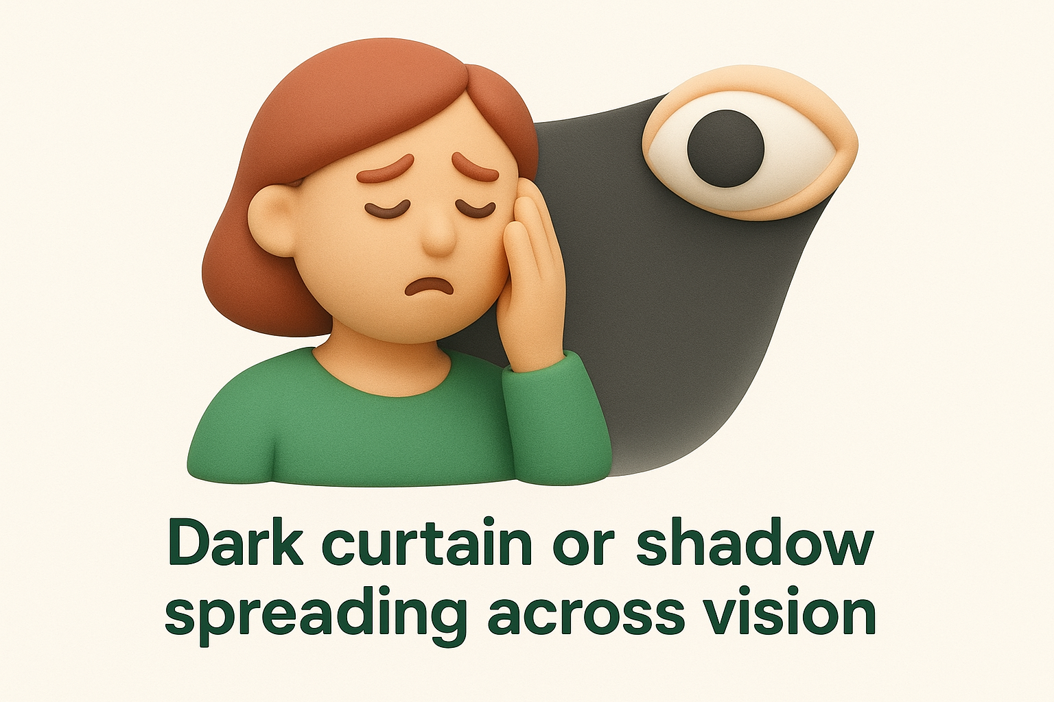
Retinal detachment constitutes an ophthalmic emergency requiring surgical repair within 24 to 48 hours to prevent permanent vision loss. The longer your retina remains detached, the lower your chances of full visual recovery, so immediate action becomes critical.
Rapid changes in spot size or appearance
Any black spot that noticeably enlarges over hours or days, darkens in intensity, or multiplies into additional spots needs urgent evaluation. Progressive changes suggest the underlying problem is actively worsening, whether from expanding haemorrhage, increasing inflammation, or spreading retinal damage. A spot that begins small but grows to block significant portions of your central or peripheral vision within 24 hours indicates serious pathology requiring immediate diagnosis and treatment.
You should also seek urgent care if mild visual disturbances suddenly worsen, new symptoms develop alongside the existing spot, or you experience any pain, redness, or discharge from the affected eye. These additional signs can point to inflammatory conditions or infections that threaten your vision if not controlled quickly.
Diagnosis and tests your eye specialist may use
Your eye specialist begins by gathering a detailed account of your symptoms and performing a thorough clinical examination to identify the cause of your black spot in vision of one eye. This diagnostic process combines patient history, direct visualisation of your eye’s internal structures, and specialised imaging tests that reveal subtle changes invisible to the naked eye. The specific tests your ophthalmologist chooses depend on your symptom description, medical background, and findings from the initial examination. Most diagnostic procedures cause minimal discomfort and provide immediate or near-immediate results that guide your treatment plan.
Patient history and symptom assessment
Your specialist asks detailed questions about when you first noticed the black spot, whether it appeared suddenly or gradually, and any accompanying symptoms like flashes or floaters. Describing the spot’s size, shape, and position helps narrow the diagnostic possibilities before any physical examination begins. You will be asked about your medical history, including diabetes, hypertension, previous eye surgery, and family history of retinal conditions. Your specialist also wants to know whether the spot moves when you shift your gaze or remains fixed in one location, as stationary spots often suggest retinal or macular pathology whilst moving spots point to vitreous causes.
Dilated fundus examination
Dilating your pupil with medicated eye drops allows your ophthalmologist to examine the entire retina, vitreous, and optic nerve thoroughly. This examination involves shining bright light into your eye using various instruments including direct ophthalmoscopy and slit-lamp biomicroscopy with specialised lenses. Your specialist looks for retinal tears, detachment, haemorrhage, macular changes, and vitreous opacities that could explain your dark spot. The drops take 15 to 30 minutes to work fully and leave your vision blurred for several hours afterward, so arrange alternative transport home before your appointment.
Dilating your pupil remains the most reliable way to detect retinal tears before they cause detachment.
Advanced imaging techniques
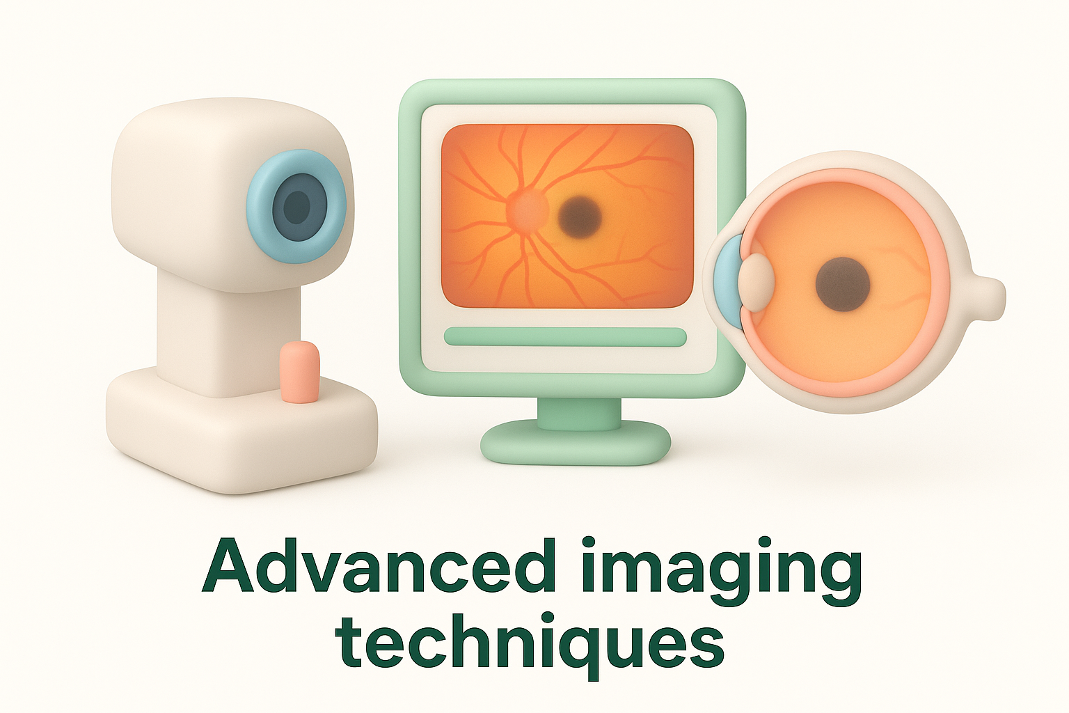
Optical coherence tomography (OCT) creates high-resolution cross-sectional images of your retina and macula, revealing subtle changes like fluid buildup, holes, or membrane formation that explain central black spots. This painless scan takes only minutes and provides detailed measurements of retinal thickness and structure. B-scan ultrasonography uses sound waves to visualise your eye’s interior when dense cataracts or vitreous bleeding obscure the view during examination. Your specialist may also perform visual field testing to map the exact location and extent of any blind spots, or fundus photography to document retinal appearance for comparison at future visits. Fluorescein angiography, where dye is injected into your arm vein to highlight retinal blood vessels, helps diagnose vascular problems and wet macular degeneration causing dark spots.
Treatment options and ongoing eye care
Your treatment plan depends entirely on what caused your black spot in vision of one eye, how severely it affects your sight, and whether the underlying condition threatens further vision loss. Some dark spots require only careful monitoring whilst others demand immediate surgical intervention to prevent permanent damage. Your ophthalmologist will explain the recommended approach based on your specific diagnosis and discuss the expected outcomes, recovery time, and potential risks of each option. Most treatments aim to preserve existing vision rather than restore areas already lost, which makes early detection and prompt treatment crucial for the best possible results.
Observation and monitoring for benign floaters
Harmless vitreous floaters that cast dark spots in your vision often need no treatment beyond regular observation. Your brain gradually adapts to these shadows over weeks or months, making them less noticeable during daily activities. You will attend follow-up appointments every six to twelve months so your specialist can confirm the floaters remain stable and have not triggered retinal tears. During this monitoring period, you should contact your eye doctor immediately if you notice sudden changes such as increased floater numbers, flashes of light, or expanding shadows that suggest complications have developed.
Laser and surgical interventions
Retinal tears require urgent laser photocoagulation or cryotherapy to seal the break before fluid accumulates and causes detachment. Your surgeon applies laser burns or freezing treatment around the tear to create scar tissue that welds the retina back into position. Established retinal detachment needs surgery, typically either vitrectomy (removing the vitreous gel and replacing it with gas or silicone oil) or scleral buckle (placing a silicone band around the eye to push the wall inward). YAG laser vitreolysis can break up large, troublesome floaters by targeting them with focused light pulses, though this procedure carries risks and suits only specific cases where floaters severely impair vision.
Laser treatment can seal retinal tears within minutes and prevent them from progressing to detachment.
Medical treatments and injections
Wet age-related macular degeneration and diabetic macular oedema respond well to intravitreal anti-VEGF injections that stop abnormal blood vessel growth and reduce fluid leakage. Your specialist administers these injections directly into your vitreous cavity every four to twelve weeks depending on your response to treatment. Inflammatory conditions causing black spots may need corticosteroid therapy delivered as eye drops, tablets, or injections to control the inflammation and clear floating debris. Central serous chorioretinopathy often resolves spontaneously, though persistent cases benefit from photodynamic therapy or focal laser to seal leaking vessels.
Long-term eye care strategies
Protecting your remaining vision requires attending all scheduled follow-up appointments and reporting any new symptoms immediately. You should monitor your vision daily using an Amsler grid to detect early changes in central vision before they become obvious. Control systemic conditions like diabetes and hypertension through medication, diet, and lifestyle changes to reduce your risk of further vascular damage. Wear UV-blocking sunglasses outdoors, maintain a diet rich in leafy greens and omega-3 fatty acids, and quit smoking to support long-term retinal health.

Key takeaways
A black spot in vision of one eye can result from harmless age-related floaters or signal serious conditions requiring immediate treatment. You should seek urgent care if you notice sudden flashes of light, multiple new floaters appearing together, or a dark curtain spreading across your vision, as these symptoms suggest retinal tears or detachment that threaten permanent vision loss. Most dark spots warrant professional assessment within 48 hours, even when they appear gradually without alarming features.
Your eye specialist uses dilated examination and advanced imaging to identify the cause and recommend appropriate treatment, from simple observation to laser therapy or surgery. Regular follow-up appointments, daily vision monitoring with an Amsler grid, and control of systemic conditions like diabetes protect your remaining sight and catch complications early. If you notice a black spot affecting one eye, book a comprehensive eye examination at Bright Sight Eye Specialists to receive expert diagnosis and personalised care that preserves your vision.