


Noticing a black spot in peripheral vision can be unsettling. Your mind might race through possibilities, is it something harmless, or could it signal a serious problem? The truth is, it could be either. While many people experience benign floaters that drift across their field of view, a new or sudden dark spot in your side vision sometimes indicates conditions requiring urgent attention, such as a retinal tear or detachment.
Understanding what’s behind this symptom matters. Some causes resolve on their own or need no treatment at all. Others demand prompt intervention to protect your sight long-term. The key lies in recognising the warning signs that distinguish a minor annoyance from a medical emergency.
At Bright Sight Eye Specialists, Dr Rahul Dubey and Dr Capucine Odouard regularly assess and treat patients presenting with peripheral vision changes. With expertise in retinal and macular conditions, our Randwick-based practice offers the diagnostic technology and surgical capabilities needed to address everything from vitreous floaters to complex retinal detachments.
This article explains what causes black spots in your peripheral vision, when to seek help, and what treatment options exist. By the end, you’ll have the information you need to make informed decisions about your eye health.
What a black spot in peripheral vision is
A black spot in peripheral vision refers to a dark area or shadow you notice to the side of where you’re directly looking. This spot appears in your side vision rather than straight ahead, and it might move when you shift your gaze or remain fixed in the same location. The spot can take different forms: a small dot, a larger patch, or even an irregular shape that blocks part of what you normally see.

Your peripheral vision covers everything outside your direct line of sight, extending roughly 90 degrees to each side, 60 degrees upward, and 70 degrees downward. When something interferes with this area, you’ll notice it most during everyday activities like driving, walking, or reading, where your side vision plays a crucial role in detecting movement and maintaining spatial awareness.
How peripheral vision works differently
The outer edges of your retina contain different light-sensitive cells compared to the centre. While your central vision handles fine detail and colour recognition, your peripheral vision excels at detecting motion and low-light conditions. This specialisation explains why a black spot in this area often appears as movement catches your attention, even though you weren’t looking directly at it.
Your brain processes peripheral information differently too. It prioritises detecting threats or movement over sharp detail, which means you might notice a shadow or dark spot before you consciously register what you’re seeing. This automatic response makes peripheral vision changes particularly noticeable and sometimes more alarming than they need to be.
What the spot looks like in practice
The appearance of a black spot in peripheral vision varies depending on its cause. Some people describe a distinct dark circle that drifts slowly across their field of view, similar to watching a speck floating in water. Others see a fixed shadow that doesn’t move, creating a consistent blind spot in the same location every time they check.
Size matters when describing these spots. A small dot might measure just a few millimetres across in your perceived vision, whilst a larger patch could obscure a significant portion of your side view. The spot’s edges can appear sharp and defined or soft and blurry, and the darkness might range from a light grey to an almost complete blackout of vision in that area.
Movement patterns provide important clues. Floaters typically drift when you move your eyes, then settle into place, following a predictable pattern. Fixed spots that never move suggest different causes, whilst spots that expand or change shape over hours or days signal more urgent concerns.
Peripheral vision changes often feel more dramatic than central vision problems because your brain isn’t expecting disruptions in this automatic monitoring system.
Normal variations versus concerning changes
Not every dark spot requires immediate action. Your eyes naturally contain small particles in the vitreous gel that can cast shadows on your retina, creating harmless floaters. These become more common after age 50 and usually appear as transparent or grey shapes rather than solid black spots.
Concerning spots behave differently. They might appear suddenly after physical impact to your head or eye, arrive alongside flashing lights, or expand progressively over time. A spot that develops into a curtain or veil spreading across your peripheral vision represents a different category of problem entirely, one that demands urgent specialist assessment.
The context surrounding the spot’s appearance matters too. Spots developing during normal daily activities with no other symptoms differ significantly from those appearing after trauma, eye surgery, or alongside headaches and vision loss in other areas. Your complete symptom picture helps distinguish between benign floaters and conditions requiring treatment.
Why it matters and what can go wrong
Your peripheral vision protects you in ways you rarely consider. It alerts you to approaching vehicles whilst crossing streets, helps you navigate crowded spaces without colliding into people, and allows you to detect movement from the corners of your eyes. When a black spot in peripheral vision appears, you’re losing a portion of this protective system, even if the spot seems small or insignificant. The real concern isn’t just what you’re experiencing now, but what that spot might signal about the underlying health of your eye.
Ignoring new or changing peripheral vision symptoms carries genuine risks. Some conditions causing dark spots progress rapidly, destroying light-sensitive cells in your retina within hours or days. Once these cells die, they cannot regenerate. The vision they provided disappears permanently, leaving you with a blind area that no surgery or treatment can restore. This irreversible nature makes early detection and intervention crucial.
The vision you can’t get back
Retinal detachment represents the most serious risk associated with peripheral black spots. When your retina separates from its supporting tissue layer, the detached area loses its blood supply and oxygen. Without prompt surgical repair, those retinal cells begin dying within 24 to 48 hours. The longer the detachment persists, the more extensive the permanent vision loss becomes.
Macular holes and retinal tears follow a similar pattern. These conditions create genuine gaps in your retina’s structure, allowing fluid to seep through and causing further separation. Each hour of delay reduces your chances of complete recovery, even with successful surgery. The peripheral vision you lose during this waiting period might never return, regardless of treatment.
Early intervention doesn’t just prevent vision loss, it often means the difference between a simple procedure and complex surgery requiring extended recovery.
How quickly things can deteriorate
Some conditions progress in measurable timeframes. A retinal tear can develop into a full detachment within days if left untreated. Vitreous haemorrhage, where blood fills the gel-like substance in your eye, might start as a few dark spots but can spread rapidly, obscuring large portions of your vision within hours.
Delayed treatment affects more than just your visual outcome. Advanced retinal detachments require more invasive surgery, longer recovery periods, and carry higher risks of complications. What might have needed a straightforward laser procedure when caught early could progress to complex vitrectomy surgery requiring face-down positioning for weeks during recovery. Your quality of life during treatment depends significantly on how quickly you act when symptoms first appear.
Common causes and what each one looks like
Several distinct conditions create black spots in peripheral vision, each with characteristic patterns you can learn to recognise. Understanding these differences helps you describe what you’re experiencing to your specialist and gives you context for how seriously to take the symptom. The appearance, movement, and accompanying symptoms vary significantly between causes, making accurate description crucial for proper diagnosis.
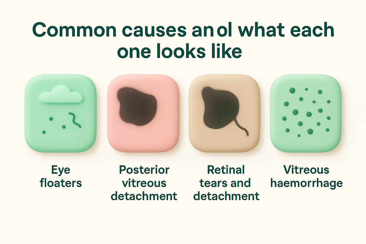
Eye floaters (vitreous floaters)
Floaters represent the most common cause of dark spots in your vision. These shadows form when tiny fibres or cells in your vitreous gel clump together, casting distinct shapes on your retina. You’ll typically see multiple floaters rather than a single spot, and they drift slowly when you move your eyes, then settle back into place following a predictable pattern.
The spots appear as grey or transparent shapes rather than solid black, and you’ll notice them most against bright backgrounds like white walls or clear skies. Common shapes include dots, circles, lines, or cobweb-like strands. Floaters become more frequent after age 50 as your vitreous gel naturally shrinks and liquefies, but they can occur at any age. Most floaters cause no harm and simply require monitoring over time rather than treatment.
Posterior vitreous detachment
Posterior vitreous detachment (PVD) occurs when your vitreous gel separates from your retina’s surface. This creates a sudden increase in floaters, often described as a shower of small dark spots or a larger cobweb drifting across your peripheral vision. You might also experience brief flashes of light, particularly in dim conditions or when moving your eyes quickly.
Posterior vitreous detachment affects roughly 75% of people over 65, making it one of the most common age-related eye changes you’ll experience.
PVD itself isn’t dangerous, but the separation process can sometimes pull on your retina hard enough to create tears, which explains why specialist assessment remains important even though most cases resolve without intervention.
Retinal tears and detachment
Retinal tears create a black spot in peripheral vision that differs from floaters. The spot tends to remain in the same location rather than drifting, and it might appear alongside a shadow or curtain spreading across your side vision. You’ll often notice sudden flashing lights before the dark spot appears, caused by your retina being pulled or torn.
Detachment produces a progressive shadow that advances across your peripheral vision like a curtain being drawn. The affected area appears darker and less defined than your normal vision, and the shadow expands over hours or days if left untreated.
Vitreous haemorrhage
Bleeding into your vitreous cavity creates numerous dark spots that appear suddenly, often described as a cloud of flies or a red haze depending on the amount of blood present. The spots move with your eye movements but don’t settle in predictable patterns like typical floaters do. Vision in the affected area becomes cloudy or dim, and you might struggle to see detail through the blood-obscured areas.
Red flags that need urgent care
Certain symptoms accompanying a black spot in peripheral vision demand immediate medical attention, not next week or when you find a convenient appointment time. These warning signs indicate conditions that can permanently damage your sight within hours or days if left untreated. Knowing which symptoms constitute true emergencies helps you protect your vision before irreversible damage occurs.
Symptoms requiring same-day assessment
You need urgent specialist evaluation if you notice a sudden increase in the number of floaters or dark spots, particularly if they appear within hours rather than gradually over weeks. This symptom often signals posterior vitreous detachment or retinal tears developing in real time. The timing matters because fresh tears can progress to full detachment rapidly, whilst early intervention with laser treatment can prevent this progression entirely.
Flashing lights accompanying your black spot represent another critical warning. These flashes appear as brief streaks or arcs of light, typically in your peripheral vision, and persist even when you close your eyes. The flashes indicate your retina is being mechanically stimulated, either through pulling, tearing, or detachment. When combined with new dark spots, flashes suggest active retinal damage requiring assessment within 24 hours.
A shadow or curtain spreading across your peripheral vision demands immediate action. This symptom indicates retinal detachment in progress, where the separated area expands as fluid accumulates behind your retina. The curtain typically advances from the periphery towards your central vision, and each hour of delay reduces your surgical success rate and final visual outcome.
Retinal detachment is a race against time, with surgical outcomes declining measurably for every day treatment is delayed beyond symptom onset.
When to call emergency services
Loss of peripheral vision across large areas within minutes or hours requires emergency department assessment, particularly if accompanied by severe headache, confusion, or vision loss in both eyes. These symptoms might indicate stroke, severe haemorrhage, or other systemic conditions affecting your brain rather than isolated eye problems.
Sudden vision loss following direct trauma to your eye or head needs immediate evaluation, regardless of whether you see a black spot in peripheral vision or other symptoms. Trauma can cause bleeding, retinal tears, or detachment that progress rapidly without proper treatment. Don’t wait to see if symptoms improve on their own after any significant impact to your eye or surrounding structures.
Pain accompanying new peripheral dark spots warrants urgent assessment, particularly sharp pain that worsens with eye movement. Whilst many retinal conditions cause minimal discomfort, pain suggests inflammation, acute glaucoma, or other conditions requiring different treatment approaches than simple observation.
How specialists find the cause
Finding the precise cause of a black spot in peripheral vision requires systematic examination combining your symptom history with specialised diagnostic tests. Your specialist can’t rely on description alone because different conditions often produce similar symptoms. The examination process follows a logical sequence, starting with non-invasive assessments before progressing to more detailed imaging when needed. Each test reveals specific information about different eye structures, allowing your specialist to pinpoint exactly what’s creating your visual disturbance.

Taking your complete visual history
Your specialist begins by documenting when you first noticed the black spot, how it behaves, and what other symptoms accompany it. Questions focus on timing patterns: did the spot appear suddenly or develop gradually? Does it move when you shift your gaze, or remain fixed in place? Understanding whether you’ve experienced flashes, floaters, or shadowy areas spreading across your vision helps narrow potential causes before any physical examination begins.
Medical history matters significantly. Your specialist needs to know about diabetes, high blood pressure, previous eye surgeries, or recent head trauma. These factors influence which conditions become more likely and guide the examination’s focus. You’ll also discuss current medications, as some drugs affect your eyes or interact with contrast agents used in certain diagnostic tests.
Direct examination and pressure testing
The physical examination starts with dilating your pupils using eye drops, which allows your specialist to view your entire retina including peripheral areas. This process takes 20 to 30 minutes to work fully, temporarily blurring your near vision and making you light-sensitive for several hours. During dilation, your specialist uses specialised lenses and bright lights to examine your vitreous gel, retina, and optic nerve directly.
Direct visualisation through dilated pupils remains the gold standard for detecting retinal tears, detachment, and vitreous abnormalities that cause peripheral dark spots.
Pressure measurement occurs using a tonometer, which determines if elevated eye pressure contributes to your symptoms. Some peripheral vision changes stem from glaucoma affecting your optic nerve, making pressure readings essential for complete assessment.
Advanced imaging when needed
Optical coherence tomography (OCT) creates detailed cross-sectional images of your retina’s layers, revealing subtle structural changes not visible during direct examination. This painless scan takes minutes and shows whether fluid has accumulated under your retina or if membrane layers have separated abnormally.
Ultrasound imaging helps when blood or dense floaters obscure your specialist’s view of your retina. Sound waves penetrate these opaque materials, creating images that reveal retinal position and identify detachments or tears hidden behind haemorrhage. Your specialist might also use fluorescein angiography, where injected dye highlights blood vessel patterns, particularly when investigating suspected circulation problems affecting your peripheral retina.
Treatment options and how to reduce your risk
Treatment for a black spot in peripheral vision depends entirely on what’s causing it. Your specialist tailors the approach to your specific diagnosis, ranging from simple observation for benign floaters to urgent surgical intervention for retinal detachment. Understanding available options helps you prepare for discussions with your specialist and set realistic expectations about outcomes and recovery timeframes.
Treatment approaches based on diagnosis
Laser photocoagulation treats retinal tears before they progress to detachment. Your specialist uses focused laser energy to create small burns around the tear, which form scar tissue that seals the retina to its underlying layer. This outpatient procedure takes minutes, requires only local anaesthetic drops, and prevents roughly 90% of tears from developing into full detachments.
Vitrectomy addresses more complex conditions like vitreous haemorrhage, large floaters, or established retinal detachment. During this surgery, your specialist removes the vitreous gel and replaces it with clear fluid or gas. Recovery requires specific positioning, sometimes face-down for days or weeks, depending on the repair location. Success rates exceed 85% for primary detachments caught early, though multiple procedures become necessary in complicated cases.
Pneumatic retinopexy offers a less invasive option for certain retinal detachments. Your specialist injects a gas bubble into your eye, which floats upward and pushes the detached retina back into place. Laser or freezing treatment then seals the tear. You’ll need to maintain specific head positions for several days whilst the bubble holds your retina in place and gradually dissolves.
Early laser treatment for retinal tears prevents detachment in the vast majority of cases, avoiding the need for more invasive surgery entirely.
Preventive measures and risk reduction
Regular eye examinations catch peripheral retinal problems before symptoms develop. Annual dilated exams become particularly important after age 50 or if you have diabetes, high myopia, or family history of retinal detachment. These preventive checks allow your specialist to identify weak areas in your peripheral retina and treat them proactively.
Managing systemic health protects your retinal blood vessels. Controlling blood sugar levels reduces diabetic retinopathy risk, whilst maintaining healthy blood pressure prevents vascular complications that cause bleeding or circulation problems in your peripheral retina. These measures won’t eliminate all risks but significantly reduce your likelihood of developing serious conditions causing peripheral vision changes.
Protecting your eyes from trauma prevents direct retinal damage. Wear appropriate safety glasses during activities involving projectiles, chemicals, or impact risks. Sports goggles protect athletes in high-contact activities where direct eye injury becomes more likely.
When observation alone suffices
Benign floaters require no treatment beyond periodic monitoring. Your specialist confirms the floaters pose no threat to your retina, then schedules follow-up examinations to ensure no new developments occur. Most people adapt to floaters over weeks or months as their brain learns to ignore the shadows.
Uncomplicated posterior vitreous detachment typically resolves without intervention. Your specialist verifies no retinal tears occurred during the separation process, then monitors you for several weeks to confirm stability. Symptoms gradually diminish as the vitreous completes its separation and settles into a new position.

A simple plan for what to do next
If you’ve noticed a black spot in peripheral vision, your immediate action depends on the accompanying symptoms. New spots appearing alongside flashes, shadows spreading across your vision, or sudden increases in floaters require same-day specialist assessment. Contact an eye specialist immediately rather than waiting for your regular optometrist appointment, as these symptoms signal conditions where hours matter for preserving your sight.
For isolated dark spots without warning signs, schedule a comprehensive eye examination within the week. Your specialist will dilate your pupils, examine your peripheral retina thoroughly, and determine whether the spot requires treatment or simply monitoring. Even benign floaters deserve professional assessment to rule out underlying problems that might progress without obvious symptoms.
Dr Rahul Dubey and Dr Capucine Odouard at Bright Sight Eye Specialists assess and treat the full spectrum of conditions causing peripheral vision changes. With advanced diagnostic technology and expertise in retinal surgery, our Randwick practice provides the specialist care you need to protect your vision. Contact our team to arrange your assessment and receive a clear diagnosis with transparent treatment recommendations.