


A black spot in my eyesight can appear suddenly or develop gradually over time. These dark spots might look like tiny dots, cobwebs, or shadowy patches that drift across your field of vision when you move your eyes. Most people experience them as harmless eye floaters caused by natural changes in the gel inside your eye. But some black spots signal serious problems like retinal tears or detachment that can lead to permanent vision loss if you ignore them.
This article explains why black spots appear in your vision and how to tell the difference between normal floaters and medical emergencies. You will learn about specific warning signs that require immediate attention from an eye specialist, the medical treatments available for different types of vision spots, and risk factors that increase your chances of developing retinal problems. Understanding these distinctions helps you protect your sight and know when to seek professional care instead of waiting for symptoms to worsen.
Why black spots appear in your line of sight
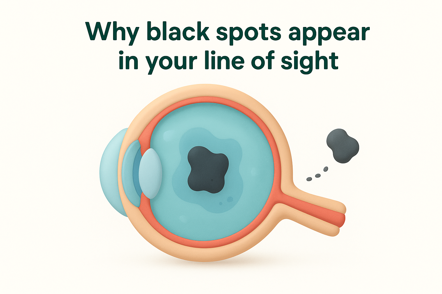
Your eyes contain a gel-like substance called the vitreous that fills the space between your lens and retina. When you notice a black spot in your eyesight, this gel has typically undergone changes that create shadows on your retina. The vitreous humour consists of 98% water and 2% collagen fibres that maintain its shape and transparency throughout most of your life. As you age, these collagen fibres clump together and the gel liquefies, casting shadows that appear as dark spots or strings floating across your vision.
Natural ageing and vitreous detachment
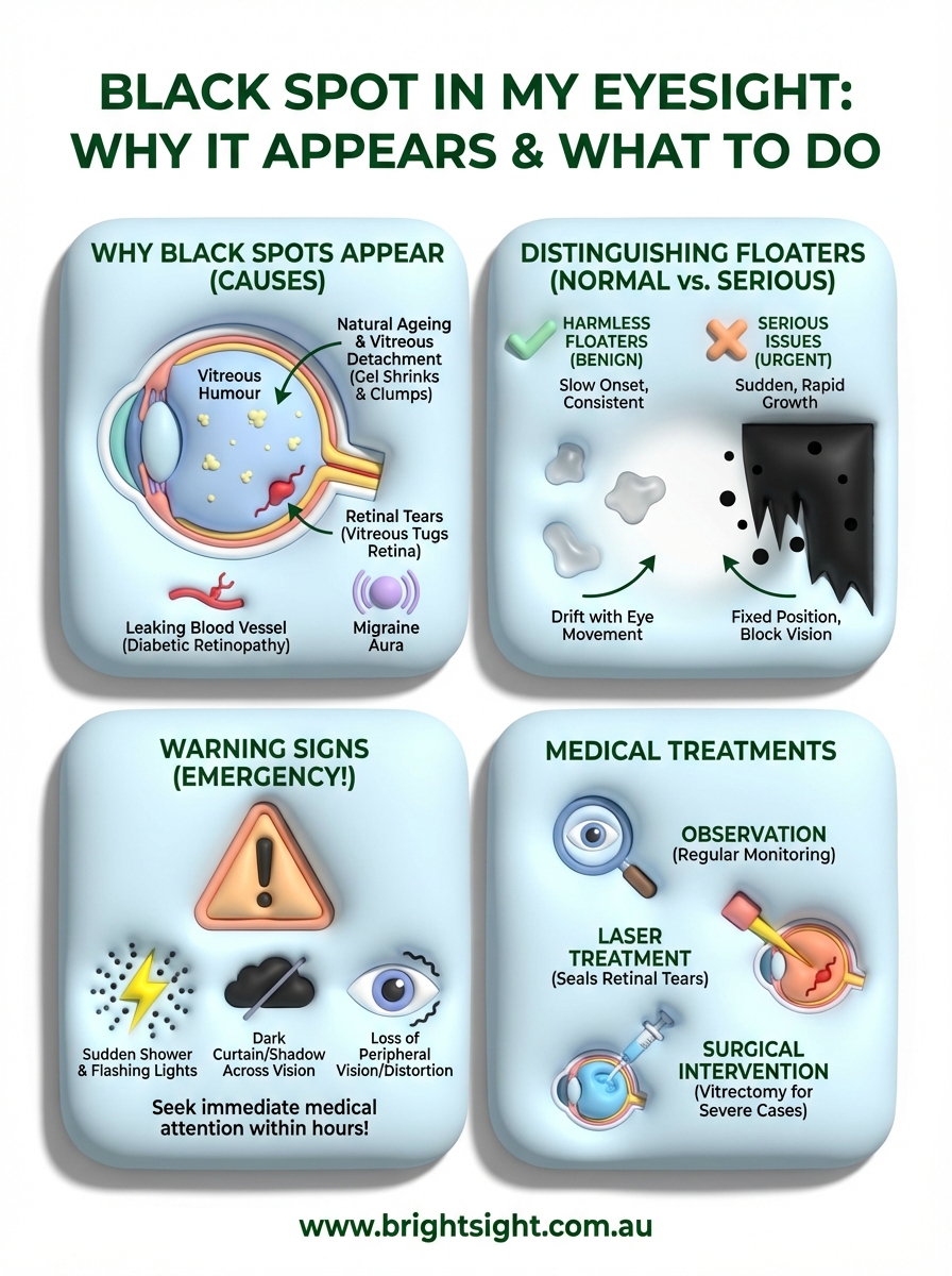
The most common cause of black spots stems from posterior vitreous detachment, which occurs when the vitreous gel shrinks and pulls away from the retina. This natural process typically begins after age 50 and affects nearly everyone by their seventies. You might notice an increase in floaters, flashes of light, or cobweb-like shapes during this transition. The vitreous gel develops pockets of liquid that allow protein strands to move freely inside your eye, creating the floating sensation when you shift your gaze.
Your brain eventually learns to ignore most floaters, though they never truly disappear. The shadows become less noticeable as the clumped collagen fibres settle below your direct line of sight. Some people adapt within weeks, whilst others remain aware of floaters for months after onset.
Retinal tears and damage
Black spots can indicate that the shrinking vitreous has torn your retina as it pulls away from the eye wall. These tears allow fluid to seep underneath the retinal tissue, potentially leading to detachment. You might see a sudden shower of new floaters, persistent flashes that resemble lightning bolts, or a dark curtain moving across your peripheral vision. Retinal tears require immediate treatment because they progress rapidly once fluid accumulation begins.
Retinal damage creates stationary dark spots called scotomas that remain fixed in your visual field, unlike floaters that drift with eye movement.
Blood vessels can break when the vitreous tugs on the retina, causing a vitreous haemorrhage that appears as dense black spots or a red haze across your vision. This bleeding obscures your sight and requires urgent evaluation to determine the underlying cause and prevent further complications.
Medical conditions affecting vision
Several health problems produce black spots through different mechanisms. Diabetic retinopathy damages blood vessels in your retina, causing them to leak fluid and blood that create dark patches in your visual field. High blood sugar levels weaken vessel walls over time, leading to swelling and abnormal new blood vessel growth that bleeds easily.
Inflammation inside your eye (uveitis) triggers white blood cells to float through the vitreous, appearing as spots or haziness that reduces clarity. Severe nearsightedness stretches your eyeball longer than normal, increasing stress on the retina and making it more susceptible to tears and detachment. Eye injuries from trauma can shake the vitreous loose or directly damage retinal tissue, creating immediate or delayed onset of black spots depending on the severity of impact.
Migraines sometimes produce temporary visual disturbances called scotomas that appear as blind spots with shimmering edges. These episodes typically last 10 to 30 minutes before resolving completely without permanent damage to your vision.
How to distinguish harmless floaters from serious issues
You can identify harmless floaters by their slow onset and consistent appearance over time. Normal floaters remain relatively stable, drifting across your vision when you move your eyes but never blocking large portions of your visual field. These benign spots typically appear translucent or grey rather than pitch black, and you notice them most clearly against bright backgrounds like white walls or blue skies. Your brain adapts to these familiar shapes within weeks or months, making them less distracting during daily activities.
Movement patterns and appearance
Harmless floaters follow your eye movements with a slight lag, creating a swimming or drifting sensation that stops when you hold your gaze steady. They move smoothly through your visual field without sudden jerks or zigzag patterns. Normal floaters maintain the same size and shape over extended periods, though they might become more visible in certain lighting conditions. You can often move them out of your central vision by shifting where you look, whereas serious issues like scotomas remain fixed in the same position regardless of eye movement.
Benign floaters cast softer shadows that appear grey or transparent, whilst urgent problems create dense black curtains or showers of new dark spots that obscure your sight.
Dangerous black spots in your eyesight appear suddenly in large numbers or grow rapidly within hours or days. These problematic floaters look darker and denser than normal age-related changes, sometimes accompanied by cobwebs, rings, or cloud-like formations that spread across your peripheral vision. You might struggle to see through them clearly, and they interfere with reading or recognising faces rather than simply floating at the edges of your awareness.
Associated symptoms and progression
Harmless floaters occur without other visual disturbances like flashing lights, blurred vision, or blind spots. Your peripheral vision remains intact, and you maintain clear sight in all directions when looking straight ahead. These normal spots develop gradually over months or years as part of natural ageing, giving you time to adjust to their presence without sudden changes in visual quality.
Serious issues combine multiple symptoms simultaneously. You might experience persistent light flashes resembling camera bursts alongside new floaters, indicating that your vitreous is tugging on the retina. Shadows spreading from your peripheral vision inward suggest retinal detachment, whilst sudden vision loss or a dark veil covering part of your sight demands immediate evaluation by an ophthalmologist.
Warning signs that require immediate medical attention
Certain symptoms associated with a black spot in my eyesight indicate medical emergencies that can cause permanent vision loss within hours or days if left untreated. Your retina has no pain receptors, so damage progresses silently without the discomfort that typically warns you of serious injuries. Recognising these warning signs allows you to seek treatment during the critical window when ophthalmologists can still save your sight through surgical intervention or laser procedures.
Symptoms requiring same-day emergency evaluation
A sudden shower of new floaters appearing within minutes or hours signals that blood or cellular debris has entered your vitreous cavity. You might see dozens or hundreds of dark spots simultaneously, often accompanied by bright flashes of light that persist even with your eyes closed. These flashes occur when the shrinking vitreous tugs on your retina, creating electrical signals your brain interprets as light. Contact an eye specialist immediately if you notice these combined symptoms, as they indicate a high risk of retinal tears that can progress to detachment.
Any dark curtain, shadow, or veil spreading across your peripheral or central vision requires emergency assessment within hours to prevent complete retinal detachment.
Loss of peripheral vision that moves like a shade being pulled down over your visual field marks advanced retinal detachment. The affected area corresponds to where your retina has separated from its blood supply, causing those photoreceptor cells to stop functioning. You might notice straight lines appearing wavy or distorted, a symptom called metamorphopsia that indicates fluid accumulation under your macula.
Progressive changes within 24 hours
Rapidly worsening vision that develops over several hours suggests vitreous haemorrhage or severe inflammation inside your eye. Your sight might become hazy or obscured by what appears as red smoke or dense black clouds that block large portions of your visual field. Sudden blindness in one eye, even if temporary, indicates complete arterial blockage or massive bleeding that demands immediate intervention.
Pain combined with new floaters, especially after eye surgery or trauma, points to infection or intraocular inflammation requiring urgent antibiotic treatment. You should visit an emergency department or eye specialist within the same day rather than waiting for a routine appointment, as delays reduce treatment success rates significantly.
Medical treatments available for vision spots
Your ophthalmologist selects treatment based on what causes your black spots and whether they threaten your vision. Most harmless floaters from natural ageing require no medical intervention, as your brain adapts to their presence over time. However, serious conditions like retinal tears, detachment, or vitreous haemorrhage demand prompt medical treatment to preserve your sight. Your eye specialist conducts a dilated eye examination using specialised imaging to determine the source of your vision spots and recommend the appropriate course of action.
Observation and monitoring approach
Doctors recommend regular monitoring when you have benign floaters without retinal damage. You attend follow-up appointments every few months to ensure no new complications develop. Your ophthalmologist photographs your retina and tracks any changes in the number or density of floaters over time. This conservative approach works well for age-related vitreous changes that pose no immediate threat to your vision, allowing your visual system to adjust naturally without unnecessary procedures.
Laser treatments for retinal problems
Laser photocoagulation seals retinal tears by creating controlled burns around the damaged area. Your surgeon directs a focused beam at the tear edges, forming scar tissue that welds the retina back to the underlying tissue. This outpatient procedure takes 15 to 30 minutes and prevents fluid from seeping underneath your retina. You receive anaesthetic eye drops before treatment, so you feel no pain during the laser application.
Laser treatment successfully prevents retinal detachment in over 90% of cases when performed before fluid accumulates behind the retina.
YAG laser vitreolysis targets large, persistent floaters by breaking them into smaller fragments that become less noticeable. Your doctor aims pulses at the vitreous opacities, vaporising the collagen clumps responsible for casting shadows. This treatment works best for specific types of floaters located away from your retina and lens, though not all patients qualify as suitable candidates.
Surgical intervention options
Vitrectomy removes the gel inside your eye when a black spot in my eyesight results from dense vitreous haemorrhage or severe floaters that block vision. Surgeons make tiny incisions in your eye wall, extract the clouded vitreous, and replace it with saline solution or gas bubble. This procedure requires local or general anaesthesia and carries risks including cataracts, infection, and retinal detachment. Recovery takes several weeks, with strict positioning requirements if your surgeon uses a gas bubble to hold your retina in place.
Factors that increase your risk of retinal issues
Understanding your personal risk factors helps you monitor your vision more carefully and schedule appropriate screening appointments. Certain characteristics make you more vulnerable to developing conditions that cause a black spot in my eyesight, ranging from age-related changes to specific medical conditions and lifestyle choices. Your ophthalmologist uses these risk factors to determine how frequently you need dilated eye examinations and whether preventive treatments might benefit you before problems develop.
Age-related risk factors
Your risk of retinal tears and detachment increases significantly after age 50 when the vitreous gel naturally shrinks and pulls away from your retina. People over 60 face the highest likelihood of experiencing posterior vitreous detachment, which creates floaters and occasionally damages retinal tissue during separation. You experience accelerated vitreous changes if you developed early cataracts or underwent cataract surgery, as these conditions alter the eye’s internal structure and chemistry.
Family history plays a substantial role in your retinal health. You inherit a higher susceptibility to retinal detachment if close relatives experienced this condition, particularly when it occurred at younger ages. Genetic factors influence your vitreous composition, retinal thickness, and how these tissues respond to normal ageing processes.
Medical conditions and lifestyle factors
Diabetes dramatically elevates your risk through progressive damage to retinal blood vessels over years of elevated blood sugar levels. You develop weakened vessel walls that leak fluid and blood, creating dark spots and vision loss if left uncontrolled. Poor glucose management accelerates these changes, whilst maintaining HbA1c levels below 7% significantly reduces your diabetic retinopathy risk.
High blood pressure damages the delicate blood vessels in your retina, increasing your likelihood of vessel blockages and bleeding that appear as sudden black spots.
Previous eye trauma, even from years ago, makes your retina more vulnerable to future tears or detachment. Injuries weaken the attachment between your retina and underlying tissue, creating areas of thinning that fail under normal vitreous stress.
Eye-specific vulnerabilities
Severe nearsightedness (myopia worse than -6.00 dioptres) stretches your eyeball into an elongated shape that thins the peripheral retina. This structural change makes you six times more likely to develop retinal tears compared to people with normal vision. Your stretched retina experiences greater tension when the vitreous shrinks, increasing the force applied to weak points during detachment.
Previous eye surgeries, particularly those involving the retina or vitreous, alter your risk profile permanently. You face elevated chances of future complications because surgical interventions change the normal anatomy and healing responses within your eye.
Summary of black spots in vision
A black spot in my eyesight can result from harmless age-related floaters or serious conditions requiring immediate treatment. Most people experience benign vitreous changes that create drifting shadows across their vision, whilst others face retinal tears or detachment that threaten permanent sight loss. You protect your vision by recognising the difference between normal floaters that develop gradually and emergency symptoms like sudden floater showers, flashing lights, or dark curtains spreading across your visual field.
Your risk factors, including age over 50, severe nearsightedness, diabetes, and previous eye injuries, determine how frequently you need professional eye examinations. Regular dilated retinal checks detect early problems before they progress to advanced stages requiring complex surgery. If you notice new or changing black spots accompanied by other visual disturbances, contact Bright Sight Eye Specialists for a comprehensive retinal evaluation and personalised treatment plan based on your specific condition.