


Those small black dots drifting across your field of vision can be unsettling. When you try to look directly at them, they seem to dart away. They become more obvious against bright backgrounds like a clear sky or white wall. These moving specks are called floaters. They appear as dots, lines, cobwebs or tiny threads that shift as your eyes move. Most floaters are harmless. They happen when microscopic fibres inside your eye cast shadows on your retina.
This guide explains what causes black dots in your vision, how to tell when floaters are normal, and which symptoms signal something more serious. You’ll learn about the diagnostic tests used to examine floaters, treatment options available if they interfere with your sight, and practical tips for managing them day to day. We’ll also cover the warning signs that require urgent medical attention. Whether you’ve noticed a single floater or several, understanding what’s happening in your eye helps you make informed decisions about your care.
Why black dots in vision that move matter
Your eye health depends on recognising when floaters signal a harmless change or a serious problem. Most black dots in vision that move are simply age-related floaters that pose no threat to your sight. They develop as the vitreous gel inside your eye naturally liquefies over time. However, a sudden increase in floaters can indicate retinal tears, retinal detachment or bleeding inside your eye. These conditions can cause permanent vision loss if left untreated.
Understanding the difference between normal floaters and warning signs protects your sight. Retinal detachment affects roughly 1 in 10,000 people each year, and early treatment dramatically improves outcomes. When you know what to watch for, you can seek help quickly if symptoms change. Many people ignore floaters because they’ve heard they’re harmless, but new floaters accompanied by flashes of light or vision loss require immediate medical attention.
The timing of treatment for retinal problems often determines whether you keep or lose vision in the affected eye.
Floaters also affect your daily comfort and visual quality. Large or numerous floaters can interfere with reading, driving and other activities that require clear vision. Knowing your options helps you manage both the medical and practical aspects of living with floaters.
How to recognise normal floaters
Normal floaters follow predictable patterns that help you distinguish them from serious problems. Most people develop floaters gradually as they age, particularly after 50 years old. These benign floaters remain stable over time and don’t multiply suddenly. You might notice one or two new floaters appearing over months or years, but they shouldn’t arrive all at once. Normal floaters drift slowly across your vision when you move your eyes, then settle as your eyes stop moving.
What normal floaters look like
Harmless black dots in vision that move typically appear as small, transparent shapes that vary in size and form. They might look like tiny dots, thin threads, or small cobweb-like strands. The shadows shift position smoothly as you move your eyes from side to side or up and down. You’ll see them most clearly against plain, bright backgrounds such as a blue sky, white wall, or computer screen. Normal floaters may appear grey, black, or semi-transparent depending on the lighting.

How normal floaters behave
Benign floaters move in the same direction as your eye movement, then drift slightly in the opposite direction when your eyes stop. They follow consistent patterns each time you look around. Your brain gradually adapts to their presence over weeks or months, making them less noticeable in daily life. Normal floaters don’t obscure your central vision or create blind spots.
Most floaters become less bothersome as your brain learns to filter them out of your awareness.
These harmless floaters remain roughly the same size and number over time, rarely interfering with your ability to read, drive, or perform detailed tasks.
Common warning signs you should never ignore
Certain symptoms indicate that black dots in vision that move signal a medical emergency rather than harmless floaters. Your retina might be tearing or detaching from the back of your eye, which can cause permanent blindness within days if left untreated. Recognising these warning signs helps you act quickly to protect your vision.
Emergency symptoms requiring immediate care
A sudden shower of new floaters appearing within hours or days represents the clearest danger sign. Unlike normal floaters that develop gradually, dozens of new black dots appearing at once often indicate retinal tears or bleeding. You should visit an emergency department or contact an ophthalmologist immediately if you notice this sudden change.
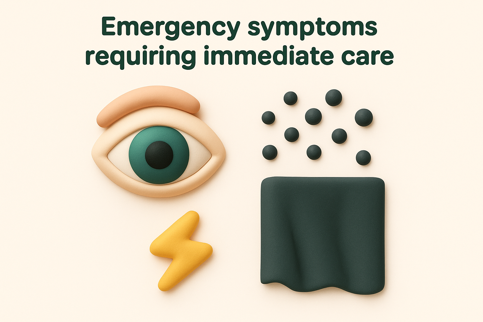
Flashes of light in your peripheral vision, particularly when they occur with new floaters, suggest your retina is being tugged or torn. These flashes might look like lightning bolts, camera flashes, or brief sparkles at the edge of your sight. They typically appear in the same eye as the new floaters and may continue occurring for several days.
Retinal detachment progresses rapidly, and treatment within 24 to 48 hours can mean the difference between saving and losing your sight.
A dark curtain, shadow or veil blocking part of your vision indicates your retina may be detaching. This shadow typically starts at the edge of your vision and gradually moves toward the centre. Sudden blurred vision, vision loss, or difficulty seeing in one eye also requires urgent assessment. These symptoms differ from normal floaters because they actively obstruct your view rather than simply drifting across it. Contact your eye specialist or visit an emergency department immediately if you experience any combination of these warning signs.
Causes, tests and diagnosis explained
The causes of black dots in vision that move range from benign age-related changes to urgent medical conditions requiring immediate intervention. Understanding what triggers floaters helps you assess your risk and know when to seek professional evaluation. Your eye specialist uses several diagnostic tests to examine the vitreous gel and retina, determining whether your floaters are harmless or signal a more serious problem. These tests are painless and provide detailed images of structures inside your eye that cannot be seen from the outside.
What causes black dots in vision that move
Age-related vitreous degeneration causes the majority of floaters in people over 50 years old. The vitreous gel filling your eye contains collagen fibres that clump together as the gel liquifies with age. These clumps cast shadows on your retina, creating the black dots you see drifting across your vision. This natural process accelerates if you have short-sightedness (myopia), which causes earlier vitreous changes and increases your floater risk.

Retinal tears occur when the vitreous gel pulls away from your retina with enough force to create a hole or tear. This separation happens more frequently in people who have had eye surgery, suffered eye trauma, or have high myopia. Vitreous haemorrhage develops when blood leaks into the vitreous cavity, often due to diabetes, blocked retinal blood vessels, or retinal tears. The blood cells appear as numerous small floaters or a general haziness in your vision.
Diabetes increases your risk of floaters by damaging the small blood vessels in your retina, which can leak blood into your vitreous gel.
Posterior uveitis creates inflammation in the back of your eye, causing white blood cells to float through the vitreous. Infections, autoimmune conditions, and inflammatory diseases can all trigger this type of inflammation, producing floaters alongside other symptoms like redness, pain, or light sensitivity.
Diagnostic tests your eye specialist will use
Your ophthalmologist begins with a dilated eye examination, placing drops in your eyes to widen your pupils. This allows complete visualisation of your vitreous and retina using specialised lenses and bright lights. The examination takes roughly 20 to 30 minutes and lets your specialist identify any tears, holes, bleeding, or detachment affecting your retina.
Optical coherence tomography (OCT) produces detailed cross-sectional images of your retina and vitreous using light waves. This painless scan takes only minutes and helps identify subtle changes invisible during standard examination. Your specialist may also use ultrasound imaging if your floaters are too dense to see through, creating sound-wave images that reveal retinal detachment or other structural problems.
Treatment options and day to day tips
Most floaters require no treatment because your brain adapts to them over time and they become less noticeable in daily life. Only floaters that significantly interfere with your vision need medical intervention. Your ophthalmologist assesses whether treatment benefits outweigh the risks, considering factors like floater size, location, and impact on your quality of life.
Medical treatments available
Vitrectomy surgery removes the vitreous gel containing floaters and replaces it with a clear saline solution. This procedure eliminates most or all floaters but carries risks including cataract formation, retinal detachment, and infection. Surgeons typically reserve vitrectomy for severe cases where floaters substantially impair vision or quality of life. Recovery takes several weeks, during which you may need to maintain specific head positions.

Laser vitreolysis breaks up larger floaters using targeted laser pulses that vaporise the collagen clumps casting shadows on your retina. This less invasive option works best for specific floater types positioned away from your retina. The procedure takes 20 to 60 minutes in your specialist’s rooms and requires no recovery time, though you may need multiple sessions.
Day to day strategies
Moving your eyes up and down shifts floaters out of your central vision temporarily when they interfere with reading or detailed work. Looking away from bright backgrounds like white walls or blue skies makes black dots in vision that move less obvious during daily activities.
Adequate lighting and contrast adjustments on screens reduce the visibility of floaters during computer work.
Wearing quality sunglasses outdoors minimises floater visibility in bright conditions. Stay well hydrated, as dehydration can worsen floater perception in some people.

What to do next
Most black dots in vision that move require no immediate action if they’ve been present for months or years without change. Monitor your floaters for any sudden increases in number or the appearance of flashes of light, which signal potential retinal problems requiring urgent care. Keep a mental note of how many floaters you typically see and where they appear in your vision. Any abrupt change demands immediate professional assessment.
Schedule regular eye examinations with an ophthalmologist to monitor your floaters and check for underlying retinal conditions, particularly if you have diabetes, short-sightedness, or a history of eye surgery. These routine checks help detect problems before they threaten your sight. If new floaters appear suddenly or you notice vision changes, don’t wait for your scheduled appointment.
Bright Sight Eye Specialists provides comprehensive retinal examinations and treatment for floaters and related conditions in Randwick, Sydney. Book a consultation with Dr Rahul Dubey if you’re concerned about changes in your vision or need specialist assessment of your eye health.