


A black dot in peripheral vision appears as a small dark spot at the edge of your sight, rather than straight ahead. It might stay in one place or drift slightly when you move your eyes. Some people describe it as a speck, a shadow or a tiny blur that sits just outside their main field of view. These dots can range from barely noticeable to genuinely distracting, and understanding what causes them helps you decide whether they’re harmless or need immediate attention.
This article explains the common and serious causes of a black dot in your peripheral vision. You’ll learn when a spot is simply a harmless floater and when it signals something urgent like a retinal tear. We’ll cover how eye specialists diagnose these dots, what treatments exist, and practical steps to protect your side vision. By the end, you’ll know exactly what to watch for and when to book an appointment with an ophthalmologist.
Why a black dot in peripheral vision matters
Your side vision does more than you might realise. It alerts you to movement, helps you navigate crowded spaces and keeps you safe on the road. When a black dot in peripheral vision appears, it can interrupt these automatic functions and make everyday tasks feel uncertain. Even a small spot might signal changes inside your eye that deserve attention. Ignoring persistent dots risks missing early warnings of conditions that could progress and affect your central sight.
Your peripheral vision protects you daily
Peripheral vision spans roughly 170 degrees of your total field of view. This wide arc lets you detect cars approaching from the side, notice when someone walks past you and judge distances without turning your head. A dark spot in this area can reduce your awareness of surroundings and make activities like driving or sport more challenging. Athletes rely on peripheral cues to track opponents, while commuters use them to cross busy streets safely.

Protecting your side vision means preserving your independence and confidence in daily life.
Early detection saves sight
Spotting a black dot early gives specialists the best chance to diagnose and treat any underlying issue before it worsens. Conditions like retinal tears or detachments often begin with subtle changes at the edges of your vision. When caught within hours or days, laser treatment can seal small tears and prevent fluid from spreading beneath the retina. Delayed care, by contrast, may allow a minor problem to escalate into permanent vision loss. Regular eye checks and prompt action after noticing new spots form the cornerstone of long-term eye health.
How to respond when you notice a black dot
Your first step after spotting a black dot in peripheral vision is to stay calm and observe the spot’s behaviour. Write down exactly when you first noticed it, whether it moves when you shift your gaze and if it appears in one eye or both. This information helps your eye specialist pinpoint the cause quickly. Avoid rubbing your eyes or straining to see the dot more clearly, as this can make assessment harder and may worsen certain conditions if they’re present.
Note when and where the spot appears
Record the exact location of the spot in your peripheral field: is it at the top, bottom, left or right edge of your vision? Document any activities you were doing when you first saw it, such as reading, exercising or waking up. If the spot seems to drift, note whether it follows your eye movements smoothly or lags behind. Check each eye separately by covering one at a time to determine if the dot appears in one eye only or both. These details become vital clues during your eye examination.
Precise observations about your spot can speed up diagnosis and ensure you receive the right treatment.
Assess whether you need urgent care
You need immediate attention if the black dot is accompanied by flashing lights, a sudden shower of new floaters or a curtain-like shadow moving across your vision. These symptoms suggest a retinal tear or detachment, which demands treatment within hours to prevent permanent sight loss. Contact an ophthalmologist or present to an emergency eye clinic straight away if you experience any of these red flags. If the spot appears alone, remains stable and causes no other symptoms, it’s usually safe to monitor it for a day or two. However, booking a routine eye appointment within the week lets a specialist confirm the spot is harmless and rule out early signs of more serious issues.
Common causes of a peripheral black dot
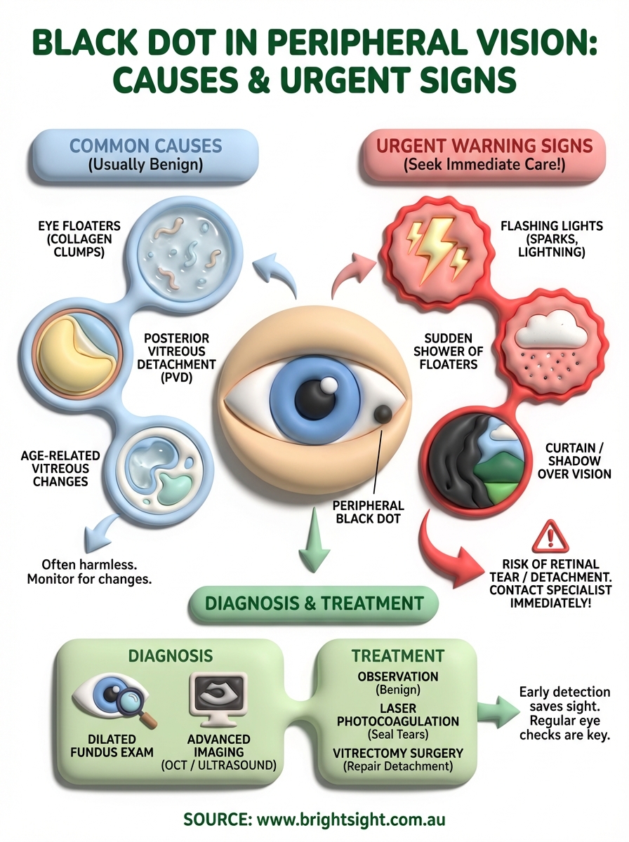
Most black dots in peripheral vision stem from changes within the vitreous gel that fills your eye. This clear, jelly-like substance sits between your lens and retina, and as you age it naturally begins to liquefy and shrink. Tiny clumps of collagen fibres can form during this process, casting shadows on your retina that appear as dark spots at the edges of your sight. While these changes sound alarming, they’re usually harmless and rarely affect your overall vision quality. Understanding the three main benign causes helps you recognise when a peripheral dot is simply part of normal ageing.
Eye floaters in your side vision
Eye floaters are the most common explanation for a peripheral black dot. These specks develop when microscopic collagen strands within the vitreous stick together, creating tiny opacities that drift through your eye. You might notice the dot moves slightly when you shift your gaze, often lagging behind your eye movements before settling into a new position. Floaters at the edges of your vision tend to be less bothersome than those in your central field, though they can still catch your attention during reading or when looking at bright, plain surfaces like a clear sky or white wall.

Floaters typically appear as single dots, multiple specks or even thread-like shapes in your peripheral field. They become more common after age 50 and affect nearly everyone to some degree by age 70. High myopia (severe short-sightedness) increases your risk of developing floaters earlier in life because your elongated eyeball places extra tension on the vitreous. Most people adapt to benign floaters within weeks or months as their brain learns to ignore these persistent shadows.
While floaters are usually harmless, a sudden increase in their number warrants immediate assessment.
Posterior vitreous detachment
Posterior vitreous detachment (PVD) occurs when the vitreous gel separates cleanly from the retinal surface at the back of your eye. This natural process affects around two-thirds of people over 70 and often announces itself with new floaters appearing in your peripheral vision. The detachment happens because the vitreous shrinks with age, eventually pulling away from the retina in a single event. You might notice a sudden onset of one or several dark spots, sometimes accompanied by brief flashes of light as the vitreous tugs on your retina during separation.
PVD itself is not dangerous and requires no treatment in most cases. However, the separation process can stress the retina, creating a small risk of tears or holes. Your eye specialist will examine your retina thoroughly to confirm the vitreous has detached cleanly without causing any damage. If a tear is found early, laser treatment can seal it before fluid seeps underneath and causes a detachment.
Age and vitreous changes
Natural ageing causes gradual changes in your vitreous that make peripheral black dots more likely. The gel-like vitreous contains roughly 99% water held together by a network of collagen fibres and hyaluronic acid molecules. As you age, this network breaks down through a process called syneresis, transforming the firm gel into pockets of liquid. These structural changes allow collagen clumps to move more freely, casting shadows that appear as dark spots at the edges of your vision.
Beyond simple ageing, certain factors accelerate vitreous changes. Previous eye surgery (such as cataract removal) can trigger earlier vitreous liquefaction, as can eye trauma or inflammation. Even routine activities like vigorous exercise or heavy lifting may temporarily shift vitreous opacities into your peripheral field, making existing dots more noticeable. These benign changes rarely progress to serious complications, but documenting when your black dot in peripheral vision first appeared helps your specialist determine whether age-related vitreous changes are the likely cause.
Serious causes and urgent warning signs
While most peripheral black dots prove harmless, certain conditions demand immediate medical attention to preserve your sight. A retinal tear or detachment can begin with a single dark spot at the edge of your vision, progressing rapidly if left untreated. These sight-threatening emergencies occur when the delicate retinal tissue pulls away from its underlying support layer, starving nerve cells of oxygen and nutrients. Understanding which symptoms signal an emergency helps you act quickly and avoid permanent vision loss.
Retinal tears and detachment
Retinal tears develop when the shrinking vitreous gel tugs hard enough on the retina to create a small break in the tissue. This tearing often happens at the periphery, where the retina is naturally thinner and more vulnerable to stress. You might notice a new black dot in peripheral vision as the first sign of a tear, particularly if it appears suddenly alongside other symptoms. The real danger emerges when fluid from the vitreous cavity seeps through the tear and accumulates beneath the retina, lifting it away from the eye wall in a detachment.
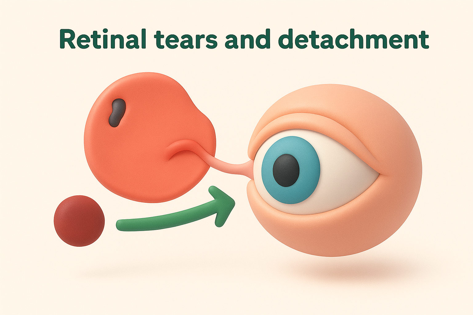
Detachment progresses differently for each person. Some experience a rapid spread across hours, while others see a gradual expansion over days. Your risk increases if you’re highly myopic, have had previous eye surgery or trauma, or have a family history of retinal problems. Prompt laser treatment can seal small tears before fluid spreads, usually completed in a single office visit. Once a detachment develops, surgical repair becomes necessary and outcomes depend heavily on how quickly you receive care.
When flashes accompany your peripheral dot
Flashing lights in your peripheral vision signal that something is physically tugging or stimulating your retina. These flashes often appear as brief sparks, lightning bolts or flickering arcs at the edge of your sight. When you see both a black dot and flashes together, the vitreous is likely pulling on your retina with enough force to risk creating a tear. The mechanical stimulation of photoreceptor cells produces these light sensations even in complete darkness.
Flashes linked to retinal problems differ from migraine-related visual disturbances. Retinal flashes typically occur in one eye only, last just seconds, and repeat irregularly throughout the day or night. They become more noticeable when you move your eyes quickly or in dim lighting. Migraine flashes, by contrast, usually affect both eyes, last 10 to 30 minutes, and often create zigzag patterns that shimmer across your central vision before fading completely.
If you notice both a peripheral black dot and flashing lights, contact an ophthalmologist immediately rather than waiting to see if symptoms resolve.
Red-flag symptoms requiring immediate care
You need urgent assessment if your peripheral black dot appears alongside any of these warning signs:
- A sudden shower of multiple new floaters, like pepper scattered across your vision
- A dark curtain, shadow or veil moving into your field of view from the side
- Rapid loss of peripheral vision, as though someone is closing a blind over the edge of your sight
- Persistent flashing lights that continue over hours or worsen over time
- A dramatic change in the size or darkness of an existing spot
These symptoms suggest active retinal damage that could progress to complete detachment within hours. Don’t wait for a routine appointment or assume symptoms will improve overnight. Present to an emergency eye clinic or contact your ophthalmologist’s after-hours service immediately. Early intervention offers the best chance of preserving your full visual field and preventing permanent blind spots in your peripheral vision.
How specialists diagnose a peripheral black dot
Diagnosing a black dot in peripheral vision starts with understanding exactly what you’ve experienced. Your eye specialist combines detailed questioning with careful examination techniques to distinguish harmless floaters from urgent retinal problems. This systematic approach ensures accurate diagnosis within a single appointment in most cases, though some conditions require follow-up imaging to confirm the underlying cause. The process typically takes 30 to 60 minutes and involves minimal discomfort.
Your consultation and symptom history
Your specialist begins by asking when you first noticed the black dot and whether it appeared suddenly or gradually over days or weeks. They’ll want to know if you’ve seen flashing lights, experienced any trauma to your eye, or noticed additional floaters appearing alongside the original spot. Questions about your medical history focus on conditions like diabetes, high blood pressure and any previous eye surgery or laser treatment. Details about your family’s eye health matter too, particularly if relatives have experienced retinal detachment or other inherited conditions.
Recording your symptoms accurately before your appointment saves valuable time during consultation. Note whether the dot moves when you shift your gaze, stays fixed in position, or disappears completely at times. Your specialist uses these details to narrow down possible causes before they even examine your eye.
Dilated fundus examination
Dilating your pupils with special eye drops allows your specialist to examine the entire retina, including the far periphery where tears and detachments often begin. The drops take 15 to 20 minutes to work fully, temporarily blurring your near vision and making you sensitive to bright light. Your specialist uses direct and indirect ophthalmoscopy to inspect the vitreous gel for floating opacities, check the retinal surface for tears or holes, and assess the optic nerve and macula for any damage. They may ask you to look in different directions, allowing them to view every corner of your peripheral retina thoroughly.
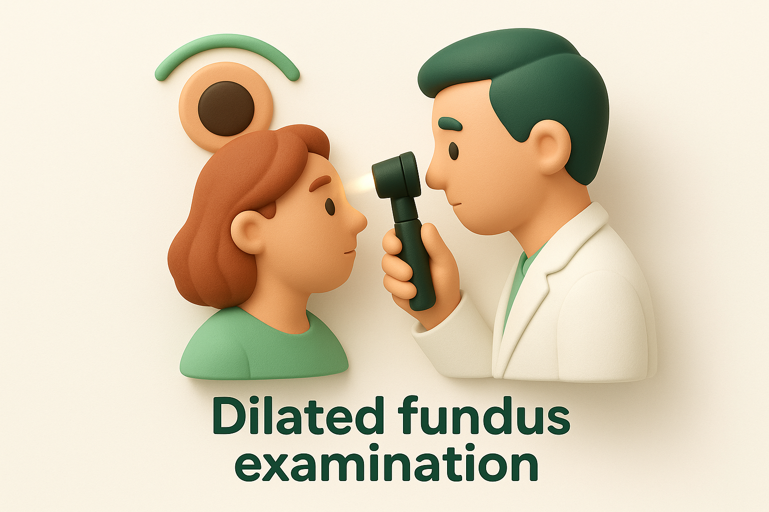
A dilated examination remains the gold standard for detecting retinal tears before they progress to detachment.
Advanced imaging when needed
Optical coherence tomography (OCT) provides detailed cross-sectional images of your retina when the specialist suspects macular involvement or subtle changes not visible during standard examination. Visual field testing maps any blind spots or areas of reduced sensitivity in your peripheral vision, helping quantify how much the black dot affects your functional sight. If dense cataracts or vitreous bleeding block the view of your retina, your specialist may order B-scan ultrasonography to visualise the posterior eye structures and rule out detachment or other serious conditions requiring urgent surgical intervention.
Treatment, risk factors and ongoing eye care
Treatment for a black dot in peripheral vision depends entirely on what’s causing it. Benign floaters from normal ageing typically require no intervention, as most people adapt within weeks and the spots become less noticeable over time. When a retinal tear is discovered early, your specialist can seal it with laser photocoagulation during a quick office procedure, preventing progression to detachment. More serious conditions like established detachments demand surgical repair, often involving vitrectomy (removal of the vitreous gel) or scleral buckling (placing a silicone band around the eye). Your ophthalmologist tailors the approach to your specific diagnosis, overall eye health and personal circumstances.
Treatment options based on cause
Laser photocoagulation works by creating tiny burns around a retinal tear, forming scar tissue that welds the retina back to the underlying eye wall. This outpatient procedure takes just minutes and prevents fluid from seeping through the tear. Vitrectomy surgery becomes necessary when floaters severely obstruct your vision or when a retinal detachment requires repair. During vitrectomy, your surgeon removes the cloudy vitreous gel and replaces it with saline solution, gas bubble or silicone oil. Recovery varies from days to weeks depending on the procedure’s complexity and whether positioning restrictions apply.
Conservative management suits most patients with simple floaters or posterior vitreous detachment. Your specialist monitors your condition through regular examinations, checking that spots remain stable and no new complications develop. Some patients find that moving their eyes quickly can shift troublesome floaters out of their direct line of sight temporarily, though this doesn’t eliminate the spots permanently.
Who faces higher risks
Age remains the primary risk factor for developing a black dot in peripheral vision, with most cases occurring after 50 as the vitreous naturally liquefies. High myopia (severe short-sightedness) significantly increases your risk because the elongated eyeball stretches the retina thinner at the periphery, making tears more likely. Previous eye surgery, particularly cataract removal, accelerates vitreous changes and raises the chance of posterior vitreous detachment occurring earlier than it might otherwise.
Family history matters too. If close relatives have experienced retinal detachment, your risk increases and warrants closer monitoring. Eye trauma, whether from sport, accidents or previous injury, creates structural weakness that can lead to peripheral vision problems years later.
Understanding your personal risk factors helps you and your specialist create an appropriate monitoring schedule.
Ongoing monitoring and prevention
Regular eye examinations form the cornerstone of long-term eye health, particularly if you’ve already experienced a black dot in your peripheral vision. Your specialist establishes a follow-up schedule based on your initial diagnosis, typically ranging from three months to yearly checks. Immediate contact remains essential if you notice sudden changes like new floaters, flashing lights or expanding shadows, even between scheduled appointments. Protecting your eyes during sport, managing diabetes and hypertension, and attending routine screenings all reduce your risk of serious peripheral vision problems developing.
Looking after your side vision
Protecting your peripheral vision starts with recognising that a black dot in peripheral vision deserves attention, not dismissal. Most spots prove harmless, but early assessment gives specialists the chance to catch serious conditions before they threaten your sight. Stay alert to sudden changes like flashing lights or curtain-like shadows, which signal urgent problems requiring immediate care. Schedule regular eye examinations based on your age and risk factors, and document any new spots carefully to help your specialist make an accurate diagnosis.
Your peripheral vision supports countless daily activities, from driving safely to navigating crowded spaces with confidence. If you’ve noticed a persistent black dot or other changes in your side vision, Bright Sight Eye Specialists offers comprehensive assessments and advanced treatments tailored to your needs. Dr Dubey and the team combine cutting-edge technology with patient-centred care to protect your vision for the long term.